ISSN Online: 2164-5396 ISSN Print: 2164-5388
DOI: 10.4236/ojbiphy.2018.83009 Jul. 3, 2018 104 Open Journal of Biophysics
The Quantum-Mechanical Sensitivity of Cell
Hydration in Mammals
Anna Nikoghosyan, Lilia Narinyan, Armenuhi Heqimyan, Sinerik Ayrapetyan
*Life Sciences International Postgraduate Educational Center, UNESCO Chair in Life Sciences, Yerevan, Armenia
Abstract
The elucidation of the mechanism on the biological effects of weak chemical and physical factors on cells and organism is one of the modern problems in life sciences. According to the Receptor Theory of Prof. Bernard Katz the im-pact of the biological substances on cells is realized through the activation of ligand-gated ion channels in the membrane. However, this theory doesn’t provide a satisfactory explanation on the similar biological effects of extremely low concentrations of different chemical substances, which are unable to acti-vate the ionic channels in the membrane and have non-linear dose-dependent effect on cells. Previously we have suggested that the metabolic control of cell hydration serves as a universal quantum-mechanical sensor for different weak physical and chemical signals. For supporting this hypothesis, in this article the comparative study of the effects of low concentrations of both cold (non-radioactive) and [3H]-ouabain (specific inhibitor for Na+/K+-ATPase) on
the hydration in different tissues of rats has been performed. The obtained data have shown that cold and [3H]-ouabain have different effects on cell
hy-dration and such a difference depends on the initial metabolic state of tissues. On the basis of our previous and present results it is suggested that such a quantum-mechanical sensitivity of cell hydration is realized through the cyc-lic-nucleotides-dependent Na+/Ca2+ exchange, having a crucial role in the
metabolic regulation of cell hydration.
Keywords
Rat, Hydration, Ouabain
1. Introduction
The elucidation of the mechanism on the biological effects of weak chemical and physical factors on cells and organism is one of the modern problems in life
How to cite this paper: Nikoghosyan, A., Narinyan, L., Heqimyan, A. and Ayrape-tyan, S. (2018) The Quantum-Mechanical Sensitivity of Cell Hydration in Mammals. Open Journal of Biophysics, 8, 104-116.
https://doi.org/10.4236/ojbiphy.2018.83009
Received: April 12, 2018 Accepted: June 30, 2018 Published: July 3, 2018
Copyright © 2018 by authors and Scientific Research Publishing Inc. This work is licensed under the Creative Commons Attribution International License (CC BY 4.0).
DOI: 10.4236/ojbiphy.2018.83009 105 Open Journal of Biophysics
sciences. At present, our knowledge on signal transduction in cells is based on the Membrane Theory which explains this transduction by the changes of cell membrane permeability for inorganic ions leading to generation of transient io-nic currents through cell membrane [1]. However, this theory based on classical thermodynamic approaches, cannot explain the biological effects of extremely low concentrations of chemical substances [2] [3] and weak physical signals [4] [5], which are unable to activate ionic channels in the membrane and have non-linear dose-dependent effect.
The main omission of the Membrane Theory is that it does not consider the direct role of cell metabolism in generation of cell membrane potential. Al-though the existence of the metabolic component of membrane potential in the living cells has been revealed in a number of studies [6] [7], there isn’t any relia-ble theory explaining the sensitivity of cells to weak physical and chemical sig-nals.
Based on our previous research data we have developed a new approach on quantum-mechanical sensitivity of living cells to different weak factors, which are realized through the metabolic control of cell hydration [8] [9] [10]. Ac-cording to this hypothesis a water molecule, having valence angle with quan-tum-mechanical sensitivity to different factors, serves as a primary messenger for signal transduction in cells. The metabolically generated water efflux from the cell balances the osmotic water uptake and has inhibitory effect on inward going ionic currents [11] [12] serving as a gate by which the weak signals mod-ulate cell metabolic activity. Therefore, the metabolic control of cell hydration has been suggested as a universal quantum-mechanical sensor through which the biological effects of extremely weak chemical and physical signals on cells and organisms are realized [9]
It is known that the Na+/K+ pump is a key mechanism through which the
me-tabolic control of cell hydration is realized. In excitable membranes three iso-forms (α1, α2, α3) of α catalytic subunit of Na+/K+-ATPase (working molecule for
Na+/K+ pump) are identified [13]. They are characterized by different affinities
to cardiac glycoside ouabain (specific inhibitor for Na+/K+-ATPase) as well as
functional roles: α1 (with low affinity) and α2 (with middle affinity) isoforms are
involved in transportation of Na+ and K+, while α
3 (with high affinity) is not
di-rectly involved in transporting Na+ and K+ and has only signaling function [14] [15]. Our previous studies have shown that these isoforms are extremely sensi-tive not only to ouabain but also to extremely low concentrations of other bio-logically active substances [2] [3] [16] as well as to weak intensity of electro-magnetic fields [17] [18]. From these data it is followed that the same chemical substances with different quantum-mechanical structures (e.g. non-radioactive ouabain and [3H]-ouabain) can have individual effects on cell hydration. It has
been shown that Na+/K+ pump is a key mechanism in regulation of cell
DOI: 10.4236/ojbiphy.2018.83009 106 Open Journal of Biophysics
in the presented article the age-dependent comparative study of the effects of pM (agonist for α3) and nM (agonist for α2) concentrations of cold (non-radioactive)
ouabain and [3H]-ouabain on the hydration of different tissues was performed.
2. Materials and Methods
2.1. Animals
All procedures performed on animals were carried out following the protocols approved by Animal Care and Use Committee of Life Sciences International Postgraduate Educational Centre (LSIPEC, Yerevan, Armenia).
The experiments were performed on young (6 weeks old) and old (18 months old) Wistar rats. They were regularly examined, kept under control of the vete-rinarians in LSIPEC and reserved in a specific pathogen-free animal room under optimum conditions of 12 h light/dark cycles, at temperature of 22˚C ± 2˚C, with a relative humidity of 50% and were fed ad libitum on a standard lab chow and water.
2.2. Chemicals
Tyrode’s Physiological solution (PS) containing (in mM) 137 NaCl, 5.4 KCl, 1.8 CaCl2, 1.05 MgCl2, 5 C6H12O6, 11.9 NaHCO3, and 0.42 NaH2PO4 and adjusted to
pH 7.4 with NaOH was used. All chemicals were obtained from “Medisar” In-dustrial Chemical Importation Company (Yerevan, Armenia). The [3H]-ouabain
with specific activity (25.34 Ci/mM) (PerkinElmer, Massachusetts, USA) and cold (non-radioactive) ouabain at pM (10−11 M) and nM (10−9 M) concentrations
dissolved in PS were used for tissue injection and incubation. All ouabain solu-tions were also adjusted to pH 7.4.
2.3. Experimental Design
It is well known that the anesthetics with different chemical and pharmacologi-cal profiles significantly affect metabolic processes, which play an important role in regulation of cell volume [20] [21]. Therefore, in the present experiments animals were sharply immobilized by freezing method (dipping their noses into liquid nitrogen for 3 - 5 sec) and decapitated [22]. After such a procedure the full absence of somatic reflexes on extra stimuli was recorded.
For in vivo experiments 15 young and 15 old animals were taken. Each animal group was divided into five subgroups (n = 3). The animals of the control group were injected with PS (according to the animal weight) and the animals of the next four subgroups were injected with 10−11 M and 10−9 M concentrations of
cold and 10−11 M and 10−9 M concentrations of [3H]-ouabain. After the 15th min
DOI: 10.4236/ojbiphy.2018.83009 107 Open Journal of Biophysics
For investigation of the water content variations and ouabain effect in in vitro
conditions 15 young and 15 old animals were taken. After their decapitation 5 samples of the above mentioned tissues were taken from each animal (similar to
in vivo experiments). These samples (n = 15) were incubated in PS (as sham), cold ouabain solution (10−11 M, 10−9 M) and [3H]-ouabain solution (10−11 M, 10−9
M) for 15 min, then washed in PS three times and weighed.
2.4. Definition of Water Content of Brain Tissues
The water content of brain cortex, heart muscle and liver tissues was determined by traditional “tissue drying” method [23]. After measuring the wet weight (w.w.) of tissue samples they were dried in oven (Factory of Medical Equipment, Odessa, Ukraine) for 24 h at 105˚C for determination of dry weight (d.w.). The quantity of water in 1 g of d.w. tissue was counted by the following equation: (w.w. − d.w.)/d.w.
2.5. Counting of [
3H]-Ouabain Receptors in the Membrane
The tissue samples from in vivo experiments, which were subjected to [3H]-ouabain, were homogenized in 50 µl of 68% HNO
3 solution after
determi-nation of wet and dry weights. Then 2 ml of Bray’s scintillation fluid was added and chemoluminescence of samples was quantified with 1450-MicroBeta liquid scintillation counter (Wallac, Turku, Finland). The number of [3H]-ouabain
molecules’ binding with cell membranes was defined per mg of dry weight of samples.
The same procedure (the definition of the number of [3H]-ouabain molecules)
was performed on the tissue samples from in vitro experiments after removing them from the oven and determining wet and dry weights.
2.6. Statistical Analysis
Microsoft Excel and Sigma-Plot (Version 8.02A, NY, USA) were used for data analyses. The statistical significance in comparison with the sham group was calculated with Student’s t-test with the following symbols (*p < 0.05; **p < 0.01; ***p < 0.001).
3. Results
3.1. Investigation of Brain Cortex Tissue Hydration
The comparative study of the hydration sensitivity of excitable (brain cortex, heart muscle) and non-excitable (liver) tissues to pM and nM concentrations of cold and [3H]-ouabain in in vivo and in in vitro experimental conditions was
performed. In in vivo experiments the animals were preliminarily i/p injected with ouabain-free PS and 10−11 M and 10−9 M concentrations of cold and
[3H]-ouabain, while in in vitro experiments the tissue samples were incubated in
the same solutions for 15 min.
DOI: 10.4236/ojbiphy.2018.83009 108 Open Journal of Biophysics
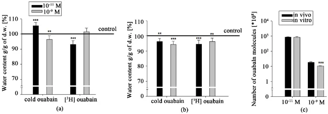
Figure 1. The water contents in cortex tissue of young rats in in vivo (a) and in in vitro
(b) experiments upon the effect of pM and nM concentrations of cold and [3H]-ouabain.
The black and gray bars on (a) and (b) indicate the mean value of water contents in the tissues upon the effect of pM and nM ouabain, respectively. The continuous line shows the control value of tissue hydration after PS injection (a) and PS incubation (b). The black and gray bars on (c) show the number of ouabain molecules binding with cell membrane in in vivo and in in vitro conditions, respectively. Each bar represents the ±SEM (n = 45). The symbols (*) and (***) indicate p < 0.05 and p < 0.001, respectively. All data were obtained from three independent experiments.
insignificant dehydration in brain cortex tissues of young animals was observed compared with the control (injected with ouabain-free PS), while 10−9 M cold
ouabain brought to significant hydration. The data on [3H]-ouabain i/p injection
(Figure 1(a)) revealed the opposite effect on brain tissue hydration, i.e.
signifi-cant hydration at 10−11 M and expressed dehydration at 10−9 M [3H]-ouabain.
In in vitro experiments (Figure 1(b)), where the metabolic activity of tissues was impaired, brain tissue hydration sensitivity to ouabain had a reverse charac-ter compared with those in vivo studies (Figure 1(a)): pM cold ouabain had hy-dration effect, while pM [3H]-ouabain had dehydration effect. In case of nM cold
ouabain there was no effect but nM [3H]-ouabain had hydration effect on the
tissues. Figure 1(c) illustrated that at each concentration of [3H]-ouabain the
number of ouabain molecules was higher in in vivo experiment compared with
in vitro one.
According to the fact that aging leads to the depression of the metabolic activ-ity, in the next series of experiments we repeated the above-mentioned protocol on old animals. As can be seen on Figure 2(a), the hydration level in brain cor-tex tissue of old animals in in vivo experiments upon the impact of 10−11 M cold
ouabain was significantly higher compared with the control, while the same concentration of [3H]-ouabain sharply dehydrated the tissues. In case of 10−9 M
cold ouabain the dehydration effect was observed, while the same concentration of [3H]-ouabain had slight hydration effect on the tissues compared with the
control.
In in vitro experiments (Figure 2(b)) the hydration levels of brain cortex samples at all concentrations of ouabain were nearly the same. Compared with the control data at both concentrations of cold and [3H]-ouabain the
DOI: 10.4236/ojbiphy.2018.83009 109 Open Journal of Biophysics
Figure 2. The water contents in cortex tissue of old rats in in vivo (a) and in in vitro (b)
experiments upon the effect of pM and nM concentrations of cold and [3H]-ouabain. The
black and gray bars on (a) and (b) indicate the mean value of water contents in tissues upon the effect of pM and nM ouabain, respectively. The continuous line shows the con-trol value of tissue hydration after PS injection (a) and PS incubation (b). The black and gray bars on (c) show the number of ouabain molecules binding with cell membrane in in vivo and in in vitro conditions, respectively. Each bar represents the ±SEM (n = 45). The symbols (**) and (***) indicate p < 0.01 and p < 0.001, respectively. All data were ob-tained from three independent experiments.
molecules at 10−9 M concentration of [3H]-ouabain was much higher in in vivo
experiment than in in vitro one.
3.2. Investigation of Heart Muscle Tissue
Considering the fact that unlike brain tissue hydration heart muscle hydration significantly depends on myosin contraction, in the next series of experiments the above mentioned protocol was performed on heart muscle tissues.The data of in vivo experiments presented on Figure 3(a) showed that the pM cold oua-bain had significant dehydration effect on heart muscle tissue of young rats, while the same concentration of [3H]-ouabain had hydration effect.
The effects of both nM cold ouabain and [3H]-ouabain were similar to the
ef-fects of pM ouabain, but nM ouabain effect was less pronounced. In in vitro ex-periments the pM cold ouabain had slight hydration effect, while the same con-centration of [3H]-ouabain had expressed hydration effect on heart muscle
tis-sue. The nM cold ouabain had more pronounced hydration effect than pM oua-bain, while nM [3H]-ouabain had less expressed hydration effect than pM
[3H]-ouabain (Figure 3(b)). As can be seen from Figure 3(c), the number of
ouabain molecules binding with cell membrane in in vivo and in in vitro expe-riments was the same.
The investigation of heart muscle tissues in old rats in in vivo condition
(Figure 4(a)) showed significant over hydration at both concentrations (10−11 M
and 10−9 M) and types of ouabain. However at 10−9 M [3H]-ouabain the
hydra-tion level was higher.
As can be seen from Figure 4(b), 10−11 M cold ouabain in in vitro experiments
led to significant dehydration in old animals, while 10−11 M [3H]-ouabain to
hy-dration. As for the effects of 10−9 M ouabain the hydration reached to the level
DOI: 10.4236/ojbiphy.2018.83009 110 Open Journal of Biophysics
Figure 3. The water contents in heart muscle tissue of young rats in in vivo (a) and in
vi-tro (b) experiments upon the effect of pM and nM concentrations of cold and [3H]-ouabain.
The black and gray bars on (a) and (b) indicate the mean value of water contents in tis-sues upon the effect of pM and nM ouabain, respectively. The continuous line shows the control value of tissue hydration after PS injection (a) and PS incubation (b). The black and gray bars on (c) show the number of ouabain molecules binding with cell membrane in in vivo and in in vitro conditions, respectively. Each bar represents the ±SEM (n = 45). The symbols (*) and (**) indicate p < 0.05 and p < 0.01, respectively. All data were ob-tained from three independent experiments.
Figure 4. The water contents in heart muscle tissue of old rats in in vivo (a) and in in
vi-tro (b) experiments upon the effect of pM and nM concentrations of cold and [3H]-ouabain. The black and gray bars on (a) and (b) indicate the mean value of water
contents in tissues upon the effect of pM and nM ouabain, respectively. The continuous line shows the control value of tissue hydration after PS injection (a) and PS incubation (b). The black and gray bars on (c) show the number of ouabain molecules binding with cell membrane in in vivo and in in vitro conditions, respectively. Each bar represents the ±SEM (n = 45). The symbols (*) and (***) indicate p < 0.05 and p < 0.001, respectively. All data were obtained from three independent experiments.
However, in spite of different effects on tissue hydration in in vivo and in in vi-tro conditions the number of ouabain molecules at [3H]-ouabain was
approx-imately the same at two different ouabain concentrations (Figure 4(c)).
3.3. Investigation of Liver Tissue
It is known that in soft tissues of healthy animals only a1 isoform of
Na+/K+-ATPase with low affinity to ouabain is expressed [24]. At the same time
our previous study has shown that nM and pM ouabain concentrations activate cyclic nucleotides-dependent Na+/Ca2+ exchange without any effect on Na+/K+
[image:7.595.213.533.307.411.2]DOI: 10.4236/ojbiphy.2018.83009 111 Open Journal of Biophysics
Although in liver cell membrane a2/a3 isoforms of Na+/K+-ATPase were not
expressed the data presented on Figure 5 indicated the modulation effect of pM and nM ouabain on its tissue hydration. Moreover, the modulation effects of low concentrations of cold and [3H]-ouabain on liver tissue hydration were different
compared with the effects on brain cortex and heart muscle tissues.
In in vivo experiments (Figure 5(a)) cold ouabain led to dose-dependent hy-dration, while [3H]-ouabain brought to dehydration. The significant hydration at
10−9 M cold ouabain turned to dehydration at 10−9 M [3H]-ouabain. In in vitro
experiments (Figure 5(b)) the incubation of liver samples at 10−11 M cold
oua-bain brought to more pronounced dehydration, while 10−11 M [3H]-ouabain had
weak dehydration effect on it. Cold ouabain at 10−9 M had no effect on tissue
hydration, while [3H]-ouabain at the same concentration had dehydration effect
on it. It is worth to note that the ouabain binding with cell membrane at both concentrations was higher in in vivo state than in in vitro one. In old animals the effects of both cold and [3H]-ouabain i/p injections on liver tissue hydration
were more pronounced than in young animals (Figure 6(a)). It is interesting to note that there was no difference between cold and isotope ouabain effects on cell hydration, while 10−9 M cold ouabain-induced hydration was more
ex-pressed than 10−9 M [3H]-ouabain-induced effect.
The incubation of the samples in cold ouabain solutions led to significant de-hydration at 10−11 M, while at the same concentration of [3H]-ouabain had
hy-dration effect (Figure 6(b)). Both cold and [3H]-ouabain at 10−9 M
concentra-tion had hydraconcentra-tion effect on tissue but [3H]-ouabain effect was less pronounced
[image:8.595.209.535.496.605.2]than the effect of the cold ouabain. It is interesting to note that the ouabain binding with cell membrane in in vivo experiment had dose-dependent increas-ing character, while in in vitro experiment it had dose-dependent weakening character (Figure 6(c)).
Figure 5. The water contents in liver tissue of young rats in in vivo (a) and in in vitro (b)
experiments upon the effect of pM and nM concentrations of cold and [3H]-ouabain. The
DOI: 10.4236/ojbiphy.2018.83009 112 Open Journal of Biophysics
Figure 6. The water contents in liver tissue of old rats in in vivo (a) and in in vitro (b)
experiments upon the effect of pM and nM concentrations of cold and [3H]-ouabain. The
black and gray bars on (a) and (b) indicate the mean value of water contents in tissues upon the effect of pM and nM ouabain, respectively. The continuous line shows the con-trol value of tissue hydration after PS injection (a) and PS incubation (b). The black and gray bars on (c) show the number of ouabain molecules binding with cell membrane in in vivo and in in vitro conditions, respectively. Each bar represents the ±SEM (n = 45). The symbols (*) and (***) indicate p < 0.05 and p < 0.001, respectively. All data were obtained from three independent experiments.
4. Discussion
Previously it was shown that in brain and heart tissues of rats the pM [3H]-ouabain stimulated the cGMP-dependent Na+/Ca2+ exchange in forward (F)
mode [27] which was accompanied by cell dehydration in young and hydration
in old animals [26]. The pM [3H]-ouabain-induced cell dehydration in young
animals was explained by the activation of Na+/K+ pump as a result of Na+/Ca2+
exchange-induced decrease of intracellular contents of [Ca2+]
i, while its
hydra-tion effect on the tissues of old animal was explained by the direct effect of F Na+/Ca2+ exchange because of high [Ca2+]i [10]. The nM [3H]-ouabain activated
the cAMP-dependent Na+/Ca2+ exchange in reverse mode (R) [28], having
age-dependent weakening character which was reversed in old animals [18]. It is worth to note that though [3H]-ouabain has been used in biological
experi-ments for 5 decades [29] [30] the comparative studies of cold and [3H]-ouabain
effects on cell metabolism have not been considered. As the metabolic control of cell hydration has quantum-mechanical sensitivity [10] it becomes possible to estimate the different effects of ouabain molecules with different quan-tum-mechanical structure, such as cold and [3H]-ouabain, on cell hydration. The
data presented in this article clearly indicate that the biological effects of cold and [3H]-ouabain are not identical.
The data presented on Figure 1(a) showed that the 10−11 M and 10−9 M cold
ouabain, as was noted above (Figure 1(a)), had dehydration and hydration ef-fects on brain tissuesin in vivo experiments, respectively, while their effects were reversed in in vitro experiments (Figure 1(b)), where the metabolic state of the slices was depressed. The pM and nM concentrations of [3H]-ouabain injection
had opposite effects on brain tissue hydration compared with cold ouabain in-jection. Moreover, the reverse of dose-dependent effect of [3H]-ouabain on
DOI: 10.4236/ojbiphy.2018.83009 113 Open Journal of Biophysics
Previously it has been shown that [3H]-ouabain binding with cell membrane
depends on both the number of ouabain receptors and their affinity: the number of receptors increases with cell swelling [30], while ouabain receptors affinity in-creases with the decrease of [Ca2+]
i [19]. The data obtained in in vivo experiment
indicating that the number of ouabain molecules at pM concentration was high-er than at nM ouabain (Figure 1(c)) can be explained by the pM ouabain-induced activation of F Na+/Ca2+ exchange leading to the decrease of [Ca2+]
i and increase
of receptors affinity to ouabain, while the decrease of ouabain binding with cell membrane in in vitro experiments can be a result of [Ca2+]
i increase.
The received data allow us to suggest that different effects of pM and nM ou-abain as well as the difference in the effects of radioactive and cold ouou-abain is due to the initial level of [Ca2+]
i. This suggestion is consistent with the data of the
same protocol of experiments performed on old rats, when the initial level of [Ca2+]
i in brain tissues was higher than in young animals [31].
The data obtained in old animals showed that in vivo experiment the pM cold ouabain led to hydration, while [3H]-ouabain had strong dehydration effect on
brain cortex tissues (Figure 2(a)). In case of the nM ouabain i/p injection we recorded opposite data: cold and [3H]-ouabain had dehydration and slight
hy-dration effect on tissue, respectively. Moreover, in in vitro experiments, in which the tissue samples had high Ca2+ contents compared with that in in vivo
experi-ments, both concentrations of cold and [3H]-ouabain ouabain had dehydration
effect on tissues. The differences between the effects of pM and nM as well as between cold and isotope ouabain were inconsistent (Figure 2(b)).
The data on dose-dependent binding with membrane in the cortex tissue of old animals indicated that there was dose-dependent increase of ouabain binding
(Figure 2(c)). These age-dependent differences can be explained by the fact that
in old animals [Ca2+]
i is higher. As a result of this, the activation of
cGMP-dependent F Na+/Ca2+ exchange had its direct hydration effect on cells [18].
Thus, the obtained data indicating that the same concentrations of cold and [3H]-ouabain had different effects on cortex tissue hydration. The fact that the
effect of pM [3H]-ouabain was approximately the same as in case of cold nM
ouabain clearly indicates that the existence of [3H] in ouabain molecules
in-creases its effect on cell hydration. This suggestion is supported by the obtained data of the similar study on heart muscle tissues.
It is known that approximately 50% of cardiomyocyte volume consists of myofibrils [32] and [Ca2+]
i-dependent contractility of the latter has a
determin-ing role in muscle tissues hydration. The data presented on Figure 3 and Figure 4 indicate that as in case of cortex tissue (Figure 1 and Figure 2) the cold and [3H]-ouabain at the same concentrations had different effects on heart tissue
hy-dration. The i/p injection of cold ouabain in young animals had dose-dependent dehydration effects on muscle compared with control, while [3H]-ouabain had
dose-dependent weakening effect on hydration in heart muscle tissue (Figure 3(a)). The data that in heart muscle tissues of old animals (containing higher [Ca2+]
DOI: 10.4236/ojbiphy.2018.83009 114 Open Journal of Biophysics
the same dose-dependent hydration effect on muscle (Figure 4(a)) indicate that their effects on cell hydration is realized by different mechanisms controlling [Ca2+]
i. The data on the difference between cold and [3H]-ouabain effects
ap-peared in heart muscle samples incubated in in vitro experiments seem ex-tremely interesting. The facts that in in vitro experiments where [Ca2+]
i is
con-sidered to be higher than in in vivo experiments and the increase of [Ca2+] i
ac-tivates cGMP-dependent F Na+/Ca2+ exchange through the activation of
Ca2+-calmodulin-induced NO production, which in its turn stimulates cGMP
formation, allow us to suggest that the modulation effect of [3H]-ouabain on cell
hydration is realized through this chain. However, to prove this suggestion more detailed investigation is needed.
It is known that all three isoforms of Na+/K+-ATPase are expressed in
excita-ble tissues (nerve and muscle membrane), while in soft tissues of healthy animals only a1 isoform is expressed [24]. At the same time by our previous experiments
performed on snail neurons [28] [30], heart muscles and brain tissues of rats
[18] it was shown that pM and nM ouabain-induced modulation of Na+/Ca2+
exchange did not depend on Na+/K+ pump and it was realized through the
changes of intracellular nucleotides [17] [18].
The data presented on Figure 5(a) indicate that after pM and nM cold oua-bain injections of young animals the liver tissue hydration increased by dose-dependent manner, while the same concentrations of [3H]-ouabain
injec-tions led to dehydration effect. These data support the above suggestion that cold and isotope had different biological effects on cells. It is worth to note that as in case of excitable tissues, the sensitivity of liver tissue hydration to low con-centrations of both cold and [3H]-ouabain was higher in old animals as well as in
the samples incubated in in vitro experiment, when the metabolic activity of tis-sues was depressed. Therefore, the data that such low concentrations of non-radioactive and radioactive ouabain effects depend on [Ca2+]
i and the data
that [3H]-ouabain binding with cell membrane is depressed in in vitro
experi-ments allow us to suggest that the modulation of tissue hydration at both con-centrations of cold and [3H]-ouabain takes place through the changes of [Ca2+]
i.
Thus, the obtained data bring us to the following conclusions:
1) Cold and [3H]-ouabain at pM and nM concentrations have different effects
on cell hydration;
2) The different effects of cold and [3H]-ouabain on cell hydration is due to their
different activities on cGMP-dependent Na+/Ca2+ exchange controlling intracellular
Ca2+ concentration. Therefore, these data allow us to conclude that in biological
ex-periments cold ouabain and radioactive ouabain effects cannot be considered as equivalent. These data indicate that Na+/Ca2+ exchange is Na+/K+ pump-independent
mechanism in the membrane having quantum-mechanical sensitivity.
References
DOI: 10.4236/ojbiphy.2018.83009 115 Open Journal of Biophysics
1148-54. https://doi.org/10.1126/science.145.3637.1148
[2] Ayrapetyan, S.N. and Carpenter, D.O. (1991) Very Low Concentrations of Acetyl-choline and GABA Modulate Transmitter Responses. Neuroreport, 2, 563-565. https://doi.org/10.1097/00001756-199110000-00002
[3] Ayrapetyan, S.N. and Carpenter, D.O. (1991b) The Modulatory Effect of Extremely Low Doses of Mediators on Functional Activity of the Neuronal Membrane. Zhur-nal Evoliutsionnoi Biokhimii, 27, 146-151. (In Russian)
[4] Devyatkov, N.D. (1973) Influence of Millimeter-Band Electromagnetic Radiation on Biological Objects. Uspekhi Fizicheskih Nauk, 110, 453-454. (In Russian)
https://doi.org/10.3367/UFNr.0110.197307l.0453
[5] Adey, W.R. (1981) Tissue Interactions with Non Ionizing Electromagnetic Fields.
Physiological Reviews, 61, 435-514. https://doi.org/10.1152/physrev.1981.61.2.435 [6] Ayrapetyan, S.N. (1969) Metabolically Dependent Fraction of Membrane Potential
and Electrode Properties of the Membrane of Giant Neurons in Mollusks. Biofizika, 14, 1027-1031. (In Russian)
[7] Thomas, R.C. (1972) Electrogenic Sodium Pump in Nerve and Muscle Cells. Physi-ological Reviews, 52, 563-594. https://doi.org/10.1152/physrev.1972.52.3.563 [8] Ayrapetyan, S.N., Nasarenko, S.A. and Sorokhina, Z.A. (1971) Dependence of
Ac-tive Ion Transport in Snail Neurons on the Ionic Composition of Extracellular Me-dium. Biofizika, 16, 1037-1042. (In Russian)
[9] Ayrapetyan, S. (2015) The Dysfunction of Metabolic Controlling of Cell Hydration Precedes Warburg Phenomenon in Carcinogenesis. Journal of Bioequivalence & Bioavailability, 7, 59. https://doi.org/10.4172/jbb.10000e59
[10] Ayrapetyan, S.N. (2016) The nM Ouabain-Induced Tissue Dehydration as a Novel Diagnostic Marker for Neuronal Pathology. Global Drugs and Therapeutics, 2, 1-2. [11] Ayrapetyan, S.N., Rychkov, G.Y. and Suleymanyan, M.A. (1988) Effects of Water
Flow on Transmembrane Ionic Currents in Neurons of Helix Pomatia and in Squid Giant Axons. Comparative Biochemistry and Physiology, 89, 179-186.
https://doi.org/10.1016/0300-9629(88)91076-6
[12] Suleymanian, M.A., Ayrapetyan, V.Y., Arakelyan, V.B., et al. (1993) The Effect of Osmotic Gradient on the Outward Potassium Current in Dialyzed Neurons of Helix Pomatia. Cellular and Molecular Neurobiology, 13, 183-190.
https://doi.org/10.1007/BF00735374
[13] Juhaszova, M. and Blaustein, M. (1997) Na+ Pump with Low and High Ouabain
Af-finity Apha Subunit Isoforms Are Differently Distributed in Cells. Proceedings of the National Academy of Sciences, 94, 1800-1805.
https://doi.org/10.1073/pnas.94.5.1800
[14] Xie, Z. and Askari, A. (2002) Na+/K+-ATPase as a Signal Transducer. European
Journal of Biochemistry, 269, 2434-2439. https://doi.org/10.1046/j.1432-1033.2002.02910.x
[15] Liu, J., Tian, J., Haas, M., Shapiro, J., Askari, A. and Xie, Z. (2000) Ouabain Interac-tion with Cardiac Na+/K+-ATPase Initiates Signal Cascades Independent of Changes
in Intracellular Na+ and Ca2+ Concentrations. The Journal of Biological Chemistry,
275, 27838-27844.
DOI: 10.4236/ojbiphy.2018.83009 116 Open Journal of Biophysics
[17] Narinyan, L., Ayrapetyan, G. and Ayrapetyan, S. (2013) Age-Dependent Magneto-sensitivity of Heart Muscle Ouabain Receptors. Bioelectromagnetics, 34, 312-322. https://doi.org/10.1002/bem.21769
[18] Heqimyan, A., Narinyan, L., Nikoghosyan, A. and Ayrapetyan, S. (2015) Age-Dependent Magnetic Sensitivity of Brain and Heart Muscles. In: Markov, M., Ed., Electromag-netic Fields in Biology and Medicine, CRC Press, Boca Raton, 217-230.
https://doi.org/10.1201/b18148-15
[19] Heqimyan, A., Narinyan, L., Nikoghosyan, A., et al. (2012) Age Dependency of High Affinity Ouabain Receptors and Their Magnetosensitivity. The Environmen-talist, 32, 228-235.https://doi.org/10.1007/s10669-011-9383-0
[20] Krnjevic, K. (1992) Cellular and Synaptic Actions of General Anaesthetics. General Pharmacology, 23, 965-975.https://doi.org/10.1016/0306-3623(92)90274-N
[21] Heqimyan, A., Deghoyan, A. and Ayrapetyan, S. (2011) Ketamine-Induced Cell Dehydration as a Mechanism of Its Analgesic and Anesthetic Effects. Journal of In-ternational Dental and Medical Research, 4, 42-49.
[22] Takahashi, R. and Aprison, M. (1964) Acetylcholine Content of Discrete Areas of the Brain Obtained by a Near-Freezing Method. Journal of Neurochemistry, 11, 887-892.https://doi.org/10.1111/j.1471-4159.1964.tb06740.x
[23] Adrian, R.H. (1956) The Effect of Internal and External Potassium Concentration on the Membrane Potential of Frog Muscle. Journal of Physiology, 133, 631-658. https://doi.org/10.1113/jphysiol.1956.sp005615
[24] Blaustein, M.P. and Lederer, W.J. (1999) Na+/Ca2+ Exchange. Its Physiological
Im-plications. Physiological Reviews, 79, 763-854. https://doi.org/10.1152/physrev.1999.79.3.763
[25] Ayrapetyan, S.N., Suleymanyan, M.A., Saghyan, A.A. and Dadalyan, S.S. (1984) Autoregulation of Electrogenic Sodium Pump. Cellular and Molecular Neurobiolo-gy, 4, 367-383.https://doi.org/10.1007/BF00733598
[26] Ayrapetyan, S., Heqimyan, A. and Nikoghosyan, A. (2012) Age-Dependent Brain Tissue Hydration, Ca Exchange and Their Dose-Dependent Ouabain Sensitivity.
Bioequivalence & Bioavailability, 4, 60-68.https://doi.org/10.4172/jbb.10000114 [27] Ayrapetyan, S. (2012) The Dysfunction of Metabolic Controlling Cell Hydration Is a
Primary Mechanism for Generation of Aging-Related Nerve Disorders. Bioequiva-lence & Bioavailability, 4, 9-12.https://doi.org/10.4172/jbb.10000e15
[28] Sagian, A.A., Ayrapetyan, S.N. and Carpenter, D.O. (1996) Low Concentrations of Ouabain Stimulate Na:Ca Exchange in Neurons. Cellular and Molecular Neurobi-ology, 16, 489-498.https://doi.org/10.1007/BF02150229
[29] Baker, P.F. and Willis, J.S. (1970) Potassium Ions and the Binding of Cardiac Gly-cosides to Mammalian Cells. Nature, 226, 521-523.
https://doi.org/10.1038/226521a0
[30] Ayrapetyan, S.N., Suleymanyan, M.A., Saghyan, A.A. and Dadalyan, S.S. (1984) Autoregulation of the Selectrogenic Na Pump. Cellular and Molecular Neurobiolo-gy, 4, 367-384.https://doi.org/10.1007/BF00733598
[31] Khachaturian, Z.S. (1989) The Role of Calcium Regulation in Brain Aging: Reex-amination of a Hypothesis. Aging, 1, 17-34.
[32] Nakano, S., Muramatsu, T., Nishimura, S. and Senbonmatsu, T. (2012) Cardiomyo-cyte and Heart Failure. In: Sugi, H., Ed., Current Basic and Pathological Approaches to the Function of Muscle Cells and Tissues, Intech, London, 161-182.