ISSN Online: 2325-7083 ISSN Print: 2325-7075
DOI: 10.4236/crcm.2018.74024 Apr. 18, 2018 259 Case Reports in Clinical Medicine
Cervical Cytology: A Review of 597 Cases in a
Tertiary Health Centre in Nigeria
S. Singh
1*, D. C. Nnadi
1, R. F. Anas
1, I. G. Ango
1, A. G. Umar
1, U. Mohammed
2, Y. K. Mohammed
11Department of Obstetrics and Gynaecology, Usmanu Danfodiyo University Teaching Hospital, Sokoto, Nigeria 2Department of Histopathology, Usmanu Danfodiyo University Teaching Hospital, Sokoto, Nigeria
Abstract
Background: Cervical cancer is the most common gynaecological cancer among women in the developing countries. It is preventable by early detection and treatment of the precursor lesions. Cervical cytology became the standard screening test for cervical cancer and premalignant lesions with the introduc-tion of the Papanicolau (Pap) smear. Aim: to determine the uptake, indica-tions and results of cervical cytology in a tertiary health center in Nigeria. Materials and Methods: a 5-year descriptive study of the results of cervical cytology among women who attended the gynaecological outpatient clinic of the Hospital. Results: a total of 597 cases of cervical Pap smears were received at the histopathology department during the study period out of which 20 (3.4%) were inadequate for cytological evaluation. The patients age ranged from 15 to 80 (mean 38.8 SD = 11.4) years. The average annual uptake of the Cervical smear was 9.3% of the total number of patients seen in the gynaeco-logical clinic during the study period and the main indications were routine medical check-up, 256 (42.9%) and cervicitis 76 (12.7%). About 398 (66.7%) were normal smears while 152 (26.3%) were abnormal. Non-specific inflam-matory changes were present in 113 (74.3%) cases. Cytology was positive for intraepithelial lesions in 6.8% (39/577) cases. Eighteen percent (7/39) of them were high-grade lesions while squamous cell carcinoma was detected in 12.8% (5/39) cases. Conclusion: the uptake of cervical cytology of 8.7% is low and the main indication was routine check up. Premalignant lesions were detected in 6.8% of cases.
Keywords
Cervical Cytology, Cervical Cancer, Nigeria
1. Introduction
Cervical cancer is the main cancer among women in sub-Saharan Africa, India, How to cite this paper: Singh, S., Nnadi,
D.C., Anas, R.F., Ango, I.G., Umar, A.G., Mohammed, U. and Mohammed, Y.K. (2018) Cervical Cytology: A Review of 597 Cases in a Tertiary Health Centre in Nige-ria. Case Reports in Clinical Medicine, 7, 259-268.
https://doi.org/10.4236/crcm.2018.74024
Received: February 18, 2018 Accepted: April 15, 2018 Published: April 18, 2018
Copyright © 2018 by authors and Scientific Research Publishing Inc. This work is licensed under the Creative Commons Attribution International License (CC BY 4.0).
http://creativecommons.org/licenses/by/4.0/
DOI: 10.4236/crcm.2018.74024 260 Case Reports in Clinical Medicine and other parts of the developing world [1]. India accounts for a quarter of world’s burden of the disease [1]. The country accounted for 23.2% (123,000) of new global cervical cancer cases and 25.1% (67,000) of cervical cancer related deaths [2]. Cervical cancer is a leading cause of cancer related death in women in most developing countries of the world and has over 500,000 new cases and more than 300,000 deaths globally each year [3]. It is the second leading cause of cancer-related deaths among women in Jamaica [4]. In Nigeria, cervical cancer accounted for 67.8% and 77% of genital tract malignancies in Sokoto and Zaria respectively [5] [6]. Most of the patients present with advanced stages of the dis-ease when treatment for cure is not feasible. This ugly scenario is due to ignor-ance, poor health seeking behaviour, presence of risk factors such as sexual promiscuity, polygamy, early sexual debut and large number of pregnancies [3] [5]. Similarly, there is no organized screening practice to detect the early forms of the disease. According to the World Health Organization (WHO), the highest burden of cervical cancer occurs in developing nations where there is a lack of effective screening programs and low uptake of Pap smear or pelvic examination [7].
pop-DOI: 10.4236/crcm.2018.74024 261 Case Reports in Clinical Medicine ulation based. Similarly, the uptake for the Pap test is low even when it is availa-ble [13]. Despite these limitations; the conventional Pap smear is still the most widely available screening method for cervical cancer especially in low resource settings. The liquid-based cytology (LBC) aims to minimize errors of omission and commission inherent in the conventional Pap smear. It is now the standard test used by the National Health System (NHS) of the United Kingdom for cer-vical screening program [11]. However it is yet to take root in many low re-source settings. Recently, HPV DNA testing has been recommended by the WHO as a standard screening method for cervical cancer. This is based on the fact that HPV DNA is found in over 95% of cervical cancers and precursor le-sions worldwide [8]. This non-cytological method of screening is not readily available in most centres in Nigeria. Despite the high prevalence of cervical can-cer in Nigeria, there is no organized national HPV vaccine immunization pro-gramme to provide for primary prevention of the disease. We present the result of a 5-year screening programme for cervical cancer by the conventional Pap smear in tertiary health institution in Nigeria.
2. Aims and Objectives
The study aimed to determine the indications, uptake and the results of cervical cytology in a tertiary health institution in Nigeria
3. Subjects and Methods
This is a review of all cases of cervical smears that were assessed at the Histopa-thology department of the hospital over a 5-year period (1st of January 2011 to 31st of December 2015). The smears were from women attending the gynaeco-logical outpatient department and in whom pap was advised by the treating gy-naecologist for clinical indications. The hospital is a tertiary health institution located in the region of Nigeria. It has 886 bed spaces and provides tertiary and secondary health care services to neighbouring states. It also runs residency training program for doctors in the various sub-specialists including Surgery, Obstetrics & Gynaecology, Internal Medicine, Paediatrics, Histopathology among others.
col-DOI: 10.4236/crcm.2018.74024 262 Case Reports in Clinical Medicine lected data was performed using the SPSS IBM version 20 (IBM version 20.0. Armouk, NY: IBM Corp.). The results were expressed in frequencies, means, percentages, tables, figures and charts. The Bethesda system TBS (2004) with standardized terminologies for reporting cervical cytology was adopted in this study.
4. Results
[image:4.595.210.539.405.519.2]A total of 597 cases of cervical smears were received in the histopathology labor-atory during the period of study. About 3.4% (20/597) were inadequate or unsa-tisfactory for cytological evaluation according to the Bethesda criteria and were thus discarded. The rest 577 smears were analyzed and thus constituted the study population. The age of the patients ranged from 15 to 80 years with a mean of 38.82 [SD = 11.42] years. The annual uptake of the Pap smear shows an increasing trend as shown in Table 1. The modal year was in 2013. The average annual uptake of the conventional Pap smear was 9.28% as shown in Table 1. Cytology was performed mostly for routine indications in 42.9% (256/597), cer-vicitis 12.7% (76/597), abnormal uterine bleeding (AUB) in 12.1% (72/597), suspected cervical cancer 7.7% (46/597) while the least indications were post menopausal bleeding (PMB) 3.2% and utero-vaginal prolapse 2.8% (17/597) re-spectively as shown in Table 2. Thus in about 57.1% (341/597) of cases, the
Table 1. Annual uptake of cervical cytology in relation to overall patient load.
Year Total no. patients registered in Gynaecological clinic Number of cervical smears Annual %
2011 1771 18 1.0
2012 1439 124 8.6
2013 1364 190 13.9
2014 1140 115 10.1
2015 1172 150 12.8
Total 6886 597 9.3
Table 2. Indications for cervical smear.
Indications for Pap smear Frequency Percentage
Routine 256 42.9
Cervicitis 76 12.7
AUB 72 12.1
PCB 47 7.9
Suspected cervical cancer 46 7.7
Inflammatory 33 5.5
Uterine polyps 31 5.2
PMB 19 3.2
UV prolapse 17 2.8
[image:4.595.211.538.550.738.2]DOI: 10.4236/crcm.2018.74024 263 Case Reports in Clinical Medicine indication for cytology was due to gynaecological problems.
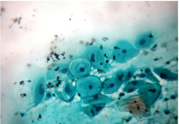
[image:5.595.230.528.263.470.2]About 66.7% (398) of the smears revealed normal findings (NILM) while 26.3% (152/577) were abnormal cytology (including epithelial anomalies, reac-tive cellular changes of inflammation and infections). Out of the 152 abnormal cases, 113 (74.3%) were reported to have non-specific inflammatory changes, 16 (10.5%) showed low grade squamous intraepithelial lesions (LSIL) as in Figure 1, 7 (4.6%) HSIL (moderate or severe dysplasia) as in Figure 2, while 5 (3.3%) revealed squamous cell carcinoma (SCC). There were 11 cases of atypical squamous cells out of which 8 (5.3%) were ASC-US and 3 (2%) were ASC-H as shown in Table 3. The LSIL showed cells with koilocytic changes suggestive of HPV infection and mild dysplasia. Thus cervical intraepithelial cell lesions were observed in 39/577 (6.8%) of the Pap smears.
Figure 1. Photomicrograph of low-grade squamous intraepithelial lesion (LSIL).
[image:5.595.252.507.510.706.2]DOI: 10.4236/crcm.2018.74024 264 Case Reports in Clinical Medicine Table 3. Cytological diagnosis of pap smears.
Cytological diagnosis Frequency Percentage
NILM 398 66.7
Inflammatory 113 18.9
Atrophic 27 4.5
Inadequate 20 3.4
LSIL 16 2.7
ASC-US 8 1.3
HSIL 7 1.2
SCC 5 0.8
ASC-H 3 0.5
Total 597 100.0
NILM = negative for intraepithelial lesion or malignancy, SCC = squamous cell carcinoma, HSIL = high-grade squamous intra-epithelial lesion, LSIL = low-grade squamous intraepithelial lesion, ASC-US = atypical squamous cell of undetermined significance, ASC-H = atypical squamous cell where high grade le-sions could not be excluded.
The maximum number of patients screened in this study i.e. 62.1% (371/597) belonged to the age group of 26 - 45 years followed by 139 (23.4%) patients within the 46 - 65 age range and was least with those in the 7th and 8th decades of life. Many of the cervical smears 28.8% (172/597) were performed for routine indications in women within the active reproductive age group of 16 - 45 years. This age group is characterized by intense sexual activity, high prevalence of sexually transmitted infections (STIs) including HPV infection, abnormal ute-rine bleeding (AUB), non-specific inflammation processes and cervicitis as shown in Table 4.
Table 5 showed indications for cytology in relation to cytological diagnosis.
Abnormal epithelial changes were observed in 11 (4.6%) cases of routine cervical smears and 2 out of the 5 cases of invasive carcinoma observed in this study were seen among those who came for routine screening. Table 6 showed that 3 out of the 5 cases of SCC observed in this study were present within the 46 - 55 year age group. Cervical cancer is common within the age range of 40 - 59 years with a mean of 55.5 years. Similarly, most of the high grade cytological lesions (6 out of 7) were prevalent within the same age bracket but were absent in patients below the age of 25 years.
5. Discussion
DOI: 10.4236/crcm.2018.74024 265 Case Reports in Clinical Medicine Table 4. Indications for pap smear in relation to age.
Age
Indications
Routine AUB Vaginal discharge Suspected cervical cancer
Uterine
polyps Cervicitis PMB PCB prolapse UV
≤15 3 2 0 0 0 3 0 1 0
16 - 25 24 5 5 4 0 7 0 8 0
26 - 35 74 21 9 13 6 31 2 16 3
36 - 45 91 26 15 15 17 20 1 14 7
46 - 55 44 15 3 13 7 13 7 8 4
56 - 65 18 1 1 1 1 1 6 0 3
66 - 75 1 1 0 0 0 1 3 0 0
[image:7.595.209.536.318.525.2]76+ 0 1 0 0 0 0 0 0 0
Table 5. Cytological diagnosis in relation to indication.
Indication Cytological diagnosis
Normal Inflammatory LSIL HSIL SCC ASCH ASCUS Atrophic
Routine 194 33 5 2 2 1 1 9
AUB 56 6 3 2 1 0 0 3
Vaginal
discharge 17 13 1 0 0 0 1 1
Suspected
cervical cancer 26 12 2 1 2 1 0 2
Uterine polyps 24 3 0 0 0 0 0 1
Cervicitis 33 31 2 1 0 0 3 3
PMB 7 0 2 1 0 1 0 7
PCB 30 12 1 0 0 0 2 1
UV Prolapse 11 3 0 0 0 0 1 0
Table 6. Cytological diagnosis in relation to age.
Age Cytological diagnosis
Normal Inflammatory LSIL HSIL SCC ASCH ASCUS Atrophy
≤15 6 3 0 0 0 0 0 0
16 - 25 42 7 2 0 0 0 0 0
26 - 35 125 32 4 1 0 0 4 2
36 - 45 141 43 4 4 1 2 3 2
46 - 55 65 21 5 2 3 1 1 13
56 - 65 14 7 0 0 1 0 0 8
66 - 75 4 0 1 0 0 0 0 1
[image:7.595.210.539.555.743.2]DOI: 10.4236/crcm.2018.74024 266 Case Reports in Clinical Medicine Arabia, and 6.8% in New Delhi India respectively [16] [17] [18]. The worldwide range of positive cytology from the conventional Pap smear is 10% [19]. The rel-atively low rate of positive cytology observed in this study could be attributed to the large number of clients 256/597 (42.9%) that had cytology performed for routine indications and not based on gynaecological symptoms. In the previous study from this centre, most of the indications for cytology were symptom-based (79.4%), while in Ibadan it was 53.8% [13] [15]. We also observed from the pre-vious study from this institution that the Pap smear was taken for routine indi-cations in only about 21.5% of cases, while in this review cytology for routine medical check-up was performed in 42.9% of patients [13]. Thus, even though the awareness has appreciated, screening for cervical cancer is still opportunistic and not the organized population based.
The annual uptake of the Pap smear showed a rising trend from 1.3% in the previous study to 9.3% in this review [13]. This recent increase in the uptake of the conventional Pap smear in our institution could be attributed to the aware-ness campaign, health education and workshops which were carried out within the hospital complex, places of worship and through the print and electronic media during the periods of 2012 to 2013. Thus, from this study, awareness of the value of cervical cancer screening may have translated to utilization and in-crease uptake of the Pap smear. This underscores the need for a well-organized population-based national screening programme for cervical cancer.
Chronic cervicitis is a very common condition in adult females and preferen-tially affects the squamo-columnar junction and the endocervix [18]. It is often accompanied by metaplasia in the epithelium. In this study, we observed that cervicitis was an indication for cervical cytology in 12.7% (76/597) of cases and inflammatory lesions were detected in 20% (113/577) of cervical cytology. Thus cervical smear examination is a valuable tool in the diagnosis of cervical infec-tions which are very common in women of reproductive age group [18].
Atypical squamous cells of undetermined significance (ASC-US) were ob-served in 8 (1.4%) of cases in this study. Previous studies have reported the asso-ciation of ASCUS in a significant proportion of women with Trichomonas vagi-nalis infection [16] [17]. However this study did not identify any causative or-ganism. A previous review from this centre identified Trichomonas vaginalis in 4% of the smears [13].
DOI: 10.4236/crcm.2018.74024 267 Case Reports in Clinical Medicine Sahara desert. However, it was also noted that most of the patients with gynae-cological symptoms were excluded from participating in the Egyptian study [20]. The major controversy in cervical cancer screening involves the age at which to begin screening (with the Papanicolau smear). In this review, we observed that epithelial cell anomalies were rare in patients below the age of 25 years but became more prevalent thereafter. This is comparable with studies from Egypt [20]. It is thus reasonable to begin screening after the age of 25 years. However, cervical cancer has been detected in women as young as 21 years of age [5]. Ac-cording to the American Society for Colposcopy and Cervical Pathology (ASCCP), cervical cancer screening should begin at age 21 years [21]. They stated that women under the age of 21 should not be screened regardless of the age of sexual debut or other risk factors. This is because cervical cancer is rare in adolescents and young women and may not be prevented by cytology screening [21]. Cytological abnormalities are common in this age group and can lead to labelling, anxiety, extended surveillance, and invasive procedures such as col-poscopy [21].
6. Conclusion
In conclusion, this study has observed that the annual uptake of the convention-al Pap smear in our institution showed a rising trend, but most of the indications remain symptom based. The presence of epithelial cell abnormalities on cervical cytology is consistent with literature. Awareness of the value of cervical cancer screening could result in increased utilization and uptake of the Pap smear.
References
[1] Arbyn, M., Sankaranarayanan, R., Muwonge, R., Keita, N., Dolo, A., Mbalawa, C.G., et al. (2008) Pooled Analysis of the Accuracy of Five Cervical Cancer Screening Tests Assessed in Eleven Studies in Africa and India. International Journal of Can-cer, 123, 153-160. https://doi.org/10.1002/ijc.23489
[2] GLOBOCAN (2012) http://www.globocan.iarc.fr/Default.aspx
[3] Ferlay, J., Shin, H.R., Bray, F., Forman, D., Mathers, C. and Parkin, D.M. (2010) Es-timates of Worldwide Burden of Cancer in 2008: GLOBOCAN 2008. International Journal of Cancer, 127, 2893-2917. https://doi.org/10.1002/ijc.25516
[4] Situational Analysis of Cervical Cancer Prevention and Control in the Caribbean. Pan American Health Organization, 2013.
http://www.who.int/mediacentre/factsheets/fs380/en/
[5] Nnadi, D.C., Singh, S., Ahmed, Y., Siddique, S. and Bilal, Y. (2014) His-to-Pathological Features of Genital Tract Malignancies as Seen in a Tertiary Health Centre in North-Western Nigeria: A 10-Year Review. Annals of Medical and Health Sciences, 4, 213-217. https://doi.org/10.4103/2141-9248.141961
[6] Ifenne, D.I., Shittu, S.O. and Ekwenpu, C.C. (2001) Cervical Smear in Pregnancy: The Zaria Experience. Nigerian Journal of Surgical Research, 3, 81-83.
https://doi.org/10.4314/njsr.v3i2.12229
DOI: 10.4236/crcm.2018.74024 268 Case Reports in Clinical Medicine [8] Ncube, B., Bey, A., Knight, J., Bessler, P. and Jolly, P.E. (2015) Factors Associated with the Uptake of Cervical Cancer Screening among Women in Portland, Jamaica. North American Journal of Medical Sciences, 7, 104-113.
https://doi.org/10.4103/1947-2714.153922
[9] de Sanjose, S., Quint, W.G., Alemany, L., Geraets, D.T., Klaustermeier, J.E., Llove-ras, B., et al. (2010) Human Papillomavirus Genotype Attribution in Invasive Cer-vical Cancer: A Retrospective Cross-Sectional Worldwide Study. The Lancet On-cology, 11, 1048-1056. https://doi.org/10.1016/S1470-2045(10)70230-8
[10] Brown, D.R., Shew, M.L., Qadadri, B., Neptune, N., Vargas, M., Tu, W., et al. (2005) A Longitudinal Study of Genital Human Papillomavirus Infection in a Cohort of Closely Followed Adolescent Women. The Journal of Infectious Diseases, 191, 182-192. https://doi.org/10.1086/426867
[11] Symonds, I.M. (2006) Screening for Gynaecologic Conditions. Current Obstetrics & Gynaecology, 16, 337-343. https://doi.org/10.1016/j.curobgyn.2006.09.003
[12] Papanicolau, G.N. and Traut, H.F. (1941) The Diagnostic Value of Vaginal Smears in Carcinoma of the Uterus. American Journal of Obstetrics & Gynecology, 42, 193-206.
[13] Daniel, C.N., Emmanuel, I.N., Lydia, R.A., Arkilla, M. and Sahabi, S.M. (2013) Screening for Cervical Cancer: Experience from a University Hospital in North Western Nigeria (2007-2009). Journal of Basic and Clinical Reproductive, 2, 18-21. https://doi.org/10.4103/2278-960X.112576
[14] Chukwuali, L.I., Onuigbo, W.I. and Mgbor, N.C. (2003) Cervical Cancer Screening in Enugu, Nigeria. Journal of Obstetrics and Gynaecology, 20, 109-112.
[15] Ayinde, A.E., Adwole, I.F. and Babarisa, I.A. (1998) Trends in Cervical Cancer Screening in Ibadan Nigeria: A Four-Year Review. West African Journal of Medi-cine, 17, 25-30.
[16] Kapila, K., George, S.S., Al-Shaheen, A., Al Ottibi, M.S., Pathan, S.K., Sheikh, Z.A., et al. (2006) Changing Spectrum of Squamous Cell Abnormalities Observed on Pa-panicolau Smears in Mubaraka Al Kabeer Hospital, Kuwait, over a 13-Year Period. Medical Principles and Practice, 15, 253-259. https://doi.org/10.1159/000092986 [17] Elhakeem, H.A., Al-Ghamdi, A.S. and Al-Maghrabi, J.A. (2005) Cytopathological
Pattern of Cervical Pap Smear According to the Bethesda System in Southwestern Saudi Arabia. Saudi Medical Journal, 26, 588-592.
[18] Rana, S., Jairapuri, Z.S. and Jetley, S. (2013) Cervical Smear Cytology on Routine Screening in a Semi Urban Population in New Delhi: A Review of 610 Cases. Arc-hives of Medicine and Health Sciences, 1, 131-135.
https://doi.org/10.4103/2321-4848.123025
[19] Cuzick, J., Jerry, C., Ho, L., Hollingworth, T. and Anderson, M. (1994) Type-Specific Human Papillomavirus DNA in Abnormal Smear as a Predictor of High Grade Cervical Intra-Epithelial Neoplasia. British Journal of Cancer, 69, 167-171. https://doi.org/10.1038/bjc.1994.28
[20] Abdel-Hadi, M., Khalaf, A., Aboulkassem, H., Naeem, N., Baqy, M.A. and Sallam, H. (2015) Cervical Intraepithelial Lesions in Females Attending Women’s Health Clinics in Alexandria, Egypt. CytoJournal, 12, 13.
https://doi.org/10.4103/1742-6413.159240