organic papers
o3416
Liliana Dobrzan´ska C6H6O3C3H3N3 doi:10.1107/S1600536805029995 Acta Cryst.(2005). E61, o3416–o3418 Acta Crystallographica Section E
Structure Reports
Online
ISSN 1600-5368
1,2,3-Trihydroxybenzene–1,3,5-triazine (1/1)
Liliana Dobrzan´ska
Department of Chemistry, University of Stellenbosch, Private Bag X1, Matieland 7602, South Africa
Correspondence e-mail: [email protected]
Key indicators
Single-crystal X-ray study
T= 100 K
Mean(C–C) = 0.003 A˚
Rfactor = 0.050
wRfactor = 0.107
Data-to-parameter ratio = 14.5
For details of how these key indicators were automatically derived from the article, see http://journals.iucr.org/e.
#2005 International Union of Crystallography Printed in Great Britain – all rights reserved
In the title molecular co-crystal, C6H6O3C3H3N3, two hydrogen-bonded R4
4
(18) tetrameric arrangements of mol-ecules involving symmetry-related O—H N hydrogen bonds link molecules of 1,2,3-trihydroxybenzene and 1,3,5-triazine to form an infinite double-chain motif. The one-dimensional array associates with neighbouring strands, via C—H O interactions, to form supramolecular sheets that are stacked together by–interactions in a three-dimensional assembly.
Comment
Understanding the nature of non-covalent interactions is essential for the development of crystal engineering, in order to facilitate the rational design of supramolecular structures (Desiraju, 1989, 2005; Braga, 2003). As an extension of our studies of supramolecular synthons and crystal packing in molecular co-crystals formed by ‘acidic’ and ‘basic’ compo-nents, we now report the crystal structure of a (1/1) adduct, (I), of pyrogallol (1,2,3-trihydroxybenzene) and 1,3,5-triazine.
In the crystal structure of a (1/1) complex of pyrogallol and pyrimidine (Dobrzan´ska, 2005), two distinct synthons were identified, namely a heterosynthon formed via O—H N hydrogen bonds and a homosynthon formed via weaker poorly directed O—H O hydrogen bonds, coded asR4
4(18) andR2
2(10) graph sets, respectively. The crystal structure was stabilized by offset –and C—H interactions, resulting in a herringbone packing mode. The same heterosynthon was also found in another co-crystal, of pyrogallol and hexa-methylenetetramine, where a three-dimensional supramol-ecular framework was generated by C—H (arene) interactions (Tremayne & Glidewell, 2000).
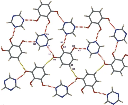
The title co-crystal was prepared in order to investigate the effect on the hydrogen-bonding motif of introducing an additional N acceptor site in the heterocyclic ring. The asymmetric unit consists of one molecule of each of pyrogallol and 1,3,5-triazine (Fig. 1). The molecules are held together by hydrogen-bonded O1—H1 N1, O2—H2 N3i and O3— H3 N2iiinteractions (symmetry codes as in Table 1). This facilitates the formation of an infinite double-chain along [010] consisting of R4
4(18) tetrameric arrangements. The one-dimensional hydrogen-bonded chains of supramolecular
heterosynthons are further linked to one another via C5— H5 O1iii[symmetry code: (iii)3
2x, 1 2+y,
3
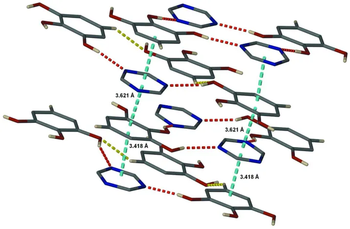
2z] interactions [C O = 3.448 (3) A˚ ], to form supramolecular sheets (Fig. 2). Moreover, benzene and triazine rings from adjacent parallel sheets interactviaoffset–interactions (centroid centroid distances 3.418 and 3.621 A˚ ), to form a three-dimensional assembly (Fig. 3).
Only R4
4(18) heterosynthons formed via O—H N hydrogen bonding are present in the structure of (I), and this motif seems to be favoured in supramolecular structures comprising pyrogallol and compounds containing two or more N acceptor sites.
Experimental
Colourless crystals of (I) suitable for single-crystal X-ray diffraction were obtained by slow evaporation of an ethanolic solution of 1,2,3-trihydroxybenzene and 1,3,5-triazine (1:1 molar ratio) at room temperature.
Crystal data
C6H6O3C3H3N3 Mr= 207.19 Monoclinic, P21=n a= 8.7299 (8) A˚ b= 10.8999 (10) A˚ c= 9.7586 (8) A˚ = 98.363 (2)
V= 918.71 (14) A˚3 Z= 4
Dx= 1.498 Mg m3 MoKradiation Cell parameters from 2011
reflections = 2.8–27.1
= 0.12 mm1 T= 100 (2) K Block, colourless 0.270.250.20 mm
Data collection
Bruker APEX CCD area-detector diffractometer
!scans
Absorption correction: multi-scan (SADABS; Sheldrick, 1997) Tmin= 0.666,Tmax= 0.977 5630 measured reflections
2011 independent reflections 1049 reflections withI> 2(I) Rint= 0.062
max= 27.1
h=8!11 k=13!11 l=11!12
Refinement
Refinement onF2 R[F2> 2(F2)] = 0.050 wR(F2) = 0.107 S= 0.89 2011 reflections 139 parameters
H-atom parameters constrained w= 1/[2
(Fo2) + (0.0395P)2] whereP= (Fo2+ 2Fc2)/3 (/)max< 0.001
max= 0.20 e A˚
3
min=0.20 e A˚3
Table 1
Hydrogen-bond geometry (A˚ ,).
D—H A D—H H A D A D—H A O1—H1 N1 0.84 2.08 2.863 (2) 155 O2—H2 N3i
0.84 2.14 2.892 (2) 149 O3—H3 N2ii
0.84 1.96 2.781 (2) 167
Symmetry codes: (i)x;yþ1;zþ1; (ii)x;yþ1;z.
H atoms were positioned geometrically, with C—H = 0.95 A˚ and O—H = 0.84 A˚ , and were constrained to ride on their parent atoms, withUiso(H) = 1.2 timesUeq(C,O).
Data collection:SMART(Bruker, 2001); cell refinement:SAINT (Bruker, 2002); data reduction:SAINT; program(s) used to solve structure:SHELXS97(Sheldrick, 1997); program(s) used to refine structure: SHELXL97 (Sheldrick, 1997); molecular graphics: X-SEED(Atwood & Barbour, 2003; Barbour, 2001); software used to prepare material for publication:X-SEED.
organic papers
Acta Cryst.(2005). E61, o3416–o3418 Liliana Dobrzan´ska C
[image:2.610.45.301.69.218.2]6H6O3C3H3N3
o3417
Figure 1
[image:2.610.313.562.73.277.2]The asymmetric unit of (I), with atom labels and 50% probability displacement ellipsoids.
Figure 2
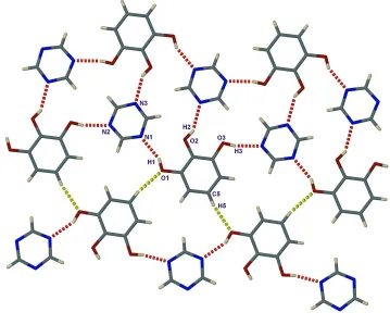
[image:2.610.315.566.326.487.2]A perspective view of a two-dimensional supramolecular sheet in the structure of (I). Hydrogen bonds are shown as dashed lines (N—H N indicated in red and C—H O in yellow).
Figure 3
The author thanks the Claude Harris Leon Foundation for financial support and further acknowledges the Nicolaus Copernicus University, Torun´, Poland, for study leave.
References
Atwood, J. L. & Barbour, L. J. (2003).Cryst. Growth Des.3, 3–8. Barbour, L. J. (2001).J. Supramol. Chem.1, 189–191.
Braga, D. (2003).Chem. Commun.pp. 2751–2754.
Bruker (2001).SMART. Version 5.625. Bruker AXS Inc., Madison, Wisconsin, USA.
Bruker (2002).SAINT. Version 6.36a. Bruker AXS Inc., Madison, Wisconsin, USA.
Desiraju, G. R. (1989).Crystal Engineering. The Design of Organic Solids. Amsterdam: Elsevier.
Desiraju, G. R. (2005).Chem. Commun.pp. 2995–3001. Dobrzan´ska, L. (2005).Acta Cryst.E61, o2981–o2983.
Sheldrick, G. M. (1997).SHELXS97,SHELXL97 andSADABS(Version 2.05). University of Go¨ttingen, Germany.
Tremayne, M. & Glidewell, C. (2000).Chem. Commun.pp. 2425–2426.
organic papers
o3418
Liliana Dobrzan´ska Csupporting information
sup-1
Acta Cryst. (2005). E61, o3416–o3418supporting information
Acta Cryst. (2005). E61, o3416–o3418 [doi:10.1107/S1600536805029995]
1,2,3-Trihydroxybenzene
–
1,3,5-triazine (1/1)
Liliana Dobrza
ń
ska
S1. Comment
Understanding the nature of non-covalent interactions is essential for the development of crystal engineering, in order to facilitate the rational design of supramolecular structures (Desiraju, 1989, 2005; Braga, 2003). As an extension of our studies of supramolecular synthons and crystal packing in molecular co-crystals formed by `acidic′ and `basic′
components, we now report the crystal structure of a (1/1) adduct, (I), of pyrogallol (1,2,3-trihydroxybenzene) and 1,3,5-triazine.
In the crystal structure of a (1/1) complex of pyrogallol and pyrimidine (Dobrzańska, 2005), two distinct synthons were identified, namely a heterosynthon formed via O—H···N hydrogen bonds and a homosynthon formed via weaker poorly directed O—H···O hydrogen bonds, coded as R4
4(18) and R22(10) graph sets, respectively. The crystal structure was
stabilized by offset π–π and C—H···π interactions, resulting in a herringbone packing mode. The same heterosynthon was also found in another co-crystal, of pyrogallol and hexamethylenetetramine, where a three-dimensional supramolecular framework was generated by C—H···π (arene) interactions (Tremayne & Glidewell, 2000).
The title co-crystal was prepared in order to investigate the effect on the hydrogen-bonding motif of introducing an additional N acceptor site in the heterocyclic ring. The asymmetric unit consists of one molecule of each of pyrogallol and 1,3,5-triazine (Fig. 1). The molecules are held together by hydrogen-bonded O1—H1···N1, O2—H2···N3i and O3—
H3···N2ii interactions (symmetry codes as in Table 1). This facilitates the formation of an infinite double-chain along
[010] consisting of R44(18) tetrameric arrangements. The one-dimensional hydrogen-bonded chains of supramolecular
heterosynthons are further linked to one another via C5—H5···O1iii [symmetry code: (iii) 3/2 − x, 1/2 + y, 3/2 − z]
interactions [C···O = 3.448 (3) Å], to form supramolecular sheets (Fig. 2). Moreover, benzene and triazine rings from adjacent parallel sheets interact via offset π–π interactions (centroid···centroid distances 3.418 and 3.621 Å), to form a three-dimensional assembly (Fig. 3).
Only R44(18) heterosynthons formed via O—H···N hydrogen bonding are present in the structure of (I), and this motif
seems to be favoured in supramolecular structures comprising pyrogallol and compounds containing two or more N acceptor sites.
S2. Experimental
Colourless crystals of (I) suitable for single-crystal X-ray diffraction were obtained by slow evaporation of an ethanolic solution of 1,2,3-trihydroxybenzene and 1,3,5-triazine (1:1 molar ratio) at room temperature.
S3. Refinement
supporting information
[image:5.610.131.485.72.278.2]sup-2
Acta Cryst. (2005). E61, o3416–o3418Figure 1
The asymmetric unit of (I), with atom labels and 50% probability displacement ellipsoids.
Figure 2
[image:5.610.124.483.317.616.2]supporting information
[image:6.610.126.484.70.305.2]sup-3
Acta Cryst. (2005). E61, o3416–o3418Figure 3
A capped-stick representation, showing the offset π–π interactions in the crystal packing of (I) (dashed blue lines). Dashed red and yellow lines represent N—H···N and C—H···O hydrogen bonds, respectively.
1,2,3-trihydroxybenzene:1,3,5-triazine (1/1)
Crystal data
C6H6O3·C3H3N3
Mr = 207.19
Monoclinic, P21/n
Hall symbol: -P2yn
a = 8.7299 (8) Å
b = 10.8999 (10) Å
c = 9.7586 (8) Å
β = 98.363 (2)°
V = 918.71 (14) Å3
Z = 4
F(000) = 432
Dx = 1.498 Mg m−3
Mo Kα radiation, λ = 0.71073 Å Cell parameters from 2011 reflections
θ = 2.8–27.1°
µ = 0.12 mm−1
T = 100 K Block, colourless 0.27 × 0.25 × 0.20 mm
Data collection
Bruker APEX CCD area-detector diffractometer
Radiation source: fine-focus sealed tube Graphite monochromator
ω scans
Absorption correction: multi-scan
(SADABS; Sheldrick, 1997)
Tmin = 0.666, Tmax = 0.977
5630 measured reflections 2011 independent reflections 1049 reflections with I > 2σ(I)
Rint = 0.062
θmax = 27.1°, θmin = 2.8°
h = −8→11
k = −13→11
l = −11→12
Refinement
Refinement on F2
Least-squares matrix: full
R[F2 > 2σ(F2)] = 0.050
wR(F2) = 0.107
S = 0.89
2011 reflections 139 parameters 0 restraints
supporting information
sup-4
Acta Cryst. (2005). E61, o3416–o3418Secondary atom site location: difference Fourier map
Hydrogen site location: inferred from neighbouring sites
H-atom parameters constrained
w = 1/[σ2(F
o2) + (0.0395P)2]
where P = (Fo2 + 2Fc2)/3
(Δ/σ)max < 0.001
Δρmax = 0.20 e Å−3
Δρmin = −0.20 e Å−3
Special details
Geometry. All e.s.d.'s (except the e.s.d. in the dihedral angle between two l.s. planes) are estimated using the full covariance matrix. The cell e.s.d.'s are taken into account individually in the estimation of e.s.d.'s in distances, angles and torsion angles; correlations between e.s.d.'s in cell parameters are only used when they are defined by crystal symmetry. An approximate (isotropic) treatment of cell e.s.d.'s is used for estimating e.s.d.'s involving l.s. planes.
Refinement. Refinement of F2 against ALL reflections. The weighted R-factor wR and goodness of fit S are based on F2,
conventional R-factors R are based on F, with F set to zero for negative F2. The threshold expression of F2 > σ(F2) is used
only for calculating R-factors(gt) etc. and is not relevant to the choice of reflections for refinement. R-factors based on F2
are statistically about twice as large as those based on F, and R- factors based on ALL data will be even larger.
Fractional atomic coordinates and isotropic or equivalent isotropic displacement parameters (Å2)
x y z Uiso*/Ueq
O3 0.25207 (18) 0.89079 (14) 0.59666 (18) 0.0346 (5)
H3 0.2643 0.9670 0.5919 0.042*
O1 0.52346 (19) 0.52246 (13) 0.72620 (18) 0.0355 (5)
H1 0.4440 0.4936 0.6781 0.043*
C2 0.3880 (3) 0.7098 (2) 0.6593 (2) 0.0237 (6) O2 0.25916 (18) 0.64060 (14) 0.61386 (18) 0.0312 (5)
H2 0.1837 0.6872 0.5885 0.037*
C4 0.5210 (3) 0.9022 (2) 0.7044 (2) 0.0303 (6)
H4 0.5213 0.9892 0.7004 0.036*
C6 0.6534 (3) 0.7129 (2) 0.7686 (2) 0.0277 (6)
H6 0.7439 0.6705 0.8090 0.033*
C3 0.3890 (3) 0.8371 (2) 0.6531 (2) 0.0247 (6) C1 0.5214 (3) 0.6477 (2) 0.7165 (2) 0.0251 (6) C5 0.6533 (3) 0.8389 (2) 0.7619 (2) 0.0286 (6)
H5 0.7444 0.8831 0.7968 0.034*
N2 0.2491 (2) 0.14320 (17) 0.5545 (2) 0.0266 (5) N3 0.0615 (2) 0.28987 (17) 0.4636 (2) 0.0275 (5) N1 0.3066 (2) 0.35516 (17) 0.5783 (2) 0.0280 (5) C8 0.1115 (3) 0.1755 (2) 0.4894 (2) 0.0285 (6)
H8 0.0417 0.1112 0.4580 0.034*
C9 0.1645 (3) 0.3751 (2) 0.5111 (2) 0.0283 (6)
H9 0.1340 0.4582 0.4958 0.034*
C7 0.3420 (3) 0.2374 (2) 0.5965 (2) 0.0286 (6)
H7 0.4422 0.2184 0.6436 0.034*
Atomic displacement parameters (Å2)
U11 U22 U33 U12 U13 U23
supporting information
sup-5
Acta Cryst. (2005). E61, o3416–o3418O2 0.0232 (10) 0.0207 (9) 0.0455 (11) 0.0031 (7) −0.0090 (9) −0.0012 (8) C4 0.0313 (16) 0.0233 (14) 0.0360 (16) −0.0035 (12) 0.0036 (13) −0.0012 (12) C6 0.0178 (14) 0.0346 (16) 0.0297 (15) 0.0028 (12) 0.0002 (12) 0.0031 (12) C3 0.0224 (14) 0.0248 (14) 0.0255 (14) 0.0021 (11) −0.0008 (11) −0.0002 (12) C1 0.0237 (15) 0.0224 (14) 0.0294 (14) 0.0027 (11) 0.0048 (12) −0.0004 (12) C5 0.0209 (14) 0.0339 (16) 0.0308 (16) −0.0072 (11) 0.0025 (12) −0.0022 (12) N2 0.0248 (12) 0.0242 (12) 0.0301 (12) 0.0040 (9) 0.0016 (10) 0.0012 (10) N3 0.0279 (13) 0.0210 (11) 0.0316 (12) 0.0040 (9) −0.0028 (10) 0.0003 (10) N1 0.0239 (12) 0.0258 (12) 0.0330 (13) 0.0033 (9) 0.0000 (10) 0.0001 (10) C8 0.0261 (15) 0.0270 (14) 0.0324 (16) −0.0015 (11) 0.0037 (12) −0.0026 (12) C9 0.0282 (15) 0.0223 (14) 0.0343 (15) 0.0027 (12) 0.0042 (12) −0.0006 (12) C7 0.0205 (15) 0.0313 (16) 0.0339 (15) 0.0031 (11) 0.0033 (12) 0.0000 (12)
Geometric parameters (Å, º)
O3—C3 1.373 (3) C6—C1 1.386 (3)
O3—H3 0.8400 C6—H6 0.9500
O1—C1 1.369 (2) C5—H5 0.9500
O1—H1 0.8400 N2—C8 1.322 (3)
C2—O2 1.373 (3) N2—C7 1.335 (3)
C2—C3 1.389 (3) N3—C9 1.329 (3)
C2—C1 1.391 (3) N3—C8 1.332 (3)
O2—H2 0.8400 N1—C7 1.326 (3)
C4—C3 1.383 (3) N1—C9 1.334 (3)
C4—C5 1.392 (3) C8—H8 0.9500
C4—H4 0.9500 C9—H9 0.9500
C6—C5 1.375 (3) C7—H7 0.9500
C3—O3—H3 109.5 C6—C1—C2 120.1 (2)
C1—O1—H1 109.5 C6—C5—C4 120.6 (2)
O2—C2—C3 123.0 (2) C6—C5—H5 119.7
O2—C2—C1 117.5 (2) C4—C5—H5 119.7
C3—C2—C1 119.5 (2) C8—N2—C7 114.3 (2)
C2—O2—H2 109.5 C9—N3—C8 113.7 (2)
C3—C4—C5 119.4 (2) C7—N1—C9 113.9 (2)
C3—C4—H4 120.3 N2—C8—N3 126.2 (2)
C5—C4—H4 120.3 N2—C8—H8 116.9
C5—C6—C1 120.0 (2) N3—C8—H8 116.9
C5—C6—H6 120.0 N3—C9—N1 126.3 (2)
C1—C6—H6 120.0 N3—C9—H9 116.9
O3—C3—C4 123.9 (2) N1—C9—H9 116.9
O3—C3—C2 115.6 (2) N1—C7—N2 125.7 (2)
C4—C3—C2 120.5 (2) N1—C7—H7 117.2
O1—C1—C6 118.9 (2) N2—C7—H7 117.2
O1—C1—C2 120.9 (2)
C5—C4—C3—O3 178.8 (2) O2—C2—C1—C6 −177.7 (2)
supporting information
sup-6
Acta Cryst. (2005). E61, o3416–o3418O2—C2—C3—O3 −0.7 (4) C1—C6—C5—C4 0.7 (4)
C1—C2—C3—O3 −179.2 (2) C3—C4—C5—C6 −0.3 (4) O2—C2—C3—C4 178.0 (2) C7—N2—C8—N3 0.2 (4) C1—C2—C3—C4 −0.6 (4) C9—N3—C8—N2 −0.4 (4) C5—C6—C1—O1 −179.0 (2) C8—N3—C9—N1 0.4 (4) C5—C6—C1—C2 −1.0 (4) C7—N1—C9—N3 −0.1 (4)
O2—C2—C1—O1 0.3 (3) C9—N1—C7—N2 −0.2 (3)
C3—C2—C1—O1 178.9 (2) C8—N2—C7—N1 0.2 (3)
Hydrogen-bond geometry (Å, º)
D—H···A D—H H···A D···A D—H···A
O1—H1···N1 0.84 2.08 2.863 (2) 155
O2—H2···N3i 0.84 2.14 2.892 (2) 149
O3—H3···N2ii 0.84 1.96 2.781 (2) 167