organic papers
o2380
Iain D. H. Oswaldet al. C6H6N2O2C3H6O2 doi: 10.1107/S1600536804028776 Acta Cryst.(2004). E60, o2380–o2383 Acta Crystallographica Section E
Structure Reports
Online
ISSN 1600-5368
A 1:2 co-crystal of isonicotinamide and
propionic acid
Iain D. H. Oswald,a
W. D. Sam Motherwellband
Simon Parsonsa*
aSchool of Chemistry, The University of
Edinburgh, King’s Buildings, West Mains Road, Edinburgh EH9 3JJ, Scotland, andbCambridge Crystallographic Data Centre, 12 Union Road, Cambridge CB2 1EZ, England
Correspondence e-mail: [email protected]
Key indicators
Single-crystal X-ray study
T= 150 K
Mean(C–C) = 0.005 A˚
Rfactor = 0.088
wRfactor = 0.198
Data-to-parameter ratio = 17.1
For details of how these key indicators were automatically derived from the article, see http://journals.iucr.org/e.
#2004 International Union of Crystallography
Printed in Great Britain – all rights reserved
Isonicotinamide has been shown to form many 1:1 co-crystals with monofunctional carboxylic acids, but with propionic acid it forms a co-crystal containing two acid molecules and one
isonicotinamide molecule per formula unit, C6H6N2O
-2C3H6O2. The crystal structure consists of ‘supermolecules’
made up of of one isonicotinamide molecule and two acid molecules, and the asymmetric unit contains two of these supermolecules. One of the acid molecules is hydrogen bonded to the pyridine function, and the other to the amide
function of the isonicotinamide. Further N—H O hydrogen
bonds connect these supermolecules into chains which run along the [100] direction. The chains are linked into layers
perpendicular to (010) by C—H O and -stacking
interac-tions. The layers are then linked together by further C—
H O interactions.
Comment
Isonicotinamide has been shown to crystallize with carboxylic acids in a 1:1 stoichiometry to form a robust building block or ‘supermolecule’ consisting of two amide and two acid
mol-ecules, (I) (Aakero¨yet al., 2002). When a saturated solution of
isonicotinamide in warm propionic acid was allowed to cool, colourless crystalline laths were obtained. Single-crystal X-ray diffraction revealed these to be a co-crystal consisting of
isonicotinamide and propionic acid in a 1:2 ratio,viz. (II).
Similar preparative routes with formic and acetic acids both yielded 1:1 co-crystals (Oswald, 2004). Attempts to prepare a 1:1 co-crystal with propionic acid failed. For example, a 1:1 mixture of propionic acid and isonicotinamide in ethanol yielded only crystals of (II); even in the presence of excess isonicotinamide, the only crystals obtained were isonicotin-amide itself and (II).
The crystal structure of (II) consists of supermolecules comprising two acid and one isonicotinamide molecule. One
acid forms anR2
2
(8) motif with the amide moiety (Bernsteinet
al., 1995). Another acid molecule forms a hydrogen bond to
the pyridine N atom, supported by a weaker C—H O
hydrogen bond (Fig. 1 and Table 1). There are two supermolecules in the asymmetric unit and, in the terminology
of Aakero¨y et al. (2002), both are in the trans–trans
conformation.
The independent supermolecules hydrogen-bond together using the second amide donor and the carbonyl group from the propionic acid molecules located at the pyridine end of the supermolecules. This builds up a helical chain in which
successive supermolecules are aligned approximately
perpendicular to one another (Figs. 2–4; hydrogen-bond
dimensions are listed in Table 1). The chains run along thea
direction, and they comprise all the conventional N—H O
and O—H O hydrogen bonds in the crystal structure (see
Table 1); additional C—H O interactions (C5A—
H5A O2Uand C5B—H5B O2V) are also formed within
the chains (Desiraju & Steiner, 1999).
Successive helical chains are distributed along thec
direc-tion atz=1 4,
3
4, . . .,etc. (Fig. 5). Though there are no direct
hydrogen-bonding interactions between neighbouring chains,
weak C—H O interactions are formed between chains
located one lattice-repeat away from each other (e.g.the red
and blue chains in Fig. 5; see also Fig. 6). These interactions
involve C2A—H2A O3T and C2B—H2B O3S.
Supermolecules in neighbouring chains are interleaved to
produce stacks of supermolecules along a (Fig. 5). Stacks
containing only the supermolecules based on isonicotinamide
moleculeAoccur atz=12, while stacks containing only those
organic papers
Acta Cryst.(2004). E60, o2380–o2383 Iain D. H. Oswaldet al. C
[image:2.610.312.565.73.260.2]6H6N2O2C3H6O2
o2381
Figure 1 [image:2.610.44.298.75.239.2] [image:2.610.44.298.313.500.2]The two crystallographically independent supermolecules, with the atomic numbering. Displacement ellipsoids are drawn at the 30% probability level. Conventional hydrogen bonds are shown in heavy dashes and the H O distances span 1.78 (4)–1.96 (4) A˚ (see Table 1). The C—H O hydrogen bonds (shown as open dashes) are quite weak for this type of interaction (2.73 and 2.72 A˚ ).
Figure 2
Hydrogen-bonded chains in the crystal structure of (II). Hydrogen bonds link supermolecules into chains. This view is approximately along the direct lattice direction [100]. Hydrogen bonds are shown as dashed lines, weak C—H O hydrogen bonds are shown in turquoise.
Figure 3
Hydrogen-bonded chains in the crystal structure of (II). Successive supermolecules are approximately perpendicular to each other; this view is perpendicular to (001).
Figure 4
[image:2.610.313.566.313.498.2]based on molecule B occur at z= 0, 1, . . . etc. Within the
stacks, pairs of pyridine moieties are-stacked across
inver-sion centres (Fig. 7). The stacking distances are 3.34 and
3.33 A˚ for theAandBpyridine rings, respectively.
Thus, layers are formed in the ac-plane by chains of
hydrogen-bonded supermolecules linked by weak C—H O
and-stacking interactions. The layers are connectedviaC—
H O hydrogen bonds involving pairs of C4T—H4T1 O2T
and C4V—H4V1 O2Sinteractions disposed about inversion
centres (Fig. 8).
Experimental
All materials were obtained from Aldrich and used as received. Isonicotinamide (0.50 g, 4.10 mmol) was dissolved in an excess of propionic acid (2.40 g, 32.43 mmol) and warmed until all the solid dissolved. The solution was cooled to room temperature, producing colourless laths.
Crystal data
C6H6N2O2C3H6O2 Mr= 270.28
Triclinic,P1 a= 10.038 (3) A˚ b= 11.559 (4) A˚ c= 12.740 (4) A˚
= 103.203 (6) = 90.140 (6) = 102.247 (6)
V= 1404.5 (8) A˚3
Z= 4
Dx= 1.278 Mg m
ÿ3 MoKradiation Cell parameters from 1107
reflections
= 2.6–22.2 = 0.10 mmÿ1 T= 150 (2) K Lath, colourless 0.750.200.08 mm
organic papers
o2382
Iain D. H. Oswaldet al. C [image:3.610.312.566.72.260.2]6H6N2O2C3H6O2 Acta Cryst.(2004). E60, o2380–o2383
Figure 5
Packing of hydrogen-bonded chains in the crystal structure of (II), forming a layer perpendicular tob*. Neighbouring chains are distributed along thecaxis. Different chains (as shown in Figs. 2–4) are shown in different colours. This view is alonga,cf. Fig. 2.
Figure 6
[image:3.610.45.294.72.263.2]Packing of hydrogen-bonded chains in the crystal structure of (II), forming a layer perpendicular to b*. As Fig. 5, but with the green molecule deleted to reveal C—H O hydrogen bonds formed between the blue and red chains shown in Fig. 5. C—H O hydrogen bonds within chains are shown in turquoise, those between chains are shown in magenta.
Figure 7
[image:3.610.44.297.326.517.2]Packing of hydrogen-bonded chains in the crystal structure of (II), forming a layer perpendicular to [010]. Neighbouring chains are connected by -stacking interactions. This figure shows two chains viewed along [001]. One chain is shown in ball-and-stick representation, the other as wireframe.
Figure 8
[image:3.610.317.558.330.506.2]Data collection
Bruker SMART CCD area-detector diffractometer with an Oxford Cryosystems low-temperature device (Cosier & Glazer, 1986)
’and!scans
Absorption correction: multi-scan (SADABS; Sheldrick, 2004) Tmin= 0.783,Tmax= 1.000
12519 measured reflections 6498 independent reflections 3362 reflections withI> 2(I) Rint= 0.044
max= 28.9
h=ÿ13!13 k=ÿ15!15 l=ÿ17!17
Refinement
Refinement onF2 R[F2> 2(F2)] = 0.088 wR(F2) = 0.198 S= 1.04 6498 reflections 379 parameters
H atoms treated by a mixture of independent and constrained refinement
w= 1/[2(F
o2) + (0.0681P)2
+ 0.3798P]
whereP= (Fo2+ 2Fc2)/3
(/)max< 0.001
max= 0.36 e A˚
ÿ3
min=ÿ0.21 e A˚
[image:4.610.43.297.330.483.2]ÿ3
Table 1
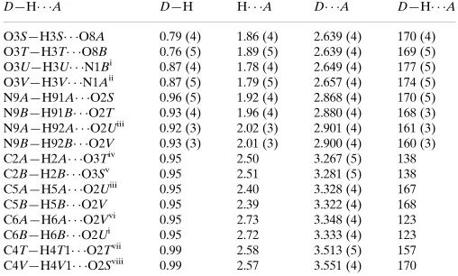
Hydrogen-bonding geometry (A˚ ,).
D—H A D—H H A D A D—H A
O3S—H3S O8A 0.79 (4) 1.86 (4) 2.639 (4) 170 (4) O3T—H3T O8B 0.76 (5) 1.89 (5) 2.639 (4) 169 (5) O3U—H3U N1Bi
0.87 (4) 1.78 (4) 2.649 (4) 177 (5) O3V—H3V N1Aii 0.87 (5) 1.79 (5) 2.657 (4) 174 (5) N9A—H91A O2S 0.96 (5) 1.92 (4) 2.868 (4) 170 (5) N9B—H91B O2T 0.93 (4) 1.96 (4) 2.880 (4) 168 (3) N9A—H92A O2Uiii
0.92 (3) 2.02 (3) 2.901 (4) 161 (3) N9B—H92B O2V 0.93 (3) 2.01 (3) 2.900 (4) 160 (3) C2A—H2A O3Tiv
0.95 2.50 3.267 (5) 138 C2B—H2B O3Sv
0.95 2.51 3.281 (5) 138 C5A—H5A O2Uiii
0.95 2.40 3.328 (4) 167 C5B—H5B O2V 0.95 2.39 3.322 (4) 168 C6A—H6A O2Vvi
0.95 2.73 3.348 (4) 123 C6B—H6B O2Ui 0.95 2.72 3.333 (4) 123 C4T—H4T1 O2Tvii
0.99 2.58 3.513 (5) 157 C4V—H4V1 O2Sviii
0.99 2.57 3.551 (4) 170 Symmetry codes: (i) 1ÿx;2ÿy;ÿz; (ii) 1þx;1þy;zÿ1; (iii) 1ÿx;1ÿy;1ÿz; (iv) xÿ1;yÿ1;z; (v) x;1þy;z; (vi) xÿ1;yÿ1;1þz; (vii) 2ÿx;1ÿy;ÿz; (viii) x;y;zÿ1.
H atoms were placed on C atoms in calculated positions [Uiso(H) =
1.2Ueq(C)] and allowed to ride on their parent atoms [C(phenyl)—
H = 0.95, C(methylene)—H = 0.99 and C(methyl)—H = 0.98 A˚ ]. Amide and hydroxyl H atoms were located in difference maps and refined freely, the former subject to the restraint N—H = 0.95 (3) A˚ . The ranges of N—H and O—H bond lengths were 0.91 (2)–0.96 (1) and 0.75 (5)–0.87 (4) A˚ , respectively.
Data collection:SMART(Bruker, 2001); cell refinement:SAINT
(Bruker, 2003); data reduction:SAINT; program(s) used to solve structure: SHELXTL (Sheldrick, 2001); program(s) used to refine structure:SHELXTL; molecular graphics:SHELXTL, MERCURY
(Taylor & Macrae, 2001) andDIAMOND (Crystal Impact, 2004); software used to prepare material for publication: SHELXTL,
EnCIFer(Allenet al., 2004) andPLATON(Spek, 2003), as incor-porated inWinGX(Farrugia, 1999)..
We thank the EPSRC, the University of Edinburgh and the Cambridge Crystallographic Data Centre for funding.
References
Aakero¨y, C. B., Beatty, A. M. & Helfrich, B. A. (2002).J. Am. Chem. Soc.124, 14425–14432.
Allen, F. H., Johnson, O., Shields, G. P., Smith, B. R. & Towler, M. (2004).J. Appl. Cryst.37, 335–338.
Bernstein, J., Davis, R. E., Shimoni, L. & Chang, N.-L. (1995).Angew. Chem. Int. Ed. Engl.34, 1555–1573.
Bruker (2001).SMART. Version 5.624. Bruker AXS Inc., Madison, Wisconsin, USA.
Bruker (2003).SAINT.Version 7. Bruker AXS Inc., Madison, Wisconsin, USA.
Cosier, J. & Glazer, A. M. (1986).J. Appl. Cryst.19, 105–107.
Crystal Impact (2004). DIAMOND. Version 3.0a. Crystal Impact GbR, Postfach 1251, 53002 Bonn, Germany. (URL: http://www.crystalimpact.com/ diamond.)
Desiraju, G. R. & Steiner, T. (1999). The Weak Hydrogen Bond. IUCr Monographs on Crystallography, No. 9. Oxford University Press. Farrugia, L. J. (1999).J. Appl. Cryst.32, 837–838.
Oswald, I. D. H. (2004) PhD thesis, The University of Edinburgh, Scotland. Sheldrick, G. M. (2001).SHELXTL.Version 6.01. University of Go¨ttingen,
Germany.
Sheldrick, G. M. (2004).SADABS.University of Go¨ttingen, Germany. Spek, A. L. (2003).J. Appl. Cryst.36, 7–13.
Taylor, R. & Macrae, C. F. (2001).Acta Cryst.B57, 815–827.
organic papers
Acta Cryst.(2004). E60, o2380–o2383 Iain D. H. Oswaldet al. C
supporting information
sup-1 Acta Cryst. (2004). E60, o2380–o2383
supporting information
Acta Cryst. (2004). E60, o2380–o2383 [https://doi.org/10.1107/S1600536804028776]
A 1:2 co-crystal of isonicotinamide and propionic acid
Iain D. H. Oswald, W. D. Sam Motherwell and Simon Parsons
Isonicotinamide–propionic acid (1:2)
Crystal data
C6H6N2O·2C3H6O2 Mr = 270.28 Triclinic, P1 Hall symbol: -P1
a = 10.038 (3) Å
b = 11.559 (4) Å
c = 12.740 (4) Å
α = 103.203 (6)°
β = 90.140 (6)°
γ = 102.247 (6)°
V = 1404.5 (8) Å3
Z = 4
F(000) = 576
Dx = 1.278 Mg m−3
Mo Kα radiation, λ = 0.71073 Å Cell parameters from 1107 reflections
θ = 2.6–22.2°
µ = 0.10 mm−1 T = 150 K Lath, colourless 0.75 × 0.20 × 0.08 mm
Data collection
Bruker SMART CCD area-detector diffractometer
Radiation source: fine-focus sealed tube Graphite monochromator
φ and ω scans
Absorption correction: multi-scan (SADABS; Sheldrick, 2004)
Tmin = 0.783, Tmax = 1.000
12519 measured reflections 6498 independent reflections 3362 reflections with I > 2σ(I)
Rint = 0.044
θmax = 28.9°, θmin = 1.6°
h = −13→13
k = −15→15
l = −17→17
Refinement
Refinement on F2
Least-squares matrix: full
R[F2 > 2σ(F2)] = 0.088 wR(F2) = 0.198 S = 1.04 6498 reflections 379 parameters 4 restraints
Primary atom site location: structure-invariant direct methods
Secondary atom site location: difference Fourier map
Hydrogen site location: inferred from neighbouring sites
H atoms treated by a mixture of independent and constrained refinement
w = 1/[σ2(Fo2) + (0.0681P)2 + 0.3798P]
where P = (Fo2 + 2Fc2)/3
(Δ/σ)max < 0.001
Δρmax = 0.36 e Å−3
supporting information
sup-2 Acta Cryst. (2004). E60, o2380–o2383
Special details
Geometry. All e.s.d.'s (except the e.s.d. in the dihedral angle between two l.s. planes) are estimated using the full covariance matrix. The cell e.s.d.'s are taken into account individually in the estimation of e.s.d.'s in distances, angles and torsion angles; correlations between e.s.d.'s in cell parameters are only used when they are defined by crystal symmetry. An approximate (isotropic)
treatment of cell e.s.d.'s is used for estimating e.s.d.'s involving l.s. planes.
Refinement. ABSTM02_ALERT_3_C The ratio of expected to reported Tmax/Tmin(RR′) is < 0.90
PLAT061_ALERT_3_C Tmax/Tmin Range Test RR′ too Large ···. 0.84 T min and Tmax reported: 0.783 1.000
Tmin′ and Tmax expected: 0.927 0.992 Noted, but no action taken. SADABS attempts to correct for all systematic errors not just absorption. The large range could represent a small amount of crystal decay for example.
PLAT029_ALERT_3_C _diffrn_measured_fraction_theta_full Low ···. 0.98
============================================================================ Resolution & Completeness Statistics (Cumulative)
============================================================================
Theta sin(th)/Lambda Complete Expected Measured Missing ———————————————————————— —- —- 20.82 0.500 0.998 2939 2932 7 23.01 0.550 0.990 3900 3861 39 25.24 0.600 0.979 5085 4978 107 ————— ——————————————— ACTA Min. Res. —- 27.51 0.650 0.961 6447 6198 249 29.84 0.700 0.875 7427 6498 929
PLAT063_ALERT_3_C Crystal Probably too Large for Beam Size ···. 0.75 mm
Gorbitz has shown that use of a large crystal does not appear to matter. See C. H. Gorbitz Acta Cryst. (1999). B55, 1090– 1098
PLAT414_ALERT_2_C Short Intra D—H.·H—X H5A.. H92A.. 1.98 A ng PLAT414_ALERT_2_C Short Intra D—H.·H
—X H5B.. H92B.. 1.96 A ng PLAT480_ALERT_4_C Long H···A H-Bond Reported H6A.. O2V.. 2.73 A ng
PLAT480_ALERT_4_C Long H···A H-Bond Reported H6B.. O2U.. 2.72 A ng See text.
PLAT222_ALERT_3_C Large Non-Solvent H Ueq(max)/Ueq(min) ··· 3.02 Ratio PLAT340_ALERT_3_C Low Bond
Precision on C—C bonds (x 1000) Ang ··· 5 PLAT720_ALERT_4_C Number of Unusual/Non-Standard Label(s) ···.. 20
PLAT790_ALERT_4_C Centre of Gravity not Within Unit Cell: Resd. # 4 C3 H6 O2 PLAT790_ALERT_4_C Centre of Gravity not Within Unit Cell: Resd. # 6 C3 H6 O2 No action taken.
Fractional atomic coordinates and isotropic or equivalent isotropic displacement parameters (Å2)
x y z Uiso*/Ueq
N1A 0.0374 (3) −0.1364 (2) 0.5184 (2) 0.0340 (6)
C2A 0.0899 (3) −0.1354 (3) 0.4224 (3) 0.0399 (9)
H2A 0.0424 −0.1899 0.3598 0.048*
C3A 0.2125 (3) −0.0566 (3) 0.4114 (3) 0.0367 (8)
H3A 0.2482 −0.0576 0.3423 0.044*
C4A 0.2807 (3) 0.0225 (3) 0.5023 (2) 0.0307 (7)
C5A 0.2266 (3) 0.0211 (3) 0.6018 (3) 0.0358 (8)
H5A 0.2724 0.0739 0.6659 0.043*
C6A 0.1023 (3) −0.0605 (3) 0.6056 (3) 0.0372 (8)
H6A 0.0635 −0.0610 0.6734 0.045*
C7A 0.4134 (3) 0.1076 (3) 0.4875 (3) 0.0300 (7)
O8A 0.4515 (2) 0.1028 (2) 0.39475 (18) 0.0422 (6)
N9A 0.4801 (3) 0.1821 (3) 0.5743 (2) 0.0400 (7)
H91A 0.559 (4) 0.239 (4) 0.561 (4) 0.13 (2)*
H92A 0.448 (4) 0.177 (4) 0.641 (2) 0.075 (14)*
N1B 0.6050 (2) 1.1345 (2) −0.0179 (2) 0.0347 (7)
supporting information
sup-3 Acta Cryst. (2004). E60, o2380–o2383
H2B 0.6352 1.1865 0.1396 0.044*
C3B 0.7394 (3) 1.0525 (3) 0.0886 (3) 0.0372 (8)
H3B 0.7738 1.0521 0.1580 0.045*
C4B 0.7705 (3) 0.9749 (3) −0.0022 (2) 0.0318 (8)
C5B 0.7187 (3) 0.9781 (3) −0.1016 (3) 0.0343 (8)
H5B 0.7395 0.9262 −0.1658 0.041*
C6B 0.6340 (3) 1.0601 (3) −0.1058 (3) 0.0358 (8)
H6B 0.5967 1.0620 −0.1738 0.043*
C7B 0.8617 (3) 0.8898 (3) 0.0125 (3) 0.0315 (8)
O8B 0.9029 (2) 0.8942 (2) 0.10510 (18) 0.0435 (6)
N9B 0.8926 (3) 0.8168 (3) −0.0743 (2) 0.0402 (7)
H91B 0.943 (4) 0.761 (3) −0.064 (3) 0.079 (14)*
H92B 0.855 (3) 0.817 (3) −0.141 (2) 0.059 (11)*
C1S 0.7475 (3) 0.3193 (3) 0.4192 (3) 0.0380 (8)
O2S 0.7115 (3) 0.3378 (2) 0.5096 (2) 0.0546 (7)
O3S 0.6802 (3) 0.2306 (2) 0.34051 (19) 0.0442 (7)
H3S 0.618 (4) 0.189 (3) 0.361 (3) 0.052 (13)*
C4S 0.8744 (4) 0.3943 (3) 0.3861 (3) 0.0473 (9)
H4S1 0.8645 0.4799 0.4013 0.057*
H4S2 0.9519 0.3916 0.4329 0.057*
C5S 0.9112 (4) 0.3599 (4) 0.2720 (3) 0.0597 (11)
H5S1 0.9249 0.2763 0.2558 0.090*
H5S2 0.9955 0.4155 0.2611 0.090*
H5S3 0.8374 0.3652 0.2241 0.090*
C1T 1.0981 (3) 0.6806 (3) 0.0803 (3) 0.0374 (8)
O2T 1.0571 (2) 0.6652 (2) −0.0116 (2) 0.0521 (7)
O3T 1.0671 (3) 0.7648 (3) 0.1601 (2) 0.0468 (7)
H3T 1.029 (5) 0.809 (4) 0.145 (4) 0.10 (2)*
C4T 1.1877 (4) 0.6048 (3) 0.1135 (3) 0.0468 (9)
H4T1 1.1369 0.5189 0.0955 0.056*
H4T2 1.2686 0.6099 0.0691 0.056*
C5T 1.2363 (4) 0.6365 (4) 0.2284 (3) 0.0621 (12)
H5T1 1.2872 0.7214 0.2480 0.093*
H5T2 1.2960 0.5829 0.2395 0.093*
H5T3 1.1579 0.6263 0.2736 0.093*
C1U 0.5951 (3) 0.7137 (3) 0.1200 (3) 0.0331 (8)
O2U 0.5695 (2) 0.7875 (2) 0.19782 (18) 0.0417 (6)
O3U 0.5494 (3) 0.7069 (2) 0.02220 (19) 0.0440 (6)
H3U 0.496 (4) 0.757 (4) 0.021 (3) 0.086 (16)*
C4U 0.6782 (3) 0.6226 (3) 0.1251 (3) 0.0442 (9)
H4U1 0.7507 0.6291 0.0730 0.053*
H4U2 0.6191 0.5400 0.1026 0.053*
C5U 0.7439 (4) 0.6374 (4) 0.2361 (3) 0.0574 (11)
H5U1 0.8006 0.7198 0.2602 0.086*
H5U2 0.8010 0.5777 0.2326 0.086*
H5U3 0.6727 0.6243 0.2873 0.086*
C1V 0.7618 (3) 0.7141 (3) −0.3789 (3) 0.0319 (8)
supporting information
sup-4 Acta Cryst. (2004). E60, o2380–o2383
O3V 0.8021 (3) 0.7075 (2) −0.47843 (18) 0.0410 (6)
H3V 0.876 (5) 0.763 (4) −0.479 (4) 0.11 (2)*
C4V 0.6318 (3) 0.6233 (3) −0.3745 (3) 0.0419 (9)
H4V1 0.6485 0.5401 −0.3985 0.050*
H4V2 0.5621 0.6322 −0.4254 0.050*
C5V 0.5763 (4) 0.6378 (4) −0.2626 (3) 0.0572 (11)
H5V1 0.6443 0.6283 −0.2118 0.086*
H5V2 0.4925 0.5756 −0.2645 0.086*
H5V3 0.5561 0.7189 −0.2393 0.086*
Atomic displacement parameters (Å2)
U11 U22 U33 U12 U13 U23
N1A 0.0336 (15) 0.0404 (17) 0.0301 (16) 0.0147 (13) 0.0006 (12) 0.0068 (13)
C2A 0.044 (2) 0.048 (2) 0.0270 (19) 0.0148 (18) 0.0026 (15) 0.0027 (16)
C3A 0.039 (2) 0.043 (2) 0.0292 (19) 0.0145 (16) 0.0062 (15) 0.0059 (16)
C4A 0.0311 (18) 0.0397 (19) 0.0268 (18) 0.0192 (15) 0.0031 (14) 0.0082 (15)
C5A 0.040 (2) 0.041 (2) 0.0266 (18) 0.0118 (16) −0.0037 (14) 0.0062 (15)
C6A 0.038 (2) 0.051 (2) 0.0258 (18) 0.0168 (17) 0.0034 (15) 0.0081 (16)
C7A 0.0331 (18) 0.0347 (19) 0.0273 (18) 0.0168 (15) 0.0033 (14) 0.0086 (15)
O8A 0.0419 (14) 0.0506 (15) 0.0308 (14) 0.0072 (11) 0.0074 (11) 0.0053 (11)
N9A 0.0385 (18) 0.053 (2) 0.0293 (17) 0.0112 (15) 0.0020 (14) 0.0098 (15)
N1B 0.0327 (15) 0.0389 (17) 0.0283 (16) −0.0003 (12) 0.0036 (12) 0.0066 (13)
C2B 0.0356 (19) 0.042 (2) 0.0260 (18) 0.0049 (16) 0.0013 (14) 0.0008 (15)
C3B 0.0333 (19) 0.045 (2) 0.0300 (19) 0.0028 (16) −0.0016 (14) 0.0074 (16)
C4B 0.0247 (17) 0.0384 (19) 0.0256 (18) −0.0044 (14) 0.0012 (13) 0.0041 (15)
C5B 0.0316 (18) 0.041 (2) 0.0261 (18) 0.0027 (15) 0.0051 (14) 0.0039 (15)
C6B 0.0305 (18) 0.047 (2) 0.0277 (19) 0.0027 (16) 0.0031 (14) 0.0097 (16)
C7B 0.0240 (17) 0.040 (2) 0.0267 (18) 0.0002 (14) 0.0046 (14) 0.0058 (15)
O8B 0.0443 (14) 0.0561 (16) 0.0298 (14) 0.0148 (12) −0.0054 (11) 0.0060 (12)
N9B 0.0369 (17) 0.057 (2) 0.0296 (17) 0.0164 (15) 0.0024 (13) 0.0105 (15)
C1S 0.037 (2) 0.036 (2) 0.041 (2) 0.0110 (16) −0.0024 (17) 0.0075 (17)
O2S 0.0603 (17) 0.0601 (18) 0.0367 (16) 0.0011 (13) 0.0122 (13) 0.0086 (13)
O3S 0.0406 (15) 0.0520 (17) 0.0380 (16) 0.0037 (13) 0.0121 (12) 0.0123 (14)
C4S 0.048 (2) 0.046 (2) 0.048 (2) 0.0055 (18) 0.0052 (18) 0.0144 (19)
C5S 0.051 (2) 0.051 (3) 0.068 (3) −0.001 (2) 0.015 (2) 0.007 (2)
C1T 0.0295 (18) 0.038 (2) 0.044 (2) 0.0010 (15) 0.0086 (16) 0.0140 (18)
O2T 0.0605 (17) 0.0616 (17) 0.0381 (16) 0.0204 (13) −0.0049 (13) 0.0128 (13)
O3T 0.0473 (16) 0.0573 (18) 0.0402 (16) 0.0202 (14) −0.0039 (12) 0.0122 (14)
C4T 0.041 (2) 0.053 (2) 0.048 (2) 0.0085 (18) −0.0012 (17) 0.0167 (19)
C5T 0.071 (3) 0.062 (3) 0.055 (3) 0.027 (2) −0.011 (2) 0.005 (2)
C1U 0.0280 (17) 0.038 (2) 0.032 (2) 0.0005 (15) 0.0009 (14) 0.0135 (16)
O2U 0.0442 (14) 0.0499 (15) 0.0316 (14) 0.0156 (12) 0.0041 (11) 0.0058 (12)
O3U 0.0492 (15) 0.0549 (17) 0.0311 (14) 0.0223 (14) 0.0019 (11) 0.0066 (12)
C4U 0.043 (2) 0.044 (2) 0.050 (2) 0.0134 (17) 0.0062 (17) 0.0182 (18)
C5U 0.055 (3) 0.055 (3) 0.069 (3) 0.018 (2) −0.008 (2) 0.023 (2)
C1V 0.0361 (19) 0.038 (2) 0.0297 (19) 0.0199 (16) 0.0084 (15) 0.0135 (16)
supporting information
sup-5 Acta Cryst. (2004). E60, o2380–o2383
O3V 0.0427 (15) 0.0500 (16) 0.0279 (13) 0.0070 (13) 0.0062 (11) 0.0070 (11)
C4V 0.041 (2) 0.039 (2) 0.048 (2) 0.0101 (17) 0.0103 (16) 0.0142 (17)
C5V 0.051 (2) 0.059 (3) 0.064 (3) 0.010 (2) 0.023 (2) 0.022 (2)
Geometric parameters (Å, º)
N1A—C6A 1.318 (4) C4S—H4S1 0.9900
N1A—C2A 1.333 (4) C4S—H4S2 0.9900
C2A—C3A 1.396 (5) C5S—H5S1 0.9800
C2A—H2A 0.9500 C5S—H5S2 0.9800
C3A—C4A 1.375 (4) C5S—H5S3 0.9800
C3A—H3A 0.9500 C1T—O2T 1.201 (4)
C4A—C5A 1.383 (4) C1T—O3T 1.325 (4)
C4A—C7A 1.519 (4) C1T—C4T 1.502 (5)
C5A—C6A 1.404 (4) O3T—H3T 0.75 (5)
C5A—H5A 0.9500 C4T—C5T 1.479 (5)
C6A—H6A 0.9500 C4T—H4T1 0.9900
C7A—O8A 1.235 (4) C4T—H4T2 0.9900
C7A—N9A 1.315 (4) C5T—H5T1 0.9800
N9A—H91A 0.96 (3) C5T—H5T2 0.9800
N9A—H92A 0.91 (2) C5T—H5T3 0.9800
N1B—C2B 1.318 (4) C1U—O2U 1.218 (4)
N1B—C6B 1.321 (4) C1U—O3U 1.307 (4)
C2B—C3B 1.401 (4) C1U—C4U 1.488 (5)
C2B—H2B 0.9500 O3U—H3U 0.87 (4)
C3B—C4B 1.375 (4) C4U—C5U 1.517 (5)
C3B—H3B 0.9500 C4U—H4U1 0.9900
C4B—C5B 1.378 (4) C4U—H4U2 0.9900
C4B—C7B 1.518 (4) C5U—H5U1 0.9800
C5B—C6B 1.411 (4) C5U—H5U2 0.9800
C5B—H5B 0.9500 C5U—H5U3 0.9800
C6B—H6B 0.9500 C1V—O2V 1.201 (4)
C7B—O8B 1.236 (4) C1V—O3V 1.323 (4)
C7B—N9B 1.310 (4) C1V—C4V 1.502 (4)
N9B—H91B 0.93 (2) O3V—H3V 0.87 (5)
N9B—H92B 0.93 (2) C4V—C5V 1.518 (5)
C1S—O2S 1.194 (4) C4V—H4V1 0.9900
C1S—O3S 1.321 (4) C4V—H4V2 0.9900
C1S—C4S 1.501 (5) C5V—H5V1 0.9800
O3S—H3S 0.79 (4) C5V—H5V2 0.9800
C4S—C5S 1.485 (5) C5V—H5V3 0.9800
C6A—N1A—C2A 119.4 (3) C4S—C5S—H5S1 109.5
N1A—C2A—C3A 121.9 (3) C4S—C5S—H5S2 109.5
N1A—C2A—H2A 119.1 H5S1—C5S—H5S2 109.5
C3A—C2A—H2A 119.1 C4S—C5S—H5S3 109.5
C4A—C3A—C2A 118.9 (3) H5S1—C5S—H5S3 109.5
supporting information
sup-6 Acta Cryst. (2004). E60, o2380–o2383
C2A—C3A—H3A 120.5 O2T—C1T—O3T 122.7 (3)
C3A—C4A—C5A 119.2 (3) O2T—C1T—C4T 122.4 (3)
C3A—C4A—C7A 117.6 (3) O3T—C1T—C4T 114.9 (3)
C5A—C4A—C7A 123.2 (3) C1T—O3T—H3T 117 (4)
C4A—C5A—C6A 118.2 (3) C5T—C4T—C1T 117.1 (3)
C4A—C5A—H5A 120.9 C5T—C4T—H4T1 108.0
C6A—C5A—H5A 120.9 C1T—C4T—H4T1 108.0
N1A—C6A—C5A 122.4 (3) C5T—C4T—H4T2 108.0
N1A—C6A—H6A 118.8 C1T—C4T—H4T2 108.0
C5A—C6A—H6A 118.8 H4T1—C4T—H4T2 107.3
O8A—C7A—N9A 124.2 (3) C4T—C5T—H5T1 109.5
O8A—C7A—C4A 118.0 (3) C4T—C5T—H5T2 109.5
N9A—C7A—C4A 117.8 (3) H5T1—C5T—H5T2 109.5
C7A—N9A—H91A 115 (3) C4T—C5T—H5T3 109.5
C7A—N9A—H92A 119 (3) H5T1—C5T—H5T3 109.5
H91A—N9A—H92A 126 (4) H5T2—C5T—H5T3 109.5
C2B—N1B—C6B 119.4 (3) O2U—C1U—O3U 122.0 (3)
N1B—C2B—C3B 122.4 (3) O2U—C1U—C4U 124.7 (3)
N1B—C2B—H2B 118.8 O3U—C1U—C4U 113.2 (3)
C3B—C2B—H2B 118.8 C1U—O3U—H3U 112 (3)
C4B—C3B—C2B 118.7 (3) C1U—C4U—C5U 113.9 (3)
C4B—C3B—H3B 120.6 C1U—C4U—H4U1 108.8
C2B—C3B—H3B 120.6 C5U—C4U—H4U1 108.8
C3B—C4B—C5B 118.9 (3) C1U—C4U—H4U2 108.8
C3B—C4B—C7B 117.8 (3) C5U—C4U—H4U2 108.8
C5B—C4B—C7B 123.3 (3) H4U1—C4U—H4U2 107.7
C4B—C5B—C6B 118.6 (3) C4U—C5U—H5U1 109.5
C4B—C5B—H5B 120.7 C4U—C5U—H5U2 109.5
C6B—C5B—H5B 120.7 H5U1—C5U—H5U2 109.5
N1B—C6B—C5B 121.9 (3) C4U—C5U—H5U3 109.5
N1B—C6B—H6B 119.0 H5U1—C5U—H5U3 109.5
C5B—C6B—H6B 119.0 H5U2—C5U—H5U3 109.5
O8B—C7B—N9B 124.1 (3) O2V—C1V—O3V 122.6 (3)
O8B—C7B—C4B 118.2 (3) O2V—C1V—C4V 125.0 (3)
N9B—C7B—C4B 117.7 (3) O3V—C1V—C4V 112.5 (3)
C7B—N9B—H91B 117 (3) C1V—O3V—H3V 110 (3)
C7B—N9B—H92B 119 (2) C1V—C4V—C5V 113.2 (3)
H91B—N9B—H92B 123 (3) C1V—C4V—H4V1 108.9
O2S—C1S—O3S 122.9 (3) C5V—C4V—H4V1 108.9
O2S—C1S—C4S 122.5 (3) C1V—C4V—H4V2 108.9
O3S—C1S—C4S 114.5 (3) C5V—C4V—H4V2 108.9
C1S—O3S—H3S 112 (3) H4V1—C4V—H4V2 107.7
C5S—C4S—C1S 117.6 (3) C4V—C5V—H5V1 109.5
C5S—C4S—H4S1 107.9 C4V—C5V—H5V2 109.5
C1S—C4S—H4S1 107.9 H5V1—C5V—H5V2 109.5
C5S—C4S—H4S2 107.9 C4V—C5V—H5V3 109.5
C1S—C4S—H4S2 107.9 H5V1—C5V—H5V3 109.5
supporting information
sup-7 Acta Cryst. (2004). E60, o2380–o2383
Hydrogen-bond geometry (Å, º)
D—H···A D—H H···A D···A D—H···A
O3S—H3S···O8A 0.79 (4) 1.86 (4) 2.639 (4) 170 (4)
O3T—H3T···O8B 0.76 (5) 1.89 (5) 2.639 (4) 169 (5)
O3U—H3U···N1Bi 0.87 (4) 1.78 (4) 2.649 (4) 177 (5)
O3V—H3V···N1Aii 0.87 (5) 1.79 (5) 2.657 (4) 174 (5)
N9A—H91A···O2S 0.96 (5) 1.92 (4) 2.868 (4) 170 (5)
N9B—H91B···O2T 0.93 (4) 1.96 (4) 2.880 (4) 168 (3)
N9A—H92A···O2Uiii 0.92 (3) 2.02 (3) 2.901 (4) 161 (3)
N9B—H92B···O2V 0.93 (3) 2.01 (3) 2.900 (4) 160 (3)
C2A—H2A···O3Tiv 0.95 2.50 3.267 (5) 138
C2B—H2B···O3Sv 0.95 2.51 3.281 (5) 138
C5A—H5A···O2Uiii 0.95 2.40 3.328 (4) 167
C5B—H5B···O2V 0.95 2.39 3.322 (4) 168
C6A—H6A···O2Vvi 0.95 2.73 3.348 (4) 123
C6B—H6B···O2Ui 0.95 2.72 3.333 (4) 123
C4T—H4T1···O2Tvii 0.99 2.58 3.513 (5) 157
C4V—H4V1···O2Sviii 0.99 2.57 3.551 (4) 170

![Figure 7Packing of hydrogen-bonded chains in the crystal structure of (II),forming a layer perpendicular to [010]](https://thumb-us.123doks.com/thumbv2/123dok_us/729346.577349/3.610.317.558.330.506/figure-packing-hydrogen-bonded-crystal-structure-forming-perpendicular.webp)