organic papers
Acta Cryst.(2006). E62, o1211–o1212 doi:10.1107/S1600536806006003 Breamet al. C
6H12
o1211
Acta Crystallographica Section EStructure Reports Online
ISSN 1600-5368
Methylcyclopentane
Richard Bream,* David Watkin and Andrew Cowley
Chemical Crystallography, Central Chemistry Laboratory, University of Oxford, Oxford OX1 3TA, England
Correspondence e-mail: [email protected]
Key indicators
Single-crystal X-ray study T= 110 K
Mean(C–C) = 0.002 A˚ Rfactor = 0.070 wRfactor = 0.097
Data-to-parameter ratio = 25.6
For details of how these key indicators were automatically derived from the article, see http://journals.iucr.org/e.
Received 13 February 2006 Accepted 17 February 2006
#2006 International Union of Crystallography All rights reserved
Methylcyclopentane, C6H12, a liquid at room temperature, was
studied as part of a project to develop a computer-controlled low-temperature crystal-growing device. A single crystal was obtained at 115 K. The ring has an envelope conformation, with a pseudo-equatorial methyl substituent on the flap atom.
Comment
The melting point of methylcyclopentane is noted by the CRC
Handbook of Chemistry and Physics as being 142.4C
(130.8 K) (Weast, 1978). A sample solidified spontaneously to a polycrystalline mass on flash-cooling to 115 K, and was then zone refined into a single crystal using tandem computer-controlled heating elements. Data collection was completed at 110 K
The molecule is in the envelope conformation (Fig. 1), with the four atoms C2—C5 almost coplanar (maximum deviation 0.04 A˚ ) and a pseudo-equatorial methyl group attached to the flap atom C1. The crystal structure consists of molecular stacks formed by unit-cell translations along theaaxis (Figs. 2 and 3). The calculated density is not unlike that of the ordered monoclinic phase of cyclohexane (0.996 Mg m3), suggesting that a low density may be a feature of small cyclic hydro-carbons (Kahnet al., 1973).
Experimental
The material was used as supplied by Acros Organics. A 2.0 mm column was flame-sealed in a 0.3 mm diameter Lindemann tube and crystallized as described above.
Crystal data
C6H12
Mr= 84.16
Monoclinic,P21=n a= 5.3934 (2) A˚
b= 11.1439 (5) A˚
c= 9.7047 (5) A˚
= 98.0288 (17)
V= 577.57 (5) A˚3
Z= 4
Dx= 0.968 Mg m
3 MoKradiation Cell parameters from 1447
reflections
= 5–28
= 0.05 mm1
T= 110 K Cylinder, colourless 1.000.20 (radius) mm
Data collection
Nonius KappaCCD diffractometer
!scans
Absorption correction: multi-scan (DENZO/SCALEPACK; Otwinowski & Minor, 1997)
Tmin= 0.69,Tmax= 0.98 7788 measured reflections
1412 independent reflections 1408 reflections withI> 3(I)
Rint= 0.063
max= 28.3
h=7!7
k=14!14
Refinement
Refinement onF2
R[F2> 2(F2)] = 0.070
wR(F2) = 0.097
S= 0.94 1408 reflections 55 parameters
H-atom parameters constrained
w= 1/[2(F2) + (0.03P)2 + 0.1P]
whereP= [max(Fo2,0) + 2Fc2]/3 (/)max< 0.001
max= 0.28 e A˚
3
min=0.22 e A˚
[image:2.610.66.272.72.192.2]3
Table 1
Selected geometric parameters (A˚ ,).
C1—C2 1.5290 (14) C1—C5 1.5274 (14) C1—C6 1.5171 (14)
C2—C3 1.5344 (14) C3—C4 1.5402 (15) C4—C5 1.5286 (14)
C2—C1—C5 101.82 (8) C2—C1—C6 114.77 (8) C5—C1—C6 115.00 (8) C1—C2—C3 105.28 (8)
C2—C3—C4 105.88 (8) C3—C4—C5 105.39 (8) C4—C5—C1 104.05 (8)
The H atoms were all located in a difference map and then repositioned geometrically. The H atoms were initially refined with soft restraints on the bond lengths and angles to regularize their geometry (C–H in the range 0.93–0.98 A˚ ) and displacement para-meters [Uiso(H) in the range 1.2–1.5 timesUeqof the parent atom],
after which they were refined with riding constraints.
Data collection: COLLECT (Nonius, 2001); cell refinement:
DENZO/SCALEPACK; data reduction: DENZO/SCALEPACK
(Otwinowski & Minor, 1997); program(s) used to solve structure:
SIR92 (Altomareet al., 1994); program(s) used to refine structure:
CRYSTALS (Betteridge et al., 2003); molecular graphics:
CAMERON(Watkinet al., 1996); software used to prepare material for publication:CRYSTALS.
References
Altomare, A., Cascarano, G., Giacovazzo, G., Guagliardi, A., Burla, M. C., Polidori, G. & Camalli, M. (1994).J. Appl. Cryst.27, 435.
Betteridge, P. W., Carruthers, J. R., Cooper, R. I., Prout, K. & Watkin, D. J. (2003).J. Appl. Cryst.36, 1487.
Kahn, R., Fourme, R., Andre, D. & Renaud, M. (1973).Acta Cryst.B29, 131– 138.
Nonius (2001).COLLECT. Nonius BV, Delft, The Netherlands.
Otwinowski, Z. & Minor, W. (1997). Methods in Enzymology, Vol. 276,
Macromolecular Crystallography, Part A, edited by C. W. Carter Jr & R. M. Sweet, pp. 307–326. New York: Academic Press.
Watkin, D. J., Prout, C. K. & Pearce, L. J. (1996).CAMERON. Chemical Crystallography Laboratory, Oxford, England.
Weast, R. C. (1978). Editor. CRC Handbook of Chemistry and Physics. Cleveland, Ohio: CRC Press.
Figure 1
The title compound with displacement ellipsoids drawn at the 50% probability level. H atoms are shown as spheres of arbitary radius.
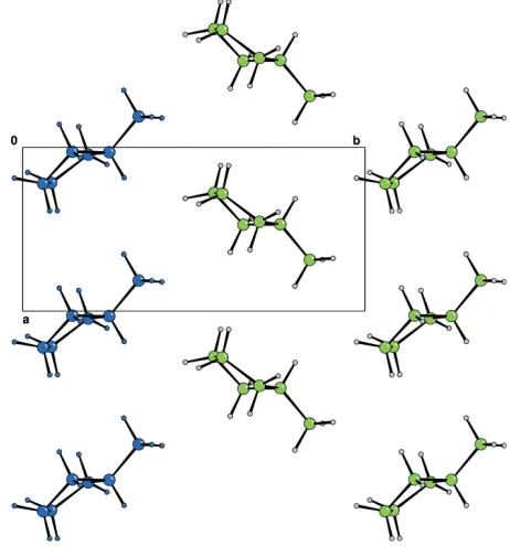
Figure 2
[image:2.610.321.552.330.578.2]Ana-axis projection of the title compound. One column of molecules has been highlighted in blue for comparison with Fig. 3.
Figure 3
A projection along thecaxis, showing the molecular stacks parallel to the
[image:2.610.43.297.356.442.2]supporting information
sup-1 Acta Cryst. (2006). E62, o1211–o1212
supporting information
Acta Cryst. (2006). E62, o1211–o1212 [https://doi.org/10.1107/S1600536806006003]
Methylcyclopentane
Richard Bream, David Watkin and Andrew Cowley
methylcyclopentane
Crystal data C6H12
Mr = 84.16
Monoclinic, P21/n
a = 5.3934 (2) Å b = 11.1439 (5) Å c = 9.7047 (5) Å β = 98.0288 (17)° V = 577.57 (5) Å3
Z = 4 F(000) = 192
Dx = 0.968 Mg m−3
Melting point: 130.8 K
Mo Kα radiation, λ = 0.71073 Å Cell parameters from 1447 reflections θ = 5–28°
µ = 0.05 mm−1
T = 110 K
Cylinder, colourless 1.00 × 0.20 (radius) mm
Data collection Nonius KappaCCD
diffractometer
Graphite monochromator ω scans
Absorption correction: multi-scan
(DENZO/SCALEPACK; Otwinowski & Minor, 1997)
Tmin = 0.69, Tmax = 0.98
7788 measured reflections 1412 independent reflections 1408 reflections with I > −3.0σ(I) Rint = 0.063
θmax = 28.3°, θmin = 5.3°
h = −7→7 k = −14→14 l = −12→12
Refinement Refinement on F2
Least-squares matrix: full R[F2 > 2σ(F2)] = 0.070
wR(F2) = 0.097
S = 0.94 1408 reflections 55 parameters 0 restraints
Primary atom site location: structure-invariant direct methods
Hydrogen site location: inferred from neighbouring sites
H-atom parameters constrained w = 1/[σ2(F2) + (0.03P)2 + 0.1P]
where P = [max(Fo2,0) + 2Fc2]/3
(Δ/σ)max < 0.001
Δρmax = 0.28 e Å−3
Δρmin = −0.22 e Å−3
Fractional atomic coordinates and isotropic or equivalent isotropic displacement parameters (Å2)
x y z Uiso*/Ueq
C1 1.03029 (18) 0.25222 (9) 0.78471 (10) 0.0284
C2 1.0448 (2) 0.19129 (9) 0.64490 (11) 0.0347
C3 1.2171 (2) 0.08259 (10) 0.67904 (11) 0.0350
C5 1.02749 (18) 0.14454 (9) 0.88184 (11) 0.0313
C6 0.81288 (19) 0.33855 (10) 0.78504 (12) 0.0360
H11 1.1857 0.2957 0.8138 0.0340*
H21 1.1039 0.2457 0.5789 0.0431*
H22 0.8742 0.1633 0.6054 0.0443*
H31 1.3876 0.1010 0.6582 0.0454*
H32 1.1534 0.0141 0.6260 0.0435*
H41 1.3888 0.0781 0.8855 0.0414*
H42 1.1882 −0.0251 0.8563 0.0421*
H51 1.0629 0.1677 0.9795 0.0402*
H52 0.8593 0.1062 0.8656 0.0385*
H61 0.8146 0.3762 0.8788 0.0540*
H62 0.8215 0.4062 0.7163 0.0519*
H63 0.6522 0.2949 0.7605 0.0531*
Atomic displacement parameters (Å2)
U11 U22 U33 U12 U13 U23
C1 0.0268 (5) 0.0270 (5) 0.0316 (6) −0.0033 (4) 0.0046 (4) 0.0004 (4)
C2 0.0411 (6) 0.0343 (6) 0.0297 (6) 0.0059 (5) 0.0078 (4) 0.0033 (4)
C3 0.0380 (6) 0.0309 (6) 0.0366 (6) 0.0042 (5) 0.0067 (4) 0.0009 (4)
C4 0.0341 (5) 0.0316 (5) 0.0364 (6) 0.0002 (4) 0.0024 (4) 0.0058 (5)
C5 0.0320 (5) 0.0333 (5) 0.0286 (6) −0.0044 (4) 0.0044 (4) 0.0021 (4)
C6 0.0348 (6) 0.0338 (6) 0.0405 (6) 0.0017 (5) 0.0085 (4) −0.0023 (5)
Geometric parameters (Å, º)
C1—C2 1.5290 (14) C3—H32 0.957
C1—C5 1.5274 (14) C4—C5 1.5286 (14)
C1—C6 1.5171 (14) C4—H41 0.984
C1—H11 0.975 C4—H42 0.981
C2—C3 1.5344 (14) C5—H51 0.975
C2—H21 0.967 C5—H52 0.995
C2—H22 0.996 C6—H61 1.001
C3—C4 1.5402 (15) C6—H62 1.013
C3—H31 0.990 C6—H63 0.993
C2—C1—C5 101.82 (8) C3—C4—C5 105.39 (8)
C2—C1—C6 114.77 (8) C3—C4—H41 109.6
C5—C1—C6 115.00 (8) C5—C4—H41 109.9
C2—C1—H11 109.2 C3—C4—H42 112.6
C5—C1—H11 107.1 C5—C4—H42 112.5
C6—C1—H11 108.5 H41—C4—H42 106.8
C1—C2—C3 105.28 (8) C4—C5—C1 104.05 (8)
C1—C2—H21 111.8 C4—C5—H51 113.6
C3—C2—H21 113.1 C1—C5—H51 112.0
C1—C2—H22 108.7 C4—C5—H52 109.5
supporting information
sup-3 Acta Cryst. (2006). E62, o1211–o1212
H21—C2—H22 108.3 H51—C5—H52 108.5
C2—C3—C4 105.88 (8) C1—C6—H61 111.0
C2—C3—H31 110.1 C1—C6—H62 111.4
C4—C3—H31 110.5 H61—C6—H62 107.0
C2—C3—H32 110.6 C1—C6—H63 109.8
C4—C3—H32 110.5 H61—C6—H63 108.7