K
9Y
3[Si
12O
32]F
2Volker Kahlenberg* and Tanja Manninger
University of Innsbruck, Institute of Mineralogy & Petrography, Innrain 52, A-6020 Innsbruck, Austria
Correspondence e-mail: [email protected]
Received 6 December 2013; accepted 21 January 2014
Key indicators: single-crystal X-ray study;T= 298 K; mean(Si–O) = 0.005 A˚;
Rfactor = 0.039;wRfactor = 0.099; data-to-parameter ratio = 10.7.
Single-crystals of the title compound, nonapotassium triyt-trium dodecasilicate difluoride, were obtained from flux synthesis experiments in the system SiO2—Y2O3—KF. The
crystal structure belongs to the group of single-layer silicates and is based on silicate sheets parallel to (110). A single layer contains secondary (Q2) and tertiary (Q3) silicate tetrahedra in the ratio 1:2 and is build up from six-, eight- and twelve-membered rings. The linkage between neighboring layers is achieved by two crystallographically independent Y3+cations, which are coordinated by six oxygen ligands in form of distorted octahedra. Charge compensation is accomplished by incorporation of additional F anions and K+ cations in the structural channels, forming anion-centred [F2K7] groups.
Apart from one K+ and one Y3+ cation (each with site symmetry 1), the 30 crystallographically independent atoms reside on general positions.
Related literature
Oxosilicates which can serve as luminescent materials containing trivalent rare earth elements, monovalent alkali cations as well as fluorine anions have already been char-acterized (Jacobsen & Meyer, 1994; Tanget al., 2008; Scha¨fer & Schleid, 2007, 2011). For structures isotypic to that of the title compound, see: Tanget al.(2008). For general aspects of
Experimental
Crystal data
K9Y3[Si12O32]F2 Mr= 1505.71 Triclinic,P1 a= 6.8187 (3) A˚ b= 11.3345 (4) A˚ c= 11.3727 (5) A˚
= 87.846 (3) = 89.747 (4)
= 80.524 (3)
V= 866.35 (6) A˚3 Z= 1
MoKradiation
= 6.60 mm 1 T= 298 K
0.120.090.05 mm
Data collection
Oxford Diffraction Xcalibur diffractometer
Absorption correction: multi-scan (CrysAlis PRO; Oxford Diffrac-tion, 2006)
Tmin= 0.801,Tmax= 1
10508 measured reflections 2842 independent reflections 2231 reflections withI> 2(I) Rint= 0.066
Refinement
R[F2> 2(F2)] = 0.039 wR(F2) = 0.099 S= 1.08 2842 reflections
265 parameters
max= 0.87 e A˚ 3
min= 0.95 e A˚ 3
Data collection: CrysAlis PRO(Oxford Diffraction, 2006); cell
refinement: CrysAlis PRO; data reduction: CrysAlis PRO;
program(s) used to solve structure: SIR2002 (Burla et al., 2003); program(s) used to refine structure:SHELXL97(Sheldrick, 2008); molecular graphics:ATOMS for Windows(Dowty, 2011); software used to prepare material for publication:publCIF(Westrip, 2010) and
WinGX(Farrugia, 2012).
Supporting information for this paper is available from the IUCr electronic archives (Reference: WM2792).
References
Brown, I. D. & Altermatt, D. (1985).Acta Cryst.B41, 244–247.
Burla, M. C., Camalli, M., Carrozzini, B., Cascarano, G. L., Giacovazzo, C., Polidori, G. & Spagna, R. (2003).J. Appl. Cryst.36, 1103.
Dowty, E. (2011).ATOMS for Windows. Shape Software, Kingsport, USA. Farrugia, L. J. (2012).J. Appl. Cryst.45, 849–854.
ICSD (2013). Inorganic Crystal Structure Database. FIZ-Karlsruhe, Germany, and the National Institute of Standards and Technology (NIST), USA. http://www.fiz-karlsruhe.de/ecid/Internet/en/DB/icsd/
Jacobsen, H. & Meyer, G. (1994).Z. Kristallogr.209, 348–350.
Liebau, F. (1985).Structural chemistry of silicates, p. 347. Berlin, Heidelberg, New York, Tokyo: Springer.
Acta Crystallographica Section E Structure Reports Online
supporting information
sup-1
Acta Cryst. (2014). E70, i11
supporting information
Acta Cryst. (2014). E70, i11 [doi:10.1107/S1600536814001470]
K
9Y
3[Si
12O
32]F
2Volker Kahlenberg and Tanja Manninger
S1. Comment
In the present paper we describe a previously unknown phase of the system KF—Y2O3—SiO2. According to Liebau's
classification (1985) the crystal structure of the title compound, K9Y3[Si12O32]F2, belongs to the group of open-branched
single-layer silicates. The more detailed crystallochemical formula can be written as K9Y3{oB,12∞}[5Si12O32]F2. A
single-layer of silicate tetrahedra expands parallel to (110) and is constructed from the condensation of fünfer single chains (Fig.
1). One discrete chain is running parallel to [001] and has a translation period of 11.3727 (5) Å. Each layer contains
secondary (Q2) and tertiary (Q3) SiO
4 tetrahedra in the ratio of 1:2 (Fig. 2). The Si—O distances of the six
crystallographically independent tetrahedra within the asymmetric unit range from 1.574 (5) to 1.662 (5) Å. As one
would expect, the Si—Oterminal bonds are considerably shorter than the distances between Si and the bridging O atoms. The
O—Si—O angles show a significant scatter throughout all present polyhedra. Nevertheless, the values are in the expected
limits for [SiO4] units (Liebau, 1985). Numerically, the degree of distortion can be expressed by the quadratic elongation
λ and the angle variance σ2 (Robinson et al., 1971). For the six tetrahedra, these two parameters vary between 1.003 and
1.005 (for λ) and 8.50 and 20.17 (for σ2) indicating that the deviation from regularity is not very pronounced.
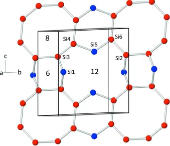
Within the corrugated silicate sheets, six-, eight- and twelve-membered rings can be identified (Fig. 2). The vertex
symbols for the [SiO4] tetrahedra are as follows: 6.8.12 (for Si2, Si3, Si4 and Si6) and 6.12 (for Si1 and Si5). A schematic
representation of the arrangement of the rings within a single layer is given in Fig. 2. Charge balance in the structure is
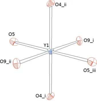
achieved by the incorporation of K+ and Y3+ cations as well as additional F- anions. Y1 resides on an inversion center and
is coordinated by six oxygen ligands belonging to six different [SiO4]-tetrahedra (Fig. 3). Within the resulting octahedron,
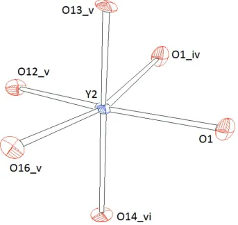
the Y—O bond lengths range from 2.237 (4) - 2.256 (4) Å. Y2 is also octahedrally coordinated (Fig. 4). However, each
two adjacent [Y2O6]-octahedra form dimers by sharing one common edge (Fig. 6). Therefore, the spread in the Y—O
bond lengths is more siginificant (2.211 (4) - 2.345 (4) Å) which is also reflected in higher values for the distortion
parameters: λ = 1.046 and σ2 = 149.49 (for Y2), and λ = 1.006 and σ2 = 20.88 (for Y1), respectively. The volumes of both
octahedra are almost identical: 14.545 Å3 (for Y2) and 14.991 Å3 (for Y1). The coordination numbers of the potassium
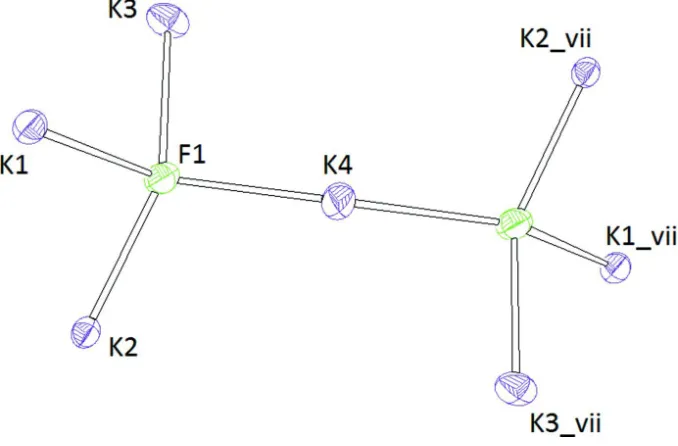
cations are as follows: K1, K2: coordinate, including one F atom; K3: 7-coordinate, including one F atom; K4:
8-coordinate, including two F atoms; K5: 7-8-coordinate, only O atoms. A slightly different understanding of the structure
can be obtained when anion-centred polyhedra are considered as well for the description. Actually, each F- has four
nearest potassium neighbors in form of a tetrahedron. Two symmetry-equivalent tetrahedra are joined by a common
corner (K4) into [F2K7]-double tetrahedra with point group symmetry 1 (Fig. 5). A side view of the whole structure is
given in Fig. 7.
Bond valence sum calculations using the parameter sets for the K—O, K—F, Y—O and Si—O bonds given by Brown
& Altermatt (1985) resulted in the following values (in v.u.) considering cation—anion interactions up to 3.4 Å: K1:
0.924, K2: 0.957, K3: 0.844, K4: 0.772, K5: 1.057, Y1: 3.242, Y2: 3.087, Si1: 4.264, Si2: 4.257, Si3: 4.347, Si4: 4.342,
The present compound is isostructural with a series of rare earth fluoride silicates: K9(REE)3[Si12O32]F2 (REE: Sm, Eu,
Gd; Tang et al., 2008). Chemically related compounds include the following phases: KEu2[Si4O10]F (Jacobsen & Meyer,
1994), Cs2Y[Si4O10]F (Schäfer & Schleid, 2007) and Rb3Sc2[Si4O10]F5 (Schäfer & Schleid, 2011).
S2. Experimental
Single-crystals of K9Y3[Si12O32]F2 were obtained during a series of flux syntheses experiments aiming on the preparation
of new K(REE)-silicate fluorides. 0.1 g of the nutrient consisting of a mixture of Y2O3:SiO2 in the molar ratio 1:4 was
homogenized in an agate mortar with 0.1 g KF, transferred into a platinum tube and welded shut. The container was fired
in a resistance heated furnace from 373 K to 1373 K with a ramp of 50 K/h. The target temperature was held for 2 h.
Subsequently, the sample was cooled down to 1073 K with a rate of 5 K/h and, finally, the temperature was reduced to
373 K with a rate of 100 K/h. The solidified melt cake was immediately crushed in an agate mortar and transferred to a
glass slide under a polarizing binocular. A first optical inspection revealed the presence of two phases: a polycrystalline
matrix of KF in which transparent birefrigent single-crystals up to 200 µm in size were embedded. However, a closer
investigation using crossed polarizers revealed that all crystals showed a fine-scale non-merohedral twinning, making it
impossible to separate a specimen consisting of only one domain state. Therefore, we finally decided to use a twinned
fragment for further structural studies. The crystal was mounted on the tip of a glass fibre using finger nail hardener as
glue.
S3. Refinement
The diffraction patterns were collected at ambient temperature using on Oxford Diffraction Gemini R Ultra single-crystal
diffractometer. They showed the expected complexity due to overlapping of two different reciprocal lattices.
Nevertheless, it was possible to index the reflections from both domains with the same triclinic unit cell but in different
orientations. From the fact that the angle β is close to 90°, the non-merohedral twinning can be readily understood.
Similar sets of lattice parameters could be found in the recent WEB-based version of the Inorganic Crystal Structure
Database (ICSD, 2013) for the chemically closely related compounds K9(REE)3[Si12O32]F2 (REE = Sm, Eu, Gd) pointing
to an isostructural relationship, which was confirmed by the subsequent structure analysis. For structure determination a
full data set (sphere) of reciprocal space was collected. Different integration strategies were tested to handle the problem
of the partially overlapping reflections of both domains, i.e. a series of data sets was produced in which the overlap
threshold was varied stepwise. Different HKLF 5 data sets produced during integration were considered for the
refinement of the structure. However, the best results concerning residuals and overall crystallochemical characteristics
of the structure were obtained when the data set of only the main twin component (representating about 70% of the total
volume) was used, i.e. the completely or partially overlapping reflections have been neglected. However, this approach
supporting information
sup-3
[image:4.610.137.475.75.379.2]Acta Cryst. (2014). E70, i11
Figure 1
Figure 2
Connectivity of the silicon atoms within a single layer. Red and blue spheres represent Q3- and Q2-connected atoms,
supporting information
sup-5
[image:6.610.136.475.73.426.2]Acta Cryst. (2014). E70, i11
Figure 3
Representation of the coordination polyhedron around Y1. Ellipsoids are drawn at the 60% level. [Symmetry codes: (i) 1
Figure 4
Representation of the coordination polyhedron around Y2. Ellipsoids are drawn at the 60% level. Symmetry codes: [(i) 1
supporting information
sup-7
[image:8.610.136.475.73.296.2]Acta Cryst. (2014). E70, i11
Figure 5
Representation of a single [F2K7]-group. Ellipsoids are drawn at the 60% level. [Symmetry codes: (i) 1 - x,-y, 1 - z; (ii) -1
Figure 6
supporting information
sup-9
[image:10.610.139.473.69.414.2]Acta Cryst. (2014). E70, i11
Figure 7
Side view of the whole crystal structure of K9Y3[Si12O32]F2. [SiO4]- and [YO6]-polyhedra are shown in light-grey and
blue. Small grey spheres represent oxygen atoms. Fluorine and potassium ions are given as larger green and pink spheres.
F—K bonds of the [F2K7]-double tetrahedra are indicated.
Nonapotassium triyttrium dodecasilicate difluoride
Crystal data
F2K9O32Si12Y3 Mr = 1505.71
Triclinic, P1 Hall symbol: -P 1
a = 6.8187 (3) Å
b = 11.3345 (4) Å
c = 11.3727 (5) Å
α = 87.846 (3)°
β = 89.747 (4)°
γ = 80.524 (3)°
V = 866.35 (6) Å3
Z = 1
F(000) = 730
Dx = 2.886 Mg m−3
Mo Kα radiation, λ = 0.71073 Å Cell parameters from 4373 reflections
θ = 3.0–29.3°
µ = 6.60 mm−1 T = 298 K
Data collection
Oxford Diffraction Xcalibur diffractometer
Graphite monochromator
Detector resolution: 10.3575 pixels mm-1 ω scans
Absorption correction: multi-scan
(CrysAlis PRO; Oxford Diffraction, 2006)
Tmin = 0.801, Tmax = 1
10508 measured reflections 2842 independent reflections 2231 reflections with I > 2σ(I)
Rint = 0.066
θmax = 25.4°, θmin = 3.3° h = −8→8
k = −13→13
l = −13→13
Refinement
Refinement on F2
Least-squares matrix: full
R[F2 > 2σ(F2)] = 0.039 wR(F2) = 0.099 S = 1.08 2842 reflections 265 parameters 0 restraints
Primary atom site location: structure-invariant direct methods
Secondary atom site location: difference Fourier map
w = 1/[σ2(F
o2) + (0.0515P)2 + 0.2439P]
where P = (Fo2 + 2Fc2)/3
(Δ/σ)max < 0.001
Δρmax = 0.87 e Å−3
Δρmin = −0.95 e Å−3
Special details
Geometry. All s.u.'s (except the s.u. in the dihedral angle between two l.s. planes) are estimated using the full covariance matrix. The cell s.u.'s are taken into account individually in the estimation of s.u.'s in distances, angles and torsion angles; correlations between s.u.'s in cell parameters are only used when they are defined by crystal symmetry. An approximate (isotropic) treatment of cell s.u.'s is used for estimating s.u.'s involving l.s. planes.
Refinement. Refinement of F2 against ALL reflections. The weighted R-factor wR and goodness of fit S are based on F2,
conventional R-factors R are based on F, with F set to zero for negative F2. The threshold expression of F2 > 2σ(F2) is
used only for calculating R-factors(gt) etc. and is not relevant to the choice of reflections for refinement. R-factors based on F2 are statistically about twice as large as those based on F, and R- factors based on ALL data will be even larger.
Fractional atomic coordinates and isotropic or equivalent isotropic displacement parameters (Å2)
x y z Uiso*/Ueq
K1 0.2651 (3) −0.38667 (12) 0.01644 (15) 0.0222 (4) K2 0.1715 (2) −0.19454 (12) −0.22364 (14) 0.0184 (4) K3 0.1624 (3) −0.15506 (14) 0.21410 (15) 0.0277 (4)
K4 0.5 0 0 0.0334 (6)
K5 0.0227 (2) 0.33078 (11) 0.51133 (13) 0.0161 (3) F1 0.2472 (7) −0.1589 (3) −0.0048 (4) 0.0256 (10)
supporting information
sup-11
Acta Cryst. (2014). E70, i11
O4 0.7743 (6) 0.1627 (3) 0.5395 (4) 0.0107 (10) O5 0.1448 (7) 0.1240 (3) 0.3837 (4) 0.0125 (10) O6 0.2791 (7) 0.2030 (3) 0.1831 (4) 0.0129 (10) O7 0.4575 (7) 0.0054 (3) 0.2739 (4) 0.0132 (10) O8 0.6920 (7) −0.1638 (3) 0.1818 (4) 0.0102 (10) O9 0.8444 (7) −0.0336 (3) 0.3338 (4) 0.0114 (10) O10 0.9033 (7) −0.3743 (3) 0.2102 (4) 0.0139 (10) O11 0.6983 (7) −0.3156 (3) 0.0189 (4) 0.0169 (11) O12 0.5030 (7) −0.3527 (3) 0.2148 (4) 0.0123 (10) O13 0.1635 (7) −0.5084 (3) 0.3462 (4) 0.0114 (10) O14 −0.1887 (6) −0.5722 (3) 0.3149 (4) 0.0121 (10) O15 0.0686 (7) −0.5849 (3) 0.1393 (4) 0.0131 (10) O16 0.4610 (7) −0.6045 (3) 0.1658 (4) 0.0123 (10)
Atomic displacement parameters (Å2)
U11 U22 U33 U12 U13 U23
Geometric parameters (Å, º)
K1—F1 2.567 (4) Si1—O3 1.631 (5)
K1—O16i 2.778 (5) Si1—O2 1.662 (5)
K1—O12 2.858 (5) Si2—O5 1.574 (5)
K1—O15ii 2.956 (5) Si2—O2 1.625 (4)
K1—O16 3.064 (5) Si2—O7 1.629 (4)
K1—O15 3.088 (5) Si2—O6 1.633 (5)
K1—O11 3.188 (5) Si3—O9 1.578 (4)
K1—O10iii 3.289 (5) Si3—O3vi 1.613 (4)
K1—O11i 3.382 (4) Si3—O7 1.623 (4)
K2—F1 2.604 (4) Si3—O8 1.628 (4)
K2—O9iv 2.816 (4) Si4—O12 1.584 (5)
K2—O5v 2.821 (5) Si4—O11 1.602 (5)
K2—O2iv 2.851 (5) Si4—O10 1.635 (5)
K2—O14ii 2.861 (4) Si4—O8 1.635 (4)
K2—O6v 3.121 (5) Si5—O13 1.579 (5)
K2—O16i 3.150 (5) Si5—O14 1.588 (4)
K2—O15ii 3.317 (4) Si5—O10iii 1.649 (4)
K3—F1 2.554 (5) Si5—O15 1.657 (5)
K3—O9iii 2.754 (5) Si6—O16 1.578 (4)
K3—O4vi 2.833 (5) Si6—O11i 1.601 (5)
K3—O12 2.949 (5) Si6—O6xi 1.620 (4)
K3—O7 3.021 (4) Si6—O15 1.642 (5)
K3—O8iii 3.248 (5) O1—Y2x 2.310 (4)
K3—O10iii 3.279 (4) O1—K5xii 3.321 (5)
K4—F1iv 2.692 (4) O2—K2iv 2.851 (5)
K4—F1 2.692 (4) O3—Si3vi 1.613 (4)
K4—O8iv 2.893 (4) O4—Y1xii 2.256 (4)
K4—O8 2.893 (4) O4—K5xii 2.759 (4)
K4—O7 3.129 (5) O4—K3vi 2.833 (5)
K4—O7iv 3.129 (5) O5—K2v 2.821 (5)
K4—O6iv 3.330 (4) O6—Si6vii 1.620 (4)
K4—O6 3.330 (4) O6—K2v 3.121 (5)
K5—O14vii 2.756 (5) O8—K3xii 3.248 (5)
K5—O4iii 2.759 (4) O9—Y1xii 2.247 (4)
K5—O13viii 2.771 (4) O9—K3xii 2.754 (5)
K5—O5 2.810 (4) O9—K2iv 2.816 (4)
supporting information
sup-13
Acta Cryst. (2014). E70, i11
Y2—O14ix 2.211 (4) O14—Y2xiii 2.211 (4)
Y2—O13vii 2.213 (4) O14—K5xi 2.756 (5)
Y2—O12vii 2.250 (4) O14—K2ii 2.861 (4)
Y2—O16vii 2.275 (4) O15—K1ii 2.956 (5)
Y2—O1x 2.310 (4) O15—K2ii 3.317 (4)
Y2—O1 2.345 (4) O16—Y2xi 2.275 (4)
Si1—O4 1.590 (5) O16—K1i 2.778 (5)
Si1—O1 1.603 (4) O16—K2i 3.150 (5)
O5—Y1—O5viii 180 O5—Si2—O7 112.2 (2)
O5—Y1—O9vi 96.02 (15) O2—Si2—O7 109.0 (2)
O5viii—Y1—O9vi 83.98 (15) O5—Si2—O6 110.5 (3)
O5—Y1—O9iii 83.98 (15) O2—Si2—O6 107.2 (2)
O5viii—Y1—O9iii 96.02 (15) O7—Si2—O6 102.9 (2)
O9vi—Y1—O9iii 180 O9—Si3—O3vi 111.7 (2)
O5—Y1—O4vi 94.15 (15) O9—Si3—O7 114.3 (2)
O5viii—Y1—O4vi 85.85 (15) O3vi—Si3—O7 109.8 (2)
O9vi—Y1—O4vi 93.05 (15) O9—Si3—O8 110.7 (2)
O9iii—Y1—O4vi 86.95 (15) O3vi—Si3—O8 106.9 (2)
O5—Y1—O4iii 85.85 (15) O7—Si3—O8 102.9 (2)
O5viii—Y1—O4iii 94.15 (15) O12—Si4—O11 112.3 (3)
O9vi—Y1—O4iii 86.95 (15) O12—Si4—O10 114.1 (2)
O9iii—Y1—O4iii 93.05 (15) O11—Si4—O10 106.7 (3)
O4vi—Y1—O4iii 180.0000 (10) O12—Si4—O8 113.3 (2)
O14ix—Y2—O13vii 162.32 (14) O11—Si4—O8 105.2 (2)
O14ix—Y2—O12vii 90.23 (16) O10—Si4—O8 104.5 (2)
O13vii—Y2—O12vii 100.81 (16) O13—Si5—O14 113.5 (2)
O14ix—Y2—O16vii 84.41 (16) O13—Si5—O10iii 107.8 (2)
O13vii—Y2—O16vii 83.37 (16) O14—Si5—O10iii 110.8 (2)
O12vii—Y2—O16vii 82.65 (15) O13—Si5—O15 110.3 (2)
O14ix—Y2—O1x 103.85 (16) O14—Si5—O15 109.2 (2)
O13vii—Y2—O1x 90.94 (16) O10iii—Si5—O15 104.9 (2)
O12vii—Y2—O1x 85.09 (15) O16—Si6—O11i 111.4 (3)
O16vii—Y2—O1x 165.26 (15) O16—Si6—O6xi 114.0 (2)
O14ix—Y2—O1 84.94 (16) O11i—Si6—O6xi 107.7 (2)
O13vii—Y2—O1 90.07 (16) O16—Si6—O15 111.7 (2)
O12vii—Y2—O1 156.35 (15) O11i—Si6—O15 104.5 (2)
O16vii—Y2—O1 119.73 (14) O6xi—Si6—O15 106.9 (2)
O1x—Y2—O1 73.70 (14) Si2—O2—Si1 137.7 (3)
O4—Si1—O1 115.9 (2) Si3vi—O3—Si1 147.0 (3)
O4—Si1—O3 111.7 (2) Si6vii—O6—Si2 137.6 (3)
O1—Si1—O3 108.3 (2) Si3—O7—Si2 142.8 (3)
O4—Si1—O2 110.8 (2) Si3—O8—Si4 129.8 (3)
O3—Si1—O2 105.4 (2) Si4—O11—Si6i 177.1 (3)
O5—Si2—O2 114.3 (2) Si6—O15—Si5 127.5 (3)

![Figure 6The dimer formed from the condensation of two edge-sharing [Y2O6]-octahedra.](https://thumb-us.123doks.com/thumbv2/123dok_us/481213.546593/9.610.196.411.71.496/figure-the-dimer-formed-condensation-edge-sharing-octahedra.webp)
![Figure 7Side view of the whole crystal structure of K9Y3[Si12O32]F2. [SiO4]- and [YO6]-polyhedra are shown in light-grey and](https://thumb-us.123doks.com/thumbv2/123dok_us/481213.546593/10.610.139.473.69.414/figure-side-view-crystal-structure-polyhedra-shown-light.webp)