Crystal structure of (7-methyl-2-oxo-2
H
-chromen-4-yl)methyl
piperidine-1-carbo-dithioate
K. R. Roopashree,aT. G. Meenakshi,bK. Mahesh Kumar,c O. Kotreshcand H. C. Devarajegowdaa*
aDepartment of Physics, Yuvaraja’s College (Constituent College), University of
Mysore, Mysore 570 005, Karnataka, India,bDepartment of Physics, Y.Y.D. Govt.
First Grade College, Belur 573 115 Hassan, Karnataka, India, andcDepartment of
Chemistry, Karnatak University’s Karnatak Science College, Dharwad, Karnataka 580 001, India. *Correspondence e-mail: devarajegowda@yahoo.com
Received 31 May 2015; accepted 20 July 2015
Edited by M. Zeller, Youngstown State University, USA
In the title compound, C17H19NO2S2, the 2H-chromene ring system is nearly planar, with a maximum deviation of 0.0383 (28) A˚ , and the piperidine ring adopts a chair conformation. The 2H-chromene ring makes dihedral angles of 32.89 (16) and 67.33 (8), respectively, with the mean planes
of the piperidine ring and the carbodithioate group. In the crystal, C—H O and weak C—H S hydrogen bonds link the molecules into chains along [001]. The crystal structure also features C—H and–interactions, with a centroid– centroid distance of 3.7097 (17) A˚ .
Keywords:crystal structure; 2H-chromene; hydrogen bonding; C—H
interactions;–interactions.
CCDC reference:1413856
1. Related literature
For biological applications of coumarins, see: Stiefel et al. (1995); Murrayet al. (1982); Khanet al.(2004); Kawaiiet al. (2001); Yuet al.(2003). For biological applications of dithia-carbamates, see: D’hooghe & de Kime (2006); Thorn & Ludwig (1962); Caoet al.(2005). For a related structure, see: Kumaret al.(2013).
2. Experimental
2.1. Crystal data
C17H19NO2S2 Mr= 333.45
Monoclinic,Pc a= 4.9641 (2) A˚ b= 11.4351 (3) A˚ c= 14.0023 (4) A˚
= 90.743 (2)
V= 794.77 (4) A˚3 Z= 2
MoKradiation
= 0.34 mm1 T= 296 K
0.240.200.12 mm
2.2. Data collection
Bruker SMART CCD area-detector diffractometer
Absorption correction: scan (SADABS; Sheldrick, 2007) Tmin= 0.770,Tmax= 1.000
4426 measured reflections 2119 independent reflections 2027 reflections withI> 2(I) Rint= 0.017
2.3. Refinement
R[F2> 2(F2)] = 0.026 wR(F2) = 0.062 S= 1.04 2119 reflections 200 parameters 2 restraints
H-atom parameters constrained
max= 0.14 e A˚
3
min=0.13 e A˚
3 Absolute structure: Flackx
determined using 704 quotients [(I+)(I
)]/[(I+)+(I
)] (Parsons et al., 2013)
Absolute structure parameter: 0.05 (3)
Table 1
Hydrogen-bond geometry (A˚ ,).
D—H A D—H H A D A D—H A
C13—H13 O4i
0.93 2.45 3.223 (4) 140 C16—H16B S2 0.97 2.70 3.152 (4) 109 C18—H18B S1 0.97 2.38 2.930 (4) 116 C22—H22A S2 0.97 2.55 3.065 (4) 113
Symmetry code: (i)x1;yþ2;zþ1 2.
Data collection: SMART (Bruker, 2001); cell refinement: SAINT
(Bruker, 2001); data reduction:SAINT; program(s) used to solve structure:SHELXS2014(Sheldrick, 2008); program(s) used to refine structure: SHELXL2014 (Sheldrick, 2015); molecular graphics:
ORTEP-3 for Windows(Farrugia, 2012); software used to prepare material for publication:SHELXL2014.
data reports
o606
Roopashreeet al. doi:10.1107/S2056989015013699 Acta Cryst.(2015).E71, o606–o607Acknowledgements
The authors thank the Universities Sophisticated Instrumental Centre, Karnatak University, Dharwad for access to their CCD X-ray facilities, the X-ray data collection, and GCMS, IR, CHNS analysis and NMR data.
Supporting information for this paper is available from the IUCr electronic archives (Reference: ZL2626).
References
Bruker (2001).SMARTandSAINT.Bruker AXS Inc., Madison, Wisconsin, USA.
Cao, S. L., Feng, Y. P., Jiang, Y. Y., Liu, S. Y., Ding, G. Y. & Li, R. T. (2005). Bioorg. Med. Chem. Lett.15, 1915–1917.
D’hooghe, M. & de Kime, N. (2006).Tetrahedron,62, 513–535. Farrugia, L. J. (2012).J. Appl. Cryst.45, 849–854.
Kawaii, S., Tomono, Y., Ogawa, K., Sugiura, M., Yano, M. & Yoshizawa, Y. (2001).Anticancer Res.21, 917–923.
Khan, K. M., Saify, Z. S., Khan, M. Z., Zia-Ullah, M. Z., Choudhary, I. M., Atta-ur-Rahman, , Perveen, S., Chohan, Z. H. & Supuran, C. T. (2004).J. Enzyme Inhib. Med. Chem.19, 373–379.
Kumar, K. M., Vinduvahini, M., Mahabhaleshwaraiah, N. M., Kotresh, O. & Devarajegowda, H. C. (2013).Acta Cryst.E69, o1683.
Murray, R. D. H., Mendez, J. & Brown, S. A. (1982).The Natural Coumarins: Occurrence, Chemistry and Biochemistry, p. 21. New York: John Wiley & Sons Ltd.
Parsons, S., Flack, H. D. & Wagner, T. (2013).Acta Cryst.B69, 249–259. Sheldrick, G. M. (2007).SADABS. Bruker AXS Inc., Madison, Wisconsin,
USA.
Sheldrick, G. M. (2008).Acta Cryst.A64, 112–122. Sheldrick, G. M. (2015).Acta Cryst.C71, 3–8.
Stiefel, E. I. & Matsumoto, K. (1995). InTransition Metal Sulfur Chemistry, ASC Symposium Series, Vol. 653. Washington, DC: American Chemical Society.
Thorn, G. D. & Ludwig, R. A. (1962). InThe Dithiocarbamates and Related Compounds. Amsterdam: Elsevier.
supporting information
sup-1
Acta Cryst. (2015). E71, o606–o607
supporting information
Acta Cryst. (2015). E71, o606–o607 [https://doi.org/10.1107/S2056989015013699]
Crystal structure of (7-methyl-2-oxo-2
H
-chromen-4-yl)methyl
piperidine-1-carbodithioate
K. R. Roopashree, T. G. Meenakshi, K. Mahesh Kumar, O. Kotresh and H. C. Devarajegowda
S1. Comment
Coumarins and their derivatives play an important role in the agricultural and pharmaceutical industries (Stiefel et al., 1995). They are widely present in higher plants such as Rutaceae, Apiaceae, Asteraceae, Leguminosae, Thymelaeaceae,
and they also occur as animal and microbial metabolites (Murray et al., 1982). Most of them show a wide spectrum of pharmacological effects, including antimicrobial (Khan et al., 2004), anti-arrhythmic, antiosteoporosis, anti-HIV, and antitumor activities (Kawaii et al., 2001; Yu et al., 2003). Accordingly, many reports have described various structures and biological evaluations of numerous coumarin analogs newly synthesized or isolated from plants.
Sulfur containing molecules are currently under study as chemoprotectants in chemotherapy. Organic substances with a
dithio functional group have been widely used in industry as rodent repellents, vulcanization additives in rubber
manufacturing, additives in lubricants, and in agriculture as fungicides on almond trees, stone fruits, and vegetables.
Among the various sulfur ligands being examined currently, dithiocarbamates have a special significance owing to their
many uses, e.g. in analytical determinations, as arrestors of human immunodeficiency virus infections such as AIDS, in pharmaceutical products, in agriculture as pesticides and fungicides, and as high-pressure lubricants (D'hooghe & de
Kime, 2006; Thorn & Ludwig, 1962; Cao et al., 2005). One molecule of (7-methyl-2-oxo-2H -chromen-4-yl)methyl-piperidine-1-carbodithioate is shown in Fig. 1. The 2Hchromene ring system (O3/C6–C13/C15) is essentially planar, with a maximum deviation of 0.0383 (28) Å for atom C10 and the piperidine (N5/C18–C22) ring adopts a chair conformation.
The dihedral angle of the 2H-chromene (O3/C6–C13/C15) ring with the piperidine (N5/C18–C22) ring and carbodithio-ate group are 32.89 (16)° and 67.33 (8)°, respectively. In addition, intermolecular C—H···O and weak C—H···S hydrogen
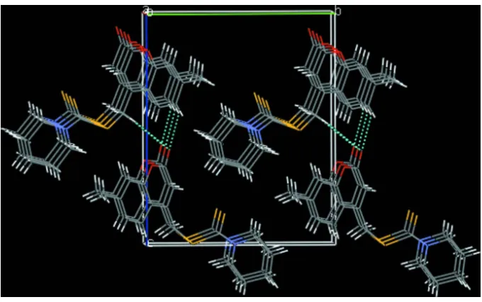
bonds (Table 1) link the components into chains along [001]. The crystal structure also features C—H···π [Cg(3) (C9– C15)] and [Cg(1) (O3/C6–C10)]π–π [Cg(3) (C9–C15)] interactions, with centroid–centroid distances of 3.7081 (15) Å that further stabilize the crystal packing, Figure 2.
S2. Experimental
The title compound compound was prepared according to a reported method (Kumar et al., 2013). Colourless needles of the title compound were grown from a mixed solution of EtOH/CHCl3 (v/v = 1/1) by slow evaporation at room
temperature. Yield: 80%, m.p. 420 K.
S3. Refinement
All H atoms were positioned geometrically, with C—H = 0.93 Å for aromatic H, C—H = 0.97 Å for methylene H and C
Figure 1
The molecular structure of the title compound. Displacement ellipsoids are drawn at the 50% probability level. Hydrogen
atoms are shown as spheres of arbitrary radius.
Figure 2
[image:4.610.135.475.446.659.2]supporting information
sup-3
Acta Cryst. (2015). E71, o606–o607
(7-Methyl-2-oxo-2H-chromen-4-yl)methyl piperidine-1-carbodithioate
Crystal data
C17H19NO2S2
Mr = 333.45
Monoclinic, Pc a = 4.9641 (2) Å
b = 11.4351 (3) Å
c = 14.0023 (4) Å
β = 90.743 (2)°
V = 794.77 (4) Å3
Z = 2
F(000) = 352
Dx = 1.393 Mg m−3 Melting point: 420 K
Mo Kα radiation, λ = 0.71073 Å Cell parameters from 2119 reflections
θ = 1.8–25.0°
µ = 0.34 mm−1
T = 296 K Plate, colourless 0.24 × 0.20 × 0.12 mm
Data collection
Bruker SMART CCD area-detector diffractometer
Radiation source: fine-focus sealed tube Graphite monochromator
Detector resolution: 10.0 pixels mm-1
ω and φ scans
Absorption correction: ψ scan (SADABS; Sheldrick, 2007)
Tmin = 0.770, Tmax = 1.000
4426 measured reflections 2119 independent reflections 2027 reflections with I > 2σ(I)
Rint = 0.017
θmax = 25.0°, θmin = 1.8°
h = −5→5
k = −13→13
l = −13→16
Refinement
Refinement on F2 Least-squares matrix: full
R[F2 > 2σ(F2)] = 0.026
wR(F2) = 0.062
S = 1.04 2119 reflections 200 parameters 2 restraints
Hydrogen site location: inferred from neighbouring sites
H-atom parameters constrained
w = 1/[σ2(F
o2) + (0.0331P)2 + 0.0991P] where P = (Fo2 + 2Fc2)/3
(Δ/σ)max < 0.001 Δρmax = 0.14 e Å−3 Δρmin = −0.13 e Å−3
Absolute structure: Flack x determined using 704 quotients [(I+)-(I-)]/[(I+)+(I-)] (Parsons et al., 2013)
Absolute structure parameter: 0.05 (3)
Special details
Geometry. All esds (except the esd in the dihedral angle between two l.s. planes) are estimated using the full covariance matrix. The cell esds are taken into account individually in the estimation of esds in distances, angles and torsion angles; correlations between esds in cell parameters are only used when they are defined by crystal symmetry. An approximate (isotropic) treatment of cell esds is used for estimating esds involving l.s. planes.
Fractional atomic coordinates and isotropic or equivalent isotropic displacement parameters (Å2)
x y z Uiso*/Ueq
H7 0.5761 0.8152 0.2518 0.045* C8 0.3150 (6) 0.9079 (2) 0.3245 (2) 0.0324 (7) C9 0.1022 (6) 0.9951 (2) 0.3146 (2) 0.0320 (7) C10 0.0312 (6) 1.0321 (2) 0.2230 (2) 0.0331 (7) C11 −0.1659 (6) 1.1147 (3) 0.2057 (2) 0.0373 (7) H11 −0.2102 1.1357 0.1433 0.045* C12 −0.2979 (6) 1.1663 (2) 0.2814 (2) 0.0370 (7) C13 −0.2277 (6) 1.1305 (3) 0.3735 (2) 0.0396 (8) H13 −0.3149 1.1640 0.4252 0.047* C14 −0.5104 (7) 1.2582 (3) 0.2648 (3) 0.0509 (9) H14A −0.5426 1.2672 0.1975 0.076* H14B −0.4501 1.3312 0.2913 0.076* H14C −0.6742 1.2348 0.2952 0.076* C15 −0.0339 (6) 1.0474 (2) 0.3902 (2) 0.0366 (7) H15 0.0076 1.0254 0.4526 0.044* C16 0.4049 (6) 0.8639 (3) 0.4206 (2) 0.0379 (7) H16A 0.4527 0.9305 0.4601 0.045* H16B 0.5665 0.8174 0.4125 0.045* C17 0.2071 (6) 0.6311 (3) 0.4400 (2) 0.0378 (7) C18 −0.1184 (8) 0.5752 (3) 0.5662 (3) 0.0561 (10) H18A −0.3046 0.5577 0.5499 0.067* H18B −0.1071 0.6570 0.5839 0.067* C19 −0.0287 (10) 0.5001 (3) 0.6495 (3) 0.0669 (11) H19A 0.1498 0.5243 0.6704 0.080* H19B −0.1507 0.5116 0.7022 0.080* C20 −0.0229 (10) 0.3718 (3) 0.6234 (3) 0.0746 (13) H20A −0.2056 0.3438 0.6136 0.090* H20B 0.0579 0.3275 0.6754 0.090* C21 0.1378 (9) 0.3527 (3) 0.5330 (3) 0.0733 (14) H21A 0.3265 0.3689 0.5461 0.088* H21B 0.1224 0.2716 0.5136 0.088* C22 0.0400 (8) 0.4302 (3) 0.4529 (3) 0.0630 (11) H22A 0.1521 0.4192 0.3974 0.076* H22B −0.1436 0.4095 0.4353 0.076*
Atomic displacement parameters (Å2)
supporting information
sup-5
Acta Cryst. (2015). E71, o606–o607
C11 0.0458 (19) 0.0343 (16) 0.0319 (18) −0.0061 (15) −0.0001 (15) 0.0054 (13) C12 0.0410 (17) 0.0295 (16) 0.0407 (19) −0.0082 (13) 0.0063 (15) 0.0018 (14) C13 0.0464 (19) 0.0342 (16) 0.038 (2) −0.0052 (15) 0.0136 (16) −0.0059 (13) C14 0.049 (2) 0.0407 (19) 0.063 (2) 0.0030 (16) 0.0030 (19) 0.0030 (16) C15 0.0479 (18) 0.0354 (16) 0.0266 (16) −0.0073 (15) 0.0026 (15) 0.0009 (13) C16 0.0450 (19) 0.0350 (16) 0.0338 (18) −0.0026 (14) 0.0008 (15) −0.0004 (14) C17 0.0439 (18) 0.0316 (16) 0.0377 (19) 0.0058 (14) −0.0067 (15) −0.0004 (13) C18 0.061 (2) 0.040 (2) 0.068 (3) 0.0003 (17) 0.014 (2) 0.0126 (17) C19 0.082 (3) 0.055 (2) 0.064 (3) −0.003 (2) −0.001 (2) 0.0077 (19) C20 0.085 (3) 0.048 (2) 0.090 (4) −0.011 (2) −0.035 (3) 0.024 (2) C21 0.073 (3) 0.038 (2) 0.109 (4) −0.001 (2) −0.026 (3) −0.002 (2) C22 0.075 (3) 0.0324 (18) 0.081 (3) −0.0030 (18) 0.003 (2) −0.0062 (18)
Geometric parameters (Å, º)
S1—C17 1.786 (3) C13—H13 0.9300 S1—C16 1.812 (3) C14—H14A 0.9600 S2—C17 1.657 (3) C14—H14B 0.9600 O3—C6 1.370 (4) C14—H14C 0.9600 O3—C10 1.378 (3) C15—H15 0.9300 O4—C6 1.202 (4) C16—H16A 0.9700 N5—C17 1.330 (4) C16—H16B 0.9700 N5—C18 1.461 (4) C18—C19 1.511 (5) N5—C22 1.475 (4) C18—H18A 0.9700 C6—C7 1.447 (4) C18—H18B 0.9700 C7—C8 1.346 (4) C19—C20 1.512 (6) C7—H7 0.9300 C19—H19A 0.9700 C8—C9 1.458 (4) C19—H19B 0.9700 C8—C16 1.499 (4) C20—C21 1.521 (6) C9—C10 1.392 (4) C20—H20A 0.9700 C9—C15 1.397 (4) C20—H20B 0.9700 C10—C11 1.379 (4) C21—C22 1.505 (6) C11—C12 1.385 (4) C21—H21A 0.9700 C11—H11 0.9300 C21—H21B 0.9700 C12—C13 1.394 (5) C22—H22A 0.9700 C12—C14 1.505 (4) C22—H22B 0.9700 C13—C15 1.371 (5)
C6—C7—H7 118.7 C19—C18—H18A 109.6 C7—C8—C9 118.7 (3) N5—C18—H18B 109.6 C7—C8—C16 119.8 (3) C19—C18—H18B 109.6 C9—C8—C16 121.5 (3) H18A—C18—H18B 108.1 C10—C9—C15 116.7 (3) C20—C19—C18 111.8 (4) C10—C9—C8 118.0 (3) C20—C19—H19A 109.3 C15—C9—C8 125.3 (3) C18—C19—H19A 109.3 C11—C10—O3 115.7 (3) C20—C19—H19B 109.3 C11—C10—C9 122.7 (3) C18—C19—H19B 109.3 O3—C10—C9 121.6 (3) H19A—C19—H19B 107.9 C10—C11—C12 120.0 (3) C19—C20—C21 110.6 (3) C10—C11—H11 120.0 C19—C20—H20A 109.5 C12—C11—H11 120.0 C21—C20—H20A 109.5 C11—C12—C13 117.9 (3) C19—C20—H20B 109.5 C11—C12—C14 121.2 (3) C21—C20—H20B 109.5 C13—C12—C14 120.9 (3) H20A—C20—H20B 108.1 C15—C13—C12 121.9 (3) C22—C21—C20 111.6 (4) C15—C13—H13 119.1 C22—C21—H21A 109.3 C12—C13—H13 119.1 C20—C21—H21A 109.3 C12—C14—H14A 109.5 C22—C21—H21B 109.3 C12—C14—H14B 109.5 C20—C21—H21B 109.3 H14A—C14—H14B 109.5 H21A—C21—H21B 108.0 C12—C14—H14C 109.5 N5—C22—C21 109.7 (3) H14A—C14—H14C 109.5 N5—C22—H22A 109.7 H14B—C14—H14C 109.5 C21—C22—H22A 109.7 C13—C15—C9 120.9 (3) N5—C22—H22B 109.7 C13—C15—H15 119.6 C21—C22—H22B 109.7 C9—C15—H15 119.6 H22A—C22—H22B 108.2 C8—C16—S1 115.4 (2)
supporting information
sup-7
Acta Cryst. (2015). E71, o606–o607
C10—C11—C12—C13 −1.1 (4) C17—N5—C22—C21 117.6 (4) C10—C11—C12—C14 178.9 (3) C18—N5—C22—C21 −60.2 (4) C11—C12—C13—C15 0.3 (4) C20—C21—C22—N5 56.3 (4)
Hydrogen-bond geometry (Å, º)
D—H···A D—H H···A D···A D—H···A
C13—H13···O4i 0.93 2.45 3.223 (4) 140 C16—H16B···S2 0.97 2.70 3.152 (4) 109 C18—H18B···S1 0.97 2.38 2.930 (4) 116 C22—H22A···S2 0.97 2.55 3.065 (4) 113