2-Nitro-1,3-dinitrooxypropane
Megan M. Breiner,aDavid E. Chavezb and Damon A. Parrishc*
a
MS C920, Los Alamos National Laboratory, Los Alamos, NM 87545, USA,
b
Technical Staff Member, MS C920, Los Alamos National Laboratory, Los Alamos, NM 87545, USA, andcCBMSE, Code 6910, Naval Research Laboratory,
Washington, DC 20375, USA
Correspondence e-mail: [email protected]
Received 28 January 2013; accepted 11 February 2013
Key indicators: single-crystal X-ray study;T= 293 K; mean(C–C) = 0.003 A˚; Rfactor = 0.038;wRfactor = 0.083; data-to-parameter ratio = 12.0.
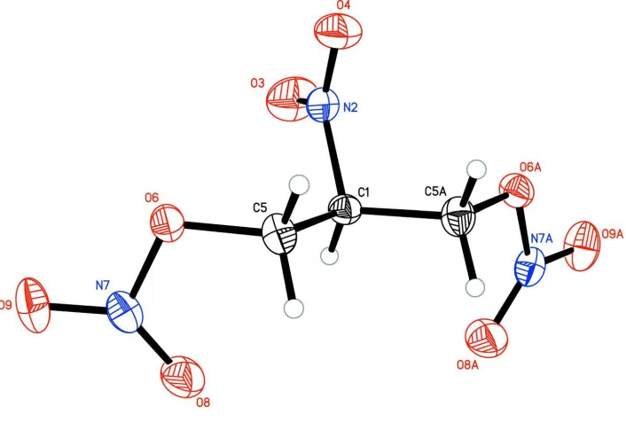
The title compound, C3H5N3O8, was synthesized by reacting
2-nitropropane-1,3-diol with acetyl nitrate. The molecule is bisected by a crystallograpic mirror plane. In the crystal, the molecules pack in a ribbon-like fashion along thecaxis, with the central nitro groups pointing in the same direction. C— H O contacts apparently provide some additional packing stabilization.
Related literature
Nitrate esters are often studied for their energetic materials properties. For example, we have reported the synthesis and crystal structure of a low melting nitrate ester (Chavez,et al.
2008)The title compound was first synthesized by Ro¨mer (1955) but no information has been reported on the crystal structure of this material. A smilar structure was reported that differs only in a nitrooxy group at the 2-position (Espenbetov
et al.1984).
Experimental
Crystal data
C3H5N3O8
Mr= 211.10 Orthorhombic,Cmc21
a= 14.046 (5) A˚
b= 9.607 (5) A˚
c= 5.903 (3) A˚
V= 796.5 (7) A˚3
Z= 4
MoKradiation
= 0.18 mm1
T= 293 K
0.380.020.01 mm
Data collection
Bruker SMART APEXII CCD diffractometer
Absorption correction: multi-scan (SADABS; Bruker, 2008)
Tmin= 0.935,Tmax= 0.998
3416 measured reflections 841 independent reflections 587 reflections withI> 2(I)
Rint= 0.052
Refinement
R[F2> 2(F2)] = 0.038
wR(F2) = 0.083
S= 1.00 841 reflections 70 parameters
1 restraint
H-atom parameters constrained max= 0.15 e A˚
3
min=0.18 e A˚3
Table 1
Hydrogen-bond geometry (A˚ ,).
D—H A D—H H A D A D—H A
C5—H5B O4i
0.97 2.56 3.405 (5) 145
Symmetry code: (i)xþ1;yþ1;z1 2.
Data collection:APEX2(Bruker, 2009); cell refinement:SAINT (Bruker, 2009); data reduction:SAINTandXPREP(Bruker, 2008); program(s) used to solve structure: SHELXTL (Sheldrick, 2008); program(s) used to refine structure:SHELXTL; molecular graphics: SHELXTL; software used to prepare material for publication: SHELXTL.
The authors would like to thank the DoD/DOE Joint Munitions Technology Development Program. Los Alamos National Laboratory is operated by Los Alamos National Security (LANS, LLC) under contract No. DE—AC52–06 N A25396 for the US Department of Energy. Crystallographic studies were supported in part by the Office of Naval Research (ONR) and the Naval Research Laboratory (NRL).
Supplementary data and figures for this paper are available from the IUCr electronic archives (Reference: LD2094).
References
Bruker (2008).SADABSandXPREP. Bruker AXS Inc., Madison, Wisconsin, USA.
Bruker (2009).APEX2andSAINT. Bruker AXS Inc., Madison, Wisconsin, USA.
Chavez, D. E., Hiskey, M. A., Naud, D. L. & Parrish, D. A. (2008).Angew. Chem. Int. Ed.23, 8307–8309.
Espenbetov, A. A., Antipin, M. Yu., Struchkov, Yu. T., Philippov, V. A., Tsirel’son, V. G., Ozerov, R. P. & Svetlov, B. S. (1984).Acta Cryst.C40, 2096– 2098.
Ro¨mer, F. (1955).Angew. Chem.67, 157. Sheldrick, G. M. (2008).Acta Cryst.A64, 112–122.
Acta Crystallographica Section E Structure Reports
Online
supporting information
Acta Cryst. (2013). E69, o384 [doi:10.1107/S1600536813004170]
2-Nitro-1,3-dinitrooxypropane
Megan M. Breiner, David E. Chavez and Damon A. Parrish
S1. Comment
In our efforts to synthesize energetic materials with novel properties, we identified the title compound,
2-nitro-1,3-di-nitrooxypropane (Fig. 1), as a nitrate ester for which very little information exists in the literature. The compound was
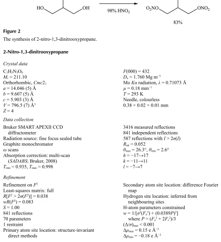
synthesized in a one- step process whereby 2-nitropropane-1,3-diol was subjected to nitration conditions using acetyl
nitrate as the substrate (Fig. 2). Suitable crystals were grown from carbon tetrachloride and subjected to X-ray analysis
for structure confirmation. The molecule lies on a mirror plane, making only half of the molecule crystallographically
unique.
S2. Experimental
Crystals suitable for X-ray crystallographic analysis were grown from carbon tetrachloride. The crystals were isolated as
white needles.
S3. Refinement
The full-matrix least-squares refinement on F2 included atomic coordinates and anisotropic thermal parameters for all
[image:2.610.130.483.433.671.2]non-H atoms. The H atoms were included using a riding model.
Figure 1
Figure 2
The synthesis of 2-nitro-1,3-dinitrooxypropane.
2-Nitro-1,3-dinitrooxypropane
Crystal data
C3H5N3O8 Mr = 211.10
Orthorhombic, Cmc21 a = 14.046 (5) Å b = 9.607 (5) Å c = 5.903 (3) Å V = 796.5 (7) Å3 Z = 4
F(000) = 432 Dx = 1.760 Mg m−3
Mo Kα radiation, λ = 0.71073 Å µ = 0.18 mm−1
T = 293 K Needle, colourless 0.38 × 0.02 × 0.01 mm
Data collection
Bruker SMART APEXII CCD diffractometer
Radiation source: fine focus sealed tube Graphite monochromator
ω scans
Absorption correction: multi-scan (SADABS; Bruker, 2008) Tmin = 0.935, Tmax = 0.998
3416 measured reflections 841 independent reflections 587 reflections with I > 2σ(I) Rint = 0.052
θmax = 26.3°, θmin = 2.6° h = −17→17
k = −11→11 l = −7→7
Refinement
Refinement on F2 Least-squares matrix: full R[F2 > 2σ(F2)] = 0.038 wR(F2) = 0.083 S = 1.00 841 reflections 70 parameters 1 restraint
Primary atom site location: structure-invariant direct methods
Secondary atom site location: difference Fourier map
Hydrogen site location: inferred from neighbouring sites
H-atom parameters constrained w = 1/[σ2(F
o2) + (0.0389P)2] where P = (Fo2 + 2Fc2)/3 (Δ/σ)max < 0.001
Δρmax = 0.15 e Å−3 Δρmin = −0.18 e Å−3
Special details
Geometry. All e.s.d.'s (except the e.s.d. in the dihedral angle between two l.s. planes) are estimated using the full covariance matrix. The cell e.s.d.'s are taken into account individually in the estimation of e.s.d.'s in distances, angles and torsion angles; correlations between e.s.d.'s in cell parameters are only used when they are defined by crystal symmetry. An approximate (isotropic) treatment of cell e.s.d.'s is used for estimating e.s.d.'s involving l.s. planes.
Refinement. Refinement of F2 against ALL reflections. The weighted R-factor wR and goodness of fit S are based on F2, conventional R-factors R are based on F, with F set to zero for negative F2. The threshold expression of F2 > σ(F2) is used only for calculating R-factors(gt) etc. and is not relevant to the choice of reflections for refinement. R-factors based on F2 are statistically about twice as large as those based on F, and R- factors based on ALL data will be even larger.
Fractional atomic coordinates and isotropic or equivalent isotropic displacement parameters (Å2)
x y z Uiso*/Ueq
C1 0.5000 0.2314 (4) 0.4290 (7) 0.0314 (8) H1 0.5000 0.1318 0.3932 0.038* N2 0.5000 0.2530 (4) 0.6828 (6) 0.0392 (8) O3 0.5000 0.1504 (4) 0.8026 (5) 0.0743 (10) O4 0.5000 0.3716 (4) 0.7543 (5) 0.0606 (9) C5 0.58746 (15) 0.3011 (3) 0.3296 (5) 0.0386 (6) H5A 0.5877 0.2874 0.1669 0.046* H5B 0.5839 0.4004 0.3581 0.046* O6 0.67514 (11) 0.24806 (16) 0.4225 (4) 0.0404 (5) N7 0.70956 (17) 0.1273 (2) 0.3130 (4) 0.0429 (6) O8 0.65815 (14) 0.07181 (19) 0.1797 (5) 0.0614 (6) O9 0.78766 (13) 0.0968 (2) 0.3745 (4) 0.0611 (7)
Atomic displacement parameters (Å2)
U11 U22 U33 U12 U13 U23
C1 0.0337 (18) 0.028 (2) 0.032 (2) 0.000 0.000 −0.0047 (17) N2 0.0324 (16) 0.045 (2) 0.040 (2) 0.000 0.000 −0.004 (2) O3 0.105 (3) 0.071 (2) 0.047 (2) 0.000 0.000 0.0159 (19) O4 0.065 (2) 0.057 (2) 0.060 (2) 0.000 0.000 −0.0235 (17) C5 0.0322 (14) 0.0359 (14) 0.0477 (17) 0.0026 (11) 0.0018 (13) 0.0015 (14) O6 0.0318 (9) 0.0415 (11) 0.0480 (11) 0.0025 (8) −0.0019 (9) −0.0094 (10) N7 0.0355 (12) 0.0428 (14) 0.0505 (14) 0.0019 (12) 0.0096 (12) 0.0041 (13) O8 0.0554 (13) 0.0563 (14) 0.0725 (15) 0.0030 (11) 0.0032 (14) −0.0254 (15) O9 0.0359 (11) 0.0679 (15) 0.0796 (17) 0.0153 (9) 0.0032 (11) 0.0177 (12)
Geometric parameters (Å, º)
C1—N2 1.512 (6) C5—O6 1.441 (3) C1—C5 1.517 (3) C5—H5A 0.9700 C1—C5i 1.517 (3) C5—H5B 0.9700 C1—H1 0.9800 O6—N7 1.413 (3) N2—O3 1.213 (4) N7—O9 1.192 (3) N2—O4 1.215 (4) N7—O8 1.194 (3)
C5—C1—C5i 108.2 (3) O6—C5—H5B 109.0 N2—C1—H1 110.3 C1—C5—H5B 109.0 C5—C1—H1 110.3 H5A—C5—H5B 107.8 C5i—C1—H1 110.3 N7—O6—C5 114.1 (2) O3—N2—O4 124.0 (4) O9—N7—O8 130.4 (2) O3—N2—C1 117.8 (3) O9—N7—O6 112.1 (2) O4—N2—C1 118.2 (3) O8—N7—O6 117.5 (2) O6—C5—C1 112.9 (2)
C5—C1—N2—O3 −121.2 (2) C5i—C1—C5—O6 176.71 (15) C5i—C1—N2—O3 121.2 (2) C1—C5—O6—N7 85.4 (3) C5—C1—N2—O4 58.8 (2) C5—O6—N7—O9 171.0 (2) C5i—C1—N2—O4 −58.8 (2) C5—O6—N7—O8 −9.7 (3) N2—C1—C5—O6 58.6 (3)
Symmetry code: (i) −x+1, y, z.
Hydrogen-bond geometry (Å, º)
D—H···A D—H H···A D···A D—H···A
C5—H5B···O4ii 0.97 2.56 3.405 (5) 145