7,7-[Ethane-1,2-diylbis(oxy)]-2-[hydroxy-
(phenyl)methyl]bicyclo[3.3.1]nonan-3-one
Jian Li, Jia Qi Ma, Wei Min Mo and Zhen Lu Shen*
College of Chemical Engineering and Materials Science, Zhejiang University of Technology, Hangzhou 310014, People’s Republic of China
Correspondence e-mail: szlzgd@sina.com
Received 28 February 2014; accepted 22 April 2014
Key indicators: single-crystal X-ray study;T= 293 K; mean(C–C) = 0.004 A˚;
Rfactor = 0.039;wRfactor = 0.096; data-to-parameter ratio = 8.5.
In the title compound, C18H22O4, the cyclohexane and
cyclohexanone rings adopt normal chair and half-chair conformations, respectively. The dioxolane ring is almost planar, with an r.m.s. deviation of 0.094 (3) A˚ . In the crystal, molecules are connected by O—H O hydrogen bonds, forming 21helical chains along thea-axis direction. The chains
are further connected by C—H O hydrogen bonds.
Related literature
For related structures having condensed cyclohexanone rings, see: Liet al.(2002); Lopez-Alvaradoet al.(2002). For a related structure with a cyclohexanone ring, see: Shallard-Brownet al. (2005). For the synthesis, see: Tomizawaet al.(2009).
Experimental
Crystal data
C18H22O4 Mr= 302.36
Orthorhombic,P212121 a= 9.1465 (3) A˚
b= 10.0346 (4) A˚
c= 16.6469 (6) A˚
V= 1527.88 (10) A˚3
Z= 4
MoKradiation
= 0.09 mm1
T= 293 K
0.350.300.29 mm
Data collection
Bruker SMART CCD area detector diffractometer
Absorption correction: multi-scan (SADABS; Bruker, 2000)
Tmin= 0.951,Tmax= 0.962
12383 measured reflections 1734 independent reflections 1560 reflections withI> 2(I)
Rint= 0.026
Refinement
R[F2> 2(F2)] = 0.039 wR(F2) = 0.096
S= 1.03 1734 reflections 203 parameters
H atoms treated by a mixture of independent and constrained refinement
max= 0.20 e A˚
3
min=0.17 e A˚
3
Table 1
Hydrogen-bond geometry (A˚ ,).
D—H A D—H H A D A D—H A
O4—H4 O3i
0.86 (4) 2.03 (4) 2.857 (3) 163 (3)
C16—H16 O3ii 0.93 2.59 3.473 (4) 159
Symmetry codes: (i)x1
2;y12;zþ1; (ii)xþ1;yþ12;zþ12.
Data collection:SMART(Bruker, 2000); cell refinement:SAINT
(Bruker, 2000); data reduction:SAINT; program(s) used to solve structure: SHELXTL (Sheldrick, 2008); program(s) used to refine structure:SHELXTL; molecular graphics:SHELXTL; software used to prepare material for publication:SHELXTL.
This work was supported by the National Natural Science Foundation of China (NSFC 21206147, 21376224).
Supporting information for this paper is available from the IUCr electronic archives (Reference: IS5345).
References
Bruker (2000).SMART,SAINTandSADABS. Bruker AXS Inc., Madison, Wisconsin, USA.
Li, W., LaCour, T. G. & Fuchs, P. L. (2002).J. Am. Chem. Soc.124, 4548–4549. Lopez-Alvarado, P., Garcia-Granda, S., Alvarez-Rua, C. & Avendano, C.
(2002).Eur. J. Org. Chem.pp. 1702–1707.
Shallard-Brown, H. A., Watkin, D. J. & Cowley, A. R. (2005).Acta Cryst.E61, o2424–o2425.
Sheldrick, G. M. (2008).Acta Cryst.A64, 112–122.
Tomizawa, M., Shibuya, M. & Iwabuchi, Y. (2009).Org. Lett.11, 1829–1831.
Acta Crystallographica Section E
Structure Reports Online
supporting information
Acta Cryst. (2014). E70, o665 [doi:10.1107/S1600536814009040]
7,7-[Ethane-1,2-diylbis(oxy)]-2-[hydroxy(phenyl)methyl]bicyclo[3.3.1]nonan-3-one
Jian Li, Jia Qi Ma, Wei Min Mo and Zhen Lu Shen
S1. Comment
2-(Hydroxyphenylmethyl)-7,7-ethylenedioxybicyclo[3.3.1]nonan-3-one is a key intermediate of the known asymmetrical
catalysis of adamantane derivatives that is used in kinetic resolution of secondary alcohols (Tomizawa et al., 2009).
Recently. it was synthesized in our lab and the structure was determined.
In the compound (Fig. 1), the C1/C4 cyclohexyl ring adopts a normal chair conformation. The O1/O2 dioxolane ring is
almost planar, with a maximum deviation 0.086 (4) Å at the O2 atom. Atoms C3, C7, C8, C9, C5 and O3 are arranged
roughly on a plane, with the maximum deviation 0.171 (4) Å at C7 atom. Therefore, the C4/C8 cyclohexanone ring
adopts a half-chair conformation, which is different from most known condensed cyclohexanone ring that is part of the
complex structure (Lopez-Alvarado et al., 2002; Li et al., 2002), and from free cyclohexanone in solid state
(Shallard-Brown et al., 2005).
In the crystal, molecules are linked by O4—H4···O3i hydrogen bonds (Fig. 2 and Table 1) to form chiral chains. In
addition, C—H···O hydrogen bonds help the stability of the crystal. No π–π packing and C—H···π contacts are observed.
S2. Experimental
2-(Hydroxyphenylmethyl)-7,7-ethylenedioxybicyclo[3.3.1]nonan-3-one was synthesized through a known procedure
(Tomizawa et al., 2009) and obtained as a white solid in a yield of 61% and a purity of 96%. Single crystals were
obtained by recrystalization from anhydrous ethanol. MS (ESI) 325.1 (M+Na+) m/z.
S3. Refinement
The hydroxyl H atom was located in a difference Fourier map and was refined freely. Other H atoms were placed in
calculated positions and allowed to ride on their parent atoms at distances 0.93 Å for phenyl, 0.97 Å for methylene and
0.98 Å for methine with Uiso(H) = 1.2Ueq(C). In the absence of significant anomalous scattering effects, Friedel pairs have
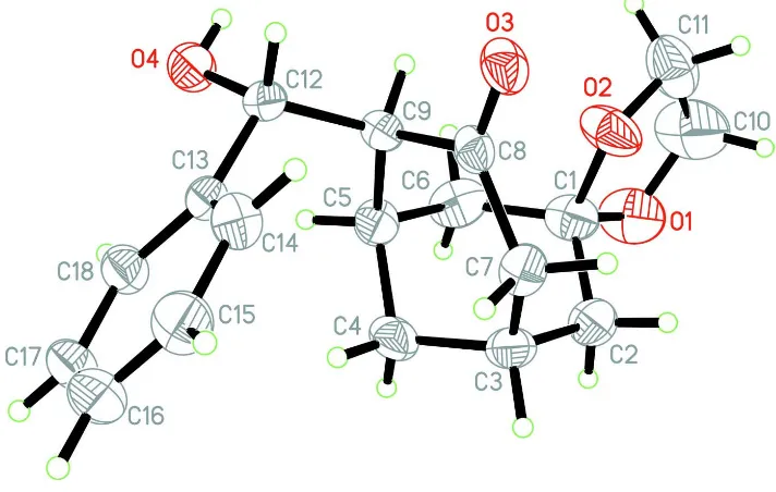
Figure 1
The molecular structure of the title compound with atom labels, showing 40% probability displacement ellipsoids.
Figure 2
The packing diagram of the title compound viewed down along the a axis. O—H···O hydrogen bonds are shown by thin
dashed lines.
7,7-[Ethane-1,2-diylbis(oxy)]-2-[hydroxy(phenyl)methyl]bicyclo[3.3.1]nonan-3-one
Crystal data
C18H22O4 Mr = 302.36
Orthorhombic, P212121 a = 9.1465 (3) Å b = 10.0346 (4) Å c = 16.6469 (6) Å V = 1527.88 (10) Å3
Z = 4 F(000) = 648 Dx = 1.314 Mg m−3
Mo Kα radiation, λ = 0.71073 Å Cell parameters from 1165 reflections θ = 2.4–24.4°
[image:3.610.129.483.341.538.2]T = 293 K Prism, colorless
0.35 × 0.30 × 0.29 mm
Data collection
Bruker SMART CCD area detector diffractometer
Radiation source: fine-focus sealed tube Graphite monochromator
φ and ω scans
Absorption correction: multi-scan (SADABS; Bruker, 2000) Tmin = 0.951, Tmax = 0.962
12383 measured reflections 1734 independent reflections 1560 reflections with I > 2σ(I) Rint = 0.026
θmax = 26.0°, θmin = 3.3° h = −10→11
k = −12→12 l = −20→20
Refinement
Refinement on F2 Least-squares matrix: full R[F2 > 2σ(F2)] = 0.039 wR(F2) = 0.096 S = 1.03 1734 reflections 203 parameters 0 restraints
Primary atom site location: structure-invariant direct methods
Secondary atom site location: difference Fourier map
Hydrogen site location: inferred from neighbouring sites
H atoms treated by a mixture of independent and constrained refinement
w = 1/[σ2(F
o2) + (0.0446P)2 + 0.4409P] where P = (Fo2 + 2Fc2)/3
(Δ/σ)max < 0.001 Δρmax = 0.20 e Å−3 Δρmin = −0.17 e Å−3
Special details
Experimental. IR (KBr) 3488, 2929, 2901, 2875, 1685, 1455, 1332, 1268, 1215, 1145, 1094, 1072, 1044, 1024, 948, 715 cm-1. 1NMR (CDCl3) 7.28–7.39 (m, 5H), 4.68–4.70(m, 1H), 3.92–3.95(m, 2H), 3.89 (d, J = 1.5 Hz, 1H), 3.80–3.87 (m, 2H), 2.59–2.63 (m, 2H), 2.51–2.55 (m, 2H), 1.96 (s, 1H), 1.80–1.83 (m, 3H), 1.64–1.67 (m, 1H), 1.52–1.57 (m, 2H) p.p.m.. 13CNMR (CDCl3) 213.2, 141.3, 128.6, 128.2, 127.1, 107.3, 74.6, 64.4, 63.4, 60.1, 45.1, 41.0, 40.1, 30.5, 29.2, 28.4 p.p.m..
Geometry. All e.s.d.'s (except the e.s.d. in the dihedral angle between two l.s. planes) are estimated using the full covariance matrix. The cell e.s.d.'s are taken into account individually in the estimation of e.s.d.'s in distances, angles and torsion angles; correlations between e.s.d.'s in cell parameters are only used when they are defined by crystal symmetry. An approximate (isotropic) treatment of cell e.s.d.'s is used for estimating e.s.d.'s involving l.s. planes.
Refinement. Refinement of F2 against ALL reflections. The weighted R-factor wR and goodness of fit S are based on F2, conventional R-factors R are based on F, with F set to zero for negative F2. The threshold expression of F2 > σ(F2) is used only for calculating R-factors(gt) etc. and is not relevant to the choice of reflections for refinement. R-factors based on F2 are statistically about twice as large as those based on F, and R- factors based on ALL data will be even larger.
Fractional atomic coordinates and isotropic or equivalent isotropic displacement parameters (Å2)
x y z Uiso*/Ueq
H3 0.7658 0.0822 0.4420 0.054* C4 0.5513 (3) 0.1339 (3) 0.45651 (16) 0.0484 (6) H4A 0.5262 0.1253 0.4001 0.058* H4B 0.5720 0.2269 0.4675 0.058* C5 0.4239 (3) 0.0869 (2) 0.50846 (16) 0.0428 (6) H5 0.3392 0.1433 0.4965 0.051* C6 0.4659 (3) 0.1088 (3) 0.59727 (17) 0.0550 (7) H6A 0.4764 0.2037 0.6069 0.066* H6B 0.3869 0.0767 0.6310 0.066* C7 0.6542 (3) −0.0947 (3) 0.45113 (15) 0.0431 (6) H7A 0.6618 −0.1010 0.3931 0.052* H7B 0.7305 −0.1503 0.4738 0.052* C8 0.5093 (3) −0.1518 (2) 0.47572 (13) 0.0351 (5) C9 0.3807 (3) −0.0595 (2) 0.49186 (13) 0.0340 (5) H9 0.3354 −0.0919 0.5415 0.041* C10 0.6360 (6) −0.0235 (4) 0.75276 (19) 0.0986 (15) H10A 0.5687 −0.0013 0.7958 0.118* H10B 0.7303 −0.0448 0.7762 0.118* C11 0.5806 (4) −0.1374 (4) 0.70659 (17) 0.0695 (10) H11A 0.6407 −0.2155 0.7160 0.083* H11B 0.4809 −0.1578 0.7222 0.083* C12 0.2635 (2) −0.0783 (2) 0.42468 (14) 0.0361 (5) H12 0.2395 −0.1734 0.4220 0.043* C13 0.3099 (2) −0.0343 (2) 0.34160 (13) 0.0351 (5) C14 0.3927 (3) −0.1185 (3) 0.29428 (14) 0.0445 (6) H14 0.4169 −0.2028 0.3133 0.053* C15 0.4402 (3) −0.0792 (3) 0.21877 (16) 0.0559 (7) H15 0.4965 −0.1369 0.1878 0.067* C16 0.4044 (3) 0.0444 (3) 0.18965 (17) 0.0598 (8) H16 0.4362 0.0709 0.1391 0.072* C17 0.3212 (3) 0.1284 (3) 0.23589 (17) 0.0541 (7) H17 0.2968 0.2123 0.2163 0.065* C18 0.2731 (3) 0.0903 (3) 0.31126 (15) 0.0437 (6) H18 0.2161 0.1481 0.3417 0.052* H4 0.078 (4) −0.061 (4) 0.474 (2) 0.085 (12)*
Atomic displacement parameters (Å2)
U11 U22 U33 U12 U13 U23
C6 0.0579 (17) 0.0506 (16) 0.0564 (16) −0.0087 (14) 0.0072 (13) −0.0189 (13) C7 0.0390 (13) 0.0511 (15) 0.0391 (12) 0.0030 (12) −0.0009 (10) −0.0044 (11) C8 0.0421 (12) 0.0352 (12) 0.0281 (10) 0.0014 (10) −0.0074 (10) 0.0013 (9) C9 0.0390 (12) 0.0311 (11) 0.0320 (10) −0.0010 (10) 0.0005 (9) 0.0025 (9) C10 0.128 (4) 0.126 (4) 0.0412 (16) −0.041 (3) −0.009 (2) 0.002 (2) C11 0.082 (2) 0.079 (2) 0.0473 (16) 0.022 (2) 0.0053 (16) 0.0114 (16) C12 0.0329 (12) 0.0333 (12) 0.0422 (12) −0.0030 (10) −0.0008 (10) 0.0050 (10) C13 0.0329 (12) 0.0359 (12) 0.0365 (11) −0.0049 (10) −0.0065 (9) 0.0017 (10) C14 0.0470 (14) 0.0447 (14) 0.0420 (12) 0.0035 (12) −0.0029 (11) −0.0007 (11) C15 0.0582 (17) 0.0692 (19) 0.0403 (14) 0.0010 (16) 0.0036 (12) −0.0045 (14) C16 0.0600 (18) 0.080 (2) 0.0393 (13) −0.0102 (17) 0.0024 (13) 0.0139 (15) C17 0.0601 (17) 0.0504 (16) 0.0518 (15) −0.0063 (14) −0.0077 (13) 0.0182 (13) C18 0.0473 (14) 0.0395 (13) 0.0445 (13) −0.0006 (11) −0.0043 (11) 0.0046 (11)
Geometric parameters (Å, º)
O1—C10 1.386 (4) C7—H7A 0.9700 O1—C1 1.429 (3) C7—H7B 0.9700 O2—C11 1.379 (3) C8—C9 1.521 (3) O2—C1 1.425 (3) C9—C12 1.561 (3) O3—C8 1.218 (3) C9—H9 0.9800 O4—C12 1.430 (3) C10—C11 1.467 (5) O4—H4 0.86 (4) C10—H10A 0.9700 C1—C2 1.508 (4) C10—H10B 0.9700 C1—C6 1.511 (4) C11—H11A 0.9700 C2—C3 1.532 (4) C11—H11B 0.9700 C2—H2A 0.9700 C12—C13 1.513 (3) C2—H2B 0.9700 C12—H12 0.9800 C3—C4 1.518 (4) C13—C14 1.381 (3) C3—C7 1.529 (4) C13—C18 1.390 (3) C3—H3 0.9800 C14—C15 1.387 (4) C4—C5 1.526 (4) C14—H14 0.9300 C4—H4A 0.9700 C15—C16 1.371 (4) C4—H4B 0.9700 C15—H15 0.9300 C5—C6 1.543 (4) C16—C17 1.372 (4) C5—C9 1.546 (3) C16—H16 0.9300 C5—H5 0.9800 C17—C18 1.384 (4) C6—H6A 0.9700 C17—H17 0.9300 C6—H6B 0.9700 C18—H18 0.9300 C7—C8 1.500 (3)
O1—C1—C6 109.9 (2) C5—C9—H9 105.9 C2—C1—C6 112.3 (2) C12—C9—H9 105.9 C1—C2—C3 113.5 (2) O1—C10—C11 107.1 (3) C1—C2—H2A 108.9 O1—C10—H10A 110.3 C3—C2—H2A 108.9 C11—C10—H10A 110.3 C1—C2—H2B 108.9 O1—C10—H10B 110.3 C3—C2—H2B 108.9 C11—C10—H10B 110.3 H2A—C2—H2B 107.7 H10A—C10—H10B 108.5 C4—C3—C7 109.0 (2) O2—C11—C10 106.7 (3) C4—C3—C2 110.3 (2) O2—C11—H11A 110.4 C7—C3—C2 113.8 (2) C10—C11—H11A 110.4 C4—C3—H3 107.8 O2—C11—H11B 110.4 C7—C3—H3 107.8 C10—C11—H11B 110.4 C2—C3—H3 107.8 H11A—C11—H11B 108.6 C3—C4—C5 109.0 (2) O4—C12—C13 109.15 (19) C3—C4—H4A 109.9 O4—C12—C9 108.93 (19) C5—C4—H4A 109.9 C13—C12—C9 115.27 (18) C3—C4—H4B 109.9 O4—C12—H12 107.7 C5—C4—H4B 109.9 C13—C12—H12 107.7 H4A—C4—H4B 108.3 C9—C12—H12 107.7 C4—C5—C6 108.0 (2) C14—C13—C18 118.4 (2) C4—C5—C9 112.8 (2) C14—C13—C12 119.8 (2) C6—C5—C9 111.7 (2) C18—C13—C12 121.8 (2) C4—C5—H5 108.1 C13—C14—C15 121.0 (3) C6—C5—H5 108.1 C13—C14—H14 119.5 C9—C5—H5 108.1 C15—C14—H14 119.5 C1—C6—C5 113.7 (2) C16—C15—C14 120.1 (3) C1—C6—H6A 108.8 C16—C15—H15 119.9 C5—C6—H6A 108.8 C14—C15—H15 119.9 C1—C6—H6B 108.8 C15—C16—C17 119.4 (3) C5—C6—H6B 108.8 C15—C16—H16 120.3 H6A—C6—H6B 107.7 C17—C16—H16 120.3 C8—C7—C3 116.9 (2) C16—C17—C18 121.0 (3) C8—C7—H7A 108.1 C16—C17—H17 119.5 C3—C7—H7A 108.1 C18—C17—H17 119.5 C8—C7—H7B 108.1 C17—C18—C13 120.1 (3) C3—C7—H7B 108.1 C17—C18—H18 119.9 H7A—C7—H7B 107.3 C13—C18—H18 119.9
C1—C2—C3—C4 53.7 (3) C8—C9—C12—O4 170.56 (19) C1—C2—C3—C7 −69.2 (3) C5—C9—C12—O4 −59.4 (3) C7—C3—C4—C5 64.1 (3) C8—C9—C12—C13 −66.4 (2) C2—C3—C4—C5 −61.5 (3) C5—C9—C12—C13 63.6 (3) C3—C4—C5—C6 62.2 (3) O4—C12—C13—C14 −154.8 (2) C3—C4—C5—C9 −61.7 (3) C9—C12—C13—C14 82.3 (3) O2—C1—C6—C5 −72.3 (3) O4—C12—C13—C18 26.0 (3) O1—C1—C6—C5 170.5 (2) C9—C12—C13—C18 −96.9 (3) C2—C1—C6—C5 49.1 (3) C18—C13—C14—C15 1.1 (4) C4—C5—C6—C1 −56.8 (3) C12—C13—C14—C15 −178.2 (2) C9—C5—C6—C1 67.8 (3) C13—C14—C15—C16 −0.5 (4) C4—C3—C7—C8 −45.7 (3) C14—C15—C16—C17 0.0 (4) C2—C3—C7—C8 77.9 (3) C15—C16—C17—C18 0.0 (4) C3—C7—C8—O3 −161.2 (2) C16—C17—C18—C13 0.6 (4) C3—C7—C8—C9 24.0 (3) C14—C13—C18—C17 −1.1 (4) O3—C8—C9—C5 165.8 (2) C12—C13—C18—C17 178.1 (2) C7—C8—C9—C5 −19.3 (3)
Hydrogen-bond geometry (Å, º)
D—H···A D—H H···A D···A D—H···A
O4—H4···O3i 0.86 (4) 2.03 (4) 2.857 (3) 163 (3) C16—H16···O3ii 0.93 2.59 3.473 (4) 159