Differentially Expressed Gene Transcripts in Wheat
and Barley Leaves upon Leaf Spot Infection
Veronika DUMALASOVÁ, Leona SVOBODOVÁ and Alena HANZALOVÁ
Crop Research Institute, Prague-Ruzyně, Czech Republic
Abstract: A cDNA library was created on the basis of transcripts that were generated during the process of infection of wheat and barley with Pyrenophora tritici-repentis or P. teres. Due to the time course of infection assays, the leaves were collected at various intervals after inoculation until 100 h after inoculation. We compared the temporal development of the two pathogens, as well as the effect of varieties of the same crop species, and differences among isolates of a single pathogen. The appressoria and vesicular structures developed more rap-idly in barley varieties inoculated by P. teres than in wheat varieties inoculated by P. tritici-repentis. The lower level of susceptibility of the wheat variety Globus and the barley variety Heris to P. tritici-repentis and P. teres, respectively, was apparent from 12 h after infection. Information on changes in expression profiles during pathogenesis and on the clones that could be functionally assigned to the known gene sequences is presented. Out of the 360 transcript derived fragments (TDFs) produced in the barley-P. teres pathosystem in our trials, 62% were specific to the plant-pathogen interaction. In the wheat-P. tritici-repentis pathosystem 291 TDFs oc-curred, 63% were specific to the plant-pathogen interaction.
Keywords: cDNA-AFLP; infection development; net blotch; Pyrenophora tritici-repentis; Pyrenophora teres; tan spot
Wheat tan spot and barley net blotch are ce-real diseases of worldwide economic importance caused by ascomycetous fungi Pyrenophora tritici-repentis (Died.) Drechs. (anamorph: Drechslera tritici-repentis /Died./ Schoem.) and Pyrenophora teres Drechs. (anamorph: Drechslera teres /Sacc./ Shoemaker). Both pathogens can cause severe yield losses due to a reduction of kernel number per ear, size of kernels, and the extent of photo-synthetically active leaf area.
Typical net blotch symptoms on leaves (form
P. teres f. teres) are distinctive dark-brown, lon-gitudinal necrotic lesions that often form a net-like pattern. Another form (Pyrenophora teres f.
maculata), known as the spot form, causes circular or elliptical brown lesions. Both forms induce sur-rounding chlorosis in the affected barley tissues. These distinct disease symptoms, the spot form
and the net form, can be explained by differences in fungal growth (Lightfoot & Able 2010).
Typical tan spot (P. tritici-repentis) symptoms include tan-coloured necrotic lesions surrounding small infection sites. The pathogen induces tan ne-crosis and/or extensive chlorosis in its wheat host.
The induction of chlorosis and necrosis symp-toms in both barley (net blotch) and wheat (tan spot) pathosystems is caused by mycotoxin activ-ity. Toxins appear to act as pathogenicity factors (Orolaza et al. 1995; Ciuffetti et al. 2010).
P. tritici-repentis ability to induce two distinct symptoms, tan necrosis and extensive chlorosis, on a set of differential wheat varieties is the basis of distinguishing eight P. tritici-repentis races (Lamari et al. 2003). Each race is differentiated by the expression of one toxin or a combination of Ptr ToxA, PtrToxB and Ptr ToxC toxins.
Plant disease resistance often plays a role in the infection process. After conidia germination, an elongating germ tube is formed. The appressorium is produced at the tip of the germ tube, followed by the formation of penetrating hyphae, vesicles, and infection hyphae. The host starts to respond to the invading pathogen. Papilla depositions, oc-casionally haloes or aggregation of cytoplasm can appear at the site of P. tritici-repentis or P. teres
penetration (Keon & Hargreaves 1983; Dush-nicky et al. 1998).
Endogenous levels of reactive oxygen intermedi-ates, nitric oxide, salicylic acid, jasmonic acid, and ethylene are increased in the incompatible reac-tion. Plant disease resistance can be triggered by specific recognition of microbial effectors by plant nucleotide binding-leucine rich repeat (NB-LRRs) receptors, and series of defence responses are acti-vated. These responses include the hypersensitive response, upregulation of phenylalanine ammonium lyase, deposition of cell wall reinforcing materials, and synthesis of antimicrobial compounds includ-ing pathogenesis related proteins and phytoalex-ins (Veronese et al. 2003; Jones & Dangl 2006; Collier & Moffett 2009; Bernoux et al. 2011).
A cDNA-AFLP (Amplified Fragment Length Polymorphism; Vos et al. 1995) technique is avail-able for the characterization of genes involved in biochemical processes. It enables to isolate genes that are differentially expressed during a process of interest. It was employed to clone cDNA copies of transcripts that are upregulated during P. teres
conidial germination (Dilger et al. 2003) to de-scribe interactions between the fungal pathogen
C. sativus and its resistant barley host (Al-Daoude & Jawhar 2009) and to identify differentially ex-pressed genes in chickpea during root infection by Fusarium oxysporum (Nimbalkar et al. 2006). Adhikari et al. (2007) analysed the expression patterns in association with resistance response of wheat to Mycosphaerella graminicola using cDNA-AFLP. Zheng et al. (2009) used cDNA-AFLP in a study on genes expressed during compatible interactions between Blumeria graminis and wheat.
The aim of our study was to determine if the cDNA-AFLP method can be usefully applied to study the infection development and host reac-tion in wheat and barley leaves during leaf spot infection. Clones with similarity to gene products described earlier from various organisms were de-tected. The function of detected gene products and their possible role in plant defence are discussed.
The temporal development of P. tritici-repentis and
P. teres pathogens, as well as the effect of varieties of the same crop species, and differences among iso-lates of a single pathogen were examined under the microscope. This information could be applied for predicting genes participating in processes of resist-ance mechanism for further, more detailed studies.
MATERIAL AND METHODS
Infection of plant materials
The barley varieties Beate (susceptible), Heris (medium resistant) and Prestige (susceptible) and wheat varieties Caphorn (susceptible), Bis-cay (susceptible), Globus (medium resistant) and Glenlea (susceptible) were chosen to study the development and degree of infection by fungi of the genus Pyrenophora (Figure 1 and 2).
The plantlets were raised in greenhouse condi-tions at the temperature of 20°C during 10 days up to the second leaf stage. These plants were inoculated with a suspension containing conidia, mycelial fragments, and sterile water.
The inoculum was prepared from isolate H602 of P. teres f. teres (PTT) that was collected in Drn-holec, Czech Republic during 1998 and from isolate H615 of Pyrenophora teres f. maculata (PTM) that was collected in Lužany during 2005. Both isolates were obtained by courtesy of Ing. Věra Minaříková, Agricultural Research Institute in Kroměříž, Ltd., Czech Republic. P. tritici-repentis isolate PTR-1 was collected in Přerov, Czech Republic in 2000. Isolate PTR-1 belongs to race 1 which is prevalent in the Czech Republic like in many countries of
P. tritici-repentis occurrence.
The detergent Tween 20 was added to the sus-pension (1μl/100 ml). The inoculum contained 20 conidia in 5 μl of the suspension. Every pot, containing approximately 30 plants, was inoculated with 10 ml of the suspension. The inoculum was dispersed evenly. The inoculum preparation was based on the method of Evans et al. (1993) and Ali and Francl (2001). The inoculated plants were incubated for 24 h in the closely covered glass cylinders. Three different isolates of P. teres
and collecting of samples were conducted simul-taneously in all involved varieties. One second leaf was always used for image analysis, and the other for molecular assay. Samples for molecular analysis were frozen in liquid nitrogen and kept at –80°C until use. Leaf segments of barley and wheat were analysed under a microscope (CX40, Olympus, Tokyo, Japan, 400× magnification) at the same time to determine the speed of formation of secondary infection structures, their amount, and the time when their development stopped. Staining with trypan blue was used for better differentiation of the structures.
RNA extraction and cDNA synthesis. Freshly harvested leaves before inoculation and 4, 23, 28, 47, 52, 71, 76, 94 and 100 h after inoculation were ground in liquid nitrogen into a powder using the mortar and pestle and suspended in TRIzol reagent (Invitrogen, Carlsbad, CA, USA). Total RNA was extracted according to the PureLinkTM RNA Mini Kit protocol (Invitrogen, Carlsbad, CA, USA). The complete removal of DNA was controlled by PCR using the primers specific to 18S rRNA (Dilger
et al. 2003) in 15-μl reaction volumes containing 200nM primers, dNTPs at 200μM each, 1U of Taq polymerase (Qiagen, Hilden, Germany) and
1× PCR buffer (Qiagen, Hilden, Germany) on a UNOII thermocycler (Biometra, Goettingen, Germany). The concentration of RNA samples was determined by spectrophotometry. Samples were diluted to the concentration of 300 ng/μl. cDNA was synthesized using the Transcriptor First Strand cDNA Synthesis Kit (Roche, Mannheim, Germany) according to the manufacturer’s protocol.
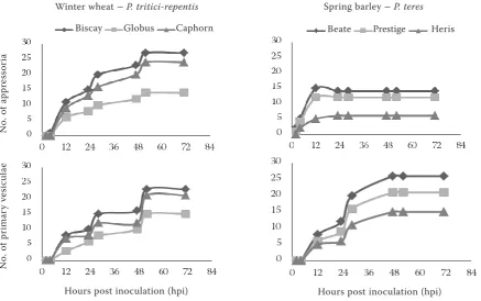
[image:3.595.65.503.83.357.2]cDNA-AFLP analysis. Generation of cDNA-AFLP amplicons was conducted with each cDNA sample prepared as described earlier using the restriction enzyme combinations MseI/EcoRI and MseI/PstI. For visualization, EcoRI and PstI prim-ers were labelled with fluorescent labels 6-fam, ned, joe or hex (Table 2). AFLP analyses were carried out according to the AFLPTM Plant Map-ping Protocol (Applied Biosystems, Foster City, CA, USA). Products of selective amplification were separated by capillary electrophoresis in ABI PRISM 310 (Perkin-Elmer, Foster City, CA, USA). Rox500 (PN 401734, Applied Biosystems) was used as the internal size standard. Chromato-grams were processed by the software GeneScan and Genotyper (Applied Biosystems). A binary matrix, reflecting the presence (1) or absence (0) of specific AFLP fragments, was generated for Figure 1. Development of fungal appressoria and vesicles on wheat leaves after inoculation by Pyrenophora tritici--repentis and on barley leaves after inoculation by Pyrenophora teres 0–72 hpi; the plants were inoculated with a suspension containing conidia, mycelial fragments and sterile water
Winter wheat – P. tritici-repentis Spring barley – P. teres
Hours post inoculation (hpi)
No. of pr
imar
y ve
sic
ul
ae
Hours post inoculation (hpi)
No. of a
ppr
ess
or
ia
Biscay Globus Caphorn Beate Prestige Heris
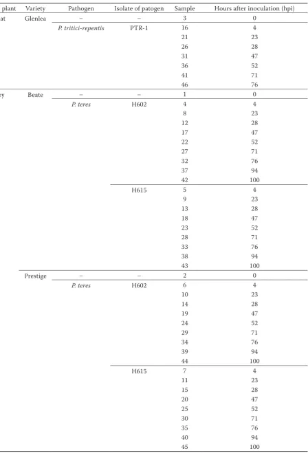
Table 1. Samples used in the study
Host plant Variety Pathogen Isolate of patogen Sample Hours after inoculation (hpi)
Wheat Glenlea – – 3 0
P. tritici-repentis PTR-1 16 4
21 23
26 28
31 47
36 52
41 71
46 76
Barley Beate – – 1 0
P. teres H602 4 4
8 23
12 28
17 47
22 52
27 71
32 76
37 94
42 100
H615 5 4
9 23
13 28
18 47
23 52
28 71
33 76
38 94
43 100
Prestige – – 2 0
P. teres H602 6 4
10 23
14 28
19 47
24 52
29 71
34 76
39 94
44 100
H615 7 4
11 23
15 28
20 47
25 52
30 71
35 76
40 94
each sample. A colormap was prepared in MS Excel software.
Sequence analysis. Selected amplicons were extracted from 2% agarose gel using a QIAquick Gel Extraction Kit (Qiagen, Hilden, Germany) and cloned into the pCR®4-TOPO plasmid vector. The resulting ligation products were used to transform
Escherichia coli TOP10 competent cells according to the manufacturer’s protocol (TOPO® TA Cloning Kit, Invitrogen, Paisley, UK). Purification of plasmids was carried out using a High Pure Plasmid Isolation Kit (Roche, Mannheim, Germany). Inserts were sequenced using a Big Dye Terminator 3.1 Sequenc-ing Kit (Applied Biosystems, Foster City, CA, USA) and universal sequencing primers, either M13+ or M13–. Extension products were separated on an ABI PRISM 3130 sequencer (Applied Biosystems,
Foster City, CA, USA). Sequence alignments were assembled using the T-Coffee software (Notredame
et al. 2000) and blasted (Zhang et al. 2000) to find homologies within sequences already deposited into databases (http://blast.ncbi.nlm.nih.gov/). Hits were considered significant if the expected value (E-value) was less than 1.0E-5.
RESULTS AND DISCUSSION
Infection of plant materials
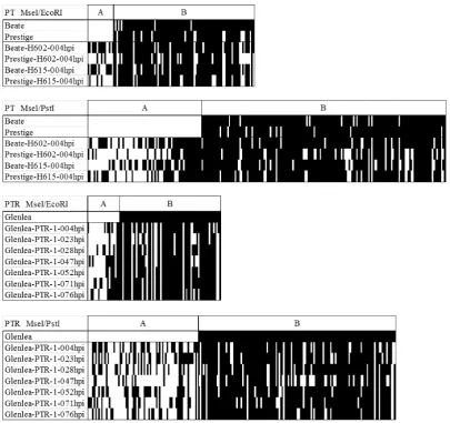
[image:5.595.92.498.81.462.2]There was no apparent germination of conidia within two hours or less after the infection in all varieties and pathogens tested. Appressoria and the penetration pegs started to form within 4–6 h. Figure 2. Early expressed profiles of the barley-P. teres pathosystem 4 h after inoculation (hpi) and profiles of the wheat-P. tritici-repentis pathosystem expressed 4–76 hpi: (A) differential cDNA AFLP bands unique to inoculated samples; (B) differential cDNA AFLP bands detected both in inoculated and uninoculated samples
Host tissue penetration and formation of vesicles occurred twelve hours after inoculation. The maxi-mum and no longer considerably changing number of appressoria was observed in the segments sampled 47 h after inoculation or later. Vesicular formation was variety specific (Figure 1). Similarly, Keon and Hargreaves (1983) described the germination of conidia, formation of appressorium and penetra-tion of host epidermal cells in P. teres within 24 h after inoculation. According to Dushnicky et al.
(1996) the penetration and infection stage of P. tritici-repentis generally occurs within the first 24 h both in susceptible and resistant varieties.
In barley varieties, a higher incidence of appres-soria followed by the secondary mycelium formation was registered in several observations in the Beate variety. The Prestige variety showed a lower occur-rence of appressoria in comparison with Beate. The difference between both varieties within 47 hpi made about 10% of the incidence of infection structures on the examined leaf segment area: 14 appressoria and 26 vesicles in Beate, 12 appressoria and 21 vesicles in Prestige. The appressoria, vesicles, and second-ary mycelium formed in both varieties at the same time. We observed fewer appressorium, vesicular, and secondary mycelium structures 28 h after the inoculation in the Heris variety than in Beate and Prestige. There were 6 appressoria and 11 vesicles in Heris, 14 appressoria and 20 vesicles in Beate and 12 appressoria and 16 vesicles in Prestige on the examined leaf segment area.
In wheat varieties, the highest proportions of appressoria and vesicular structures in 52 h post inoculation were produced in the Biscay variety, 27 appressoria and 23 vesicles on the examined leaf segment area. At the same time point, 18% less of these structures were present in the Caphorn variety, 24 appressoria and 21 vesicles. In addition, the Globus variety showed 14 appressoria and 15 vesicles at the same time point, which means 45% less of appressoria and vesicles than in Biscay.
Growth rate studies on the hosts revealed that the invasion of barley varieties by P. teres was faster than that by P. tritici-repentis. P. tritici-repentis
produced appressoria on the wheat varieties later, after 12 h. However, the influence of the particular isolates was not important for the rate and percent of infection. The image analysis revealed no cor-relation between the symptom development and the time after inoculation.
Many factors are involved in the manifestation of symptoms induced by the toxins varying not
only chemically but also in the mechanism of their activity. In addition, the phenotypic observation of the disease is influenced by the plant ability to recover impairment. When the formation of an infection peg does not lead to penetration, as may happen due to the host response, the fungus attempts to penetrate again. Papillae formation may stop the entry of infection hyphae into some epidermal cells, but not into all of them, even in resistant varieties (Dushnicky et al. 1996). In all these processes, multiple parameters may display a substantive influence, for example even a small variation in the leaf state and in light, moisture, temperature or other conditions.
cDNA-AFLP analysis
The restriction endonuclease combinations MseI/EcoRI and MseI/PstI were used for cDNA-AFLP analysis. In total, 39 primer combinations (21 for MseI/EcoRI enzyme combination, and 18 for MseI/PstI enzyme combination) were obtained for 46 cDNA samples.
The MseI primer was extended with seven com-binations of three selective nucleotides, whereas the extensions of EcoRI and PstI primers had two nucleotides. Produced fragments were in the size range of 80–500 bp.
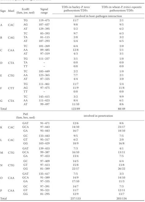
In the barley-Pyrenophora teres pathosystem, 62% of the total 360 transcript-derived fragments (TDFs) were supposed to be specifically expressed only dur-ing the plant-pathogen interaction (Table 2). MseI/ EcoRI primer combinations applied in the barley-
P. teres pathosystem produced 123 transcript-derived fragments (TDFs). Out of 123 fragments, 89 (72%) were supposed to be specific to the host-pathogen interaction. The MseI/PstI primer combination pro-vided 237 TDFs in the barley-P. teres pathosystem, out of which 133 (56%) were supposed to be specific to the host-pathogen interaction.
In the wheat-P. tritici-repentis pathosystem, 291 TDFs were identified, out of which 183 frag-ments (63%) were supposed to be specific to plant-pathogen interaction. The MseI/EcoRI primer combination used in the wheat-P. tritici-repentis
Table 2. List of selective primers used for cDNA AFLP analysis, their range of detection and the numbers of TDFs detected in plant-pathogen interactions
Sign MseI (fam, joe, ned)EcoRI Signal range TDFs in barley-pathosystem/TDFs P. teres TDFs in wheat-pathosystem/TDFsP. tritici-repentis involved in host-pathogen interaction
A CAC
TG 119–475 11/7 2/1
AG 107–437 9/8 9/5
AT 129–395 5/2 6/2
B CAG
TC 85–393 9/7 6/3
TA 81–115 2/0 3/2
AT 107–293 5/4 6/5
C CAA
TC 101–269 6/4 2/0
AA 89–485 12/8 5/1
AT 97–319 4/3 3/1
D CTA
TG 111–257 3/1 1/0
TA – 0/0 0/0
TT – 0/0 0/0
E CTG
TC 185–449 2/2 1/0
AA 123–365 7/7 2/1
AT 87–325 4/4 3/0
F CTT
TG 111–461 11/7 5/4
AG 97–475 11/9 11/8
TT – 0/0 0/0
G CTA
TC 145–415 3/2 9/9
AA 111–423 8/4 6/1
AT 99–497 11/10 8/6
Total 123/89 88/49
PstI
(fam, hex, ned) involved in penetration
K CAC
GAT 91–471 12/6 8/6
GCA 97–443 14/10 23/17
GA 93–443 16/7 18/10
L CAC
GC 133–443 9/5 7/5
GT 95–317 6/2 2/0
GG 103–429 18/9 16/8
M CTG
GAT 139–453 7/3 4/1
GCA 99–387 16/10 13/11
GA 97–453 13/4 7/5
N CTG
GC 97–409 14/5 6/4
GT 97–413 15/8 13/8
GG 81–389 22/17 26/22
O CAA
GAT 135–417 7/5 3/3
GCA 91–389 14/9 14/10
GA 97–335 17/10 11/3
P CAA
GC 97–391 14/7 7/3
GT 95–321 11/7 12/11
GG 81–295 12/9 13/7
Total 237/133 203/134
TDFs was comparable with that reported in previ-ous studies (Al-Daoude & Jawhar 2009; Zheng
et al. 2009). The results of cDNA AFLP analysis were summarized in colormaps showing the expres-sion profile of both pathosystems (Figure 2). The varieties Prestige and Beate differed significantly in their expression profiles in 4 hpi. A slower course of the infection process for some combinations of varieties and isolates is a possible explanation. We must admit that the detected fragments do not reflect the infection process, but rather some other factors such as microenvironmental differences in the greenhouse. Frequent coincidence of the pres-ence of fungal and plant bands can indicate mere presence or development of mycelium in the plant. In the barley-P. teres pathosystem, the infected plants of barley varieties Beate and Prestige were examined with primer combinations MseI/EcoRI 4 hpi. Fragments present only in the uninfected variety compared to the infected variety can correspond with differentially expressed genes involved in the plant defence. It is possible that some processes could already be displayed in some varieties, but delayed in the others, during the early stage of infection (4 hpi). Thus the infected variety Beate showed seven TDFs possibly connected with the plant defence, and two such TDFs were shown in Prestige. Another band specific to the plant infected with isolate H602, CAG/ TC-111 bp, was observed. Fragment CAG/TC-111 bp distinguished P. teres isolates H602 (PTT) and H615 (PTM). Fragment CAG/TC-261 bp distinguished the varieties Prestige and Beate regardless whether they were infected or not.
Another primer combination, MseI/PstI, distin-guished P. teres isolates H602 and H615 by means of fragments CAA/GG 195 bp, CAC/GCA 111 bp. Since the fragments are in the region where also fungal fragments are present, this unequal band reproduction could be caused by slower develop-ment of one of the fungal isolates. The MseI/PstI primer combination did not distinguish the varieties Prestige and Beate regardless whether they were infected or not. We noticed possible downregula-tion while searching for transcripts specific to the plant response to fungal infection. The fragment was present in some infected varieties, while in the uninfected varieties it was always absent.
The wheat-P. tritici-repentis pathosystem was examined with MseI/EcoRI primer combinations. Ten fragments vanished after inoculation of var. Glenlea with P. tritici-repentis. In one case, a frag-ment was detected only after inoculation. It was
probably linked to a gene expressed by the patho-gen. In addition, specific fragments were present in the region of the fungus expression only for definite time periods after inoculation.
MseI/PstI primer combinations in the wheat-
P. tritici-repentis pathosystem revealed three cases of a strong fungal band at the same time point when infecting var. Glenlea. It implies specificity to the plant infection, and more precisely for the pres-ence of the mycelium. The primer combinations resulted in the amplification of 405 differentially amplified PCR fragments.
Sequence analysis
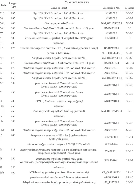
Thirty-six differential cDNA-AFLP fragments with the strongest signal were chosen for a sequence analysis. The results of the sequence comparisons are summarised in Table 3. No significant hits were detected for seven clones. Though their function is unknown, they still may represent novel transcripts associated with defence response (Nimbalkar
et al. 2006). No clones were recognized as fungal genes based on their homology to known fungal gene sequences. Generally, there are few fungal gene sequences, namely of the genus Pyrenophora,
present in the databases.
Seven clones showed homology to a hypothetical protein of unknown function. Five of them showed similarity to a sequence known from Hordeum vulgare (Matsumoto et al. 2011), the other two to a sequence from Sorghum bicolor. Both Sorghum bicolor clones showed similarity to the gene known as Sb01g031570 (Paterson et al. 2009).
Table 3. Similarities between the nucleotide and deduced amino acid sequences of the differentially expressed clones and sequences in databases; in total, 36 TDFs with the strongest signal were chosen for cloning and sequencing
Clone Length (bp) Maximum similarity
Gene product Accession No. E-value
A16 285 Rye 26S rRNA 3' end and 18S rRNA, 5' end M37231.1 3E-33
A4 285 Rye 26S rRNA 3' end and 18S rRNA, 5' end M37231.1 4E-87
A4b 480 Zea mays peroxin Pex14 NM_001155897.1 5E-51
A4d 800 Chasmanthium latifolium 16S ribosomal RNA (rrn16) gene HM363119.1 0.0
A7 285 Rye 26S rRNA 3' end and 18S rRNA, 5' end M37231.1 5E-80
B5 800 Triticum aestivum (L.) partial chloroplast 16S rRNA gene AJ239003.1 0.0
C1 200 unknown
I16 175 nucellin-like aspartic protease-like (Oryza sativa Japonica Group) BAD19623.1 2E-06
pepsin A (Zea mays) NP_001151415.1 5E-05
I26 175 Sorghum bicolor hypothetical protein, mRNA XM_002467601.1 5E-66 I4 175 Chasmanthium latifolium 16S ribosomal RNA (rrn16) gene HM363119.1 5E-158 I6 175 Hordeum vulgare subsp. vulgare mRNA for predicted protein AK363881.1 3E-09 I5 150 Hordeum vulgare subsp. vulgare mRNA for predicted protein AK358266.1 2E-29 I6 150 Sorghum bicolor hypothetical protein, mRNA XM_002467601.1 3E-69 J26 500 putative amino-acid N-acetyltransferase
(Oryza sativa Japonica Group) AAR87160.1 3E-26 J7 250 putative amino-acid N-acetyltransferase
(Oryza sativa Japonica Group) AAR87160.1 5E-22
L4 200 PP2C (Hordeum vulgare subsp. vulgare) ABO32801.1 3E-10
L4b 250 unknown
L5 250 Zea mays chlorophyll a/b binding protein 8 NM_001155126.1 1E-16
L7 250 unknown
L4c 380 putative amino-acid N-acetyltransferase
(Oryza sativa Japonica Group) AAR87160.1 3E-26 L4d 400 Hordeum vulgare subsp. vulgare mRNA for predicted protein AK369867.1 6E-20 L6 400 Fragaria × ananassa mRNA for β-galactosidase
(beta-gal2 gene) AJ278704.1 1E-14 Hordeum vulgare subsp. vulgare PP2C (PP2C) mRNA EF446603.1 3E-10 E4 111 Brachypodium pinnatum ribulose-1,5-bisphosphate carboxylase/
oxygenase large subunit (rbcL) gene AY632361.1 2E-16 E1 250 Thamnosma trifoliata partial rbcL gene
for ribulose-1,5-bisphosphate carboxylase/oxygenase large subunit FN552686.1 1E-05
E5 250 unknown
E5b 400 ATP binding protein, putative (Ricinus communis) XP_002511570.1 1E-40 putative methyltransferase (Solanum tuberosum) ABO93008.1 3E-40 dehydration-responsive family protein (Arabidopsis thaliana) NP_192782.1 1E-38
chlorophyll a/b-binding protein (Alexandrov et al. 2009) that has its role in a system for balancing the excitation energy between photosystems I and II in the light-harvesting process. The sequence of clone E5b shows homology to methyltransferase from Solanum tuberosum (Ballvora et al. 2007), an enzyme catalysing various metabolic functions through DNA methylation. The same clone showed homology also to dehydration-responsive family protein from Arabidopsis lyrata that acts mainly during cell dehydration and stress response and to ATP binding protein from Ricinus communis.
Clone E4 and E1 showed homology to ribulose-1,5-bisphosphate carboxylase/oxygenase, an enzyme involved in the Calvin cycle, which catalyses the first major step of carbon fixation. Clone B5 showed homology to 16S rRNA from T. aestivum (Kovacs
et al. 2000) as well as the clones A4d and I4 from
Chasmanthium latifolium (Yankaitis et al. 2010). The sequences indicated plastid-encoded rRNA. Clones A16, A4, A7 and clone G5 showed similar-ity to eukaryotic ribosomal RNA, clones A16, A4, A7 from rye to 26S and 18S rRNA (Appels et al.
1986), clone G5 from Lolium temulentum to 25S rRNA (Dombrowski & Martin 2009). Clone L4 and L6 showed homology to H. vulgare HvPP2C,
phosphatase 2C protein (Shen et al. 2001; Thiv
et al. 2011). Clone L6 also showed homology to
Fragaria × ananassa mRNA for β-galactosidase (Trainotti et al. 2001) catalysing the hydrolysis of β-galactosides into monosaccharides. Clone I16 was homologous with a sequence of nucellin-like, aspartic protease-like protein, hypothesized to play a role in the programmed cell death of the nucellus as aspartic protease. Another homology of the same sequence was found to pepsin (Alexandrov et al. 2009). Clone M7 showed homology to a puta-tive retroelement from Oryza sativa. Clone N16 showed similarity to putative sexual differentia-tion process protein from Oryza sativa as well as to putative oligopeptide transporter from Ricinus communis. Clone O6 showed homology to G10-like protein from Zea mays, a highly conserved protein of unknown function present in a wide range of eukaryotic species (Alexandrov et al. 2009).
[image:10.595.61.531.109.406.2]Similar transcripts have also been detected in other studies. Adhikari et al. (2007) identified genes with increased expression during the re-sistance response of wheat to Mycosphaerella graminicola. Novel genes not previously implicated in defence responses, but known from wheat plants treated with abiotic stressors such as drought were Table 3 to be continued
Clone Length (bp) Maximum similarity
Gene product Accession No. E-value
G5 500 Lolium temulentum 25S ribosomal RNA EU328537.1 4E-31
M16 450 unknown
M26 450 unknown
M7 450 Putative retroelement (Oryza sativa Japonica Group) AAN04214.1 2E-15 putative non-LTR retroelement reverse transcriptase
(Oryza sativa Japonica Group) AAT01370.1 1E-14 retrotransposon protein, putative, unclassified
(Oryza sativa Japonica Group) ABA97428.1 2E-14 N16 450 putative sexual differentiation process protein isp4
(Oryza sativa Japonica Group) BAD67732.1 4E-18 Oligopeptide transporter, putative (Ricinus communis) XP_002518892.1 2E-13 O16 300 Wheat (clone p80k-34) initiation factor isozyme 4F p82 subunit M95747.1 2E-91
O4 300 unknown
O6 300 G10-like protein (Zea mays) NP_001148940.1 2E-08
detected. Similarly, homology to a dehydration-responsive family protein was found in our study. Al-Daoude and Jawhar (2009) randomly isolated sequences of several disease stress-responsive genes including phosphatases. Two clones from our study also showed homology to phosphatase, namely PP2C (protein phosphatase 2C). The expression of ribulose-1,5-bisphosphate carboxylase/oxygenase was induced in response to wheat infection with powdery mildew (Zheng et al. 2009). Retroele-ments appeared to be differentially expressed dur-ing the chickpea reaction to Fusarium oxysporum
infection (Nimbalkar et al. 2006). Although plant retrotransposons have been found to be activated by abiotic and biotic stresses, their presence may be a mere evidence of the amplification of abundant RNA fragments by cDNA-AFLP method while some minor specific involved gene transcripts could be missed (Reijans et al. 2003).
We have described several differentially expressed transcript derived fragments (TDFs) in this study. Some of them showed homology to sequences of genes that may be involved in resistance mecha-nisms. The differentially expressed TDFs can provide information on changes in expression during patho-genesis including classical pathopatho-genesis-related (PR) genes that play a role in the signal transduction pathway. Our cDNA-AFLP approach may allow the identification of the gene function in defence mechanisms. However, many sequences identified by cDNA-AFLP have homology to genes that may be part of the plant’s general response to stress rather than having a specific role in defence. The charac-terisation of genes that play a role in biochemical processes during host-pathogen interactions could be useful for development of new methods for de-tection of resistant and susceptible lines in research programs. Thus the knowledge of changes induced in expression during pathogenesis can contribute to effective pest management.
Acknowledgements. We thank Dr. N. Champouret and Dr. B. Shaw for the critical reading of our ma-nuscript. The project was supported by Ministry of Ag-riculture of the Czech Republic, Project No. QH71242.
References
Adhikari T.B., Balaji B., Breeden J., Goodwin S.B. (2007): Resistance of wheat to Mycosphaerella grami-nicola involves early and late peaks of gene
expres-sion. Physiological and Molecular Plant Pathology, 71: 55–68.
Al-Daoude A., Jawhar M. (2009): Transcriptional changes in barley – Cochliobolus sativus interaction. Australasian Plant Pathology, 38: 467–471.
Alexandrov N.N., Brover V.V., Freidin S., Troukhan M.E., Tatarinova T.V., Zhang H., Swaller T.J., Lu Y.P., Bouck J., Flavell R.B., Feldman K.A. (2009): Insights into corn genes derived from large-scale cDNA sequencing. Plant Molecular Biology, 69: 179–194. Ali S., Francl L.J. (2001): Recovery of Pyrenophora
tritici-repentis from barley and reaction of 12 culti-vars to five races and two host-specific toxins. Plant Disease, 85: 580–584.
Allen M.L., Metz A.M., Timmer R.T., Rhoads R.E., Browning K.S. (1992): Isolation and sequence of the cDNAs encoding the subunits of the isozyme form of wheat protein synthesis initiation factor 4F. Journal of Biological Chemistry, 267: 23232–23236.
Appels R., Moran L.B., Gustafson J.P. (1986): The structure of DNA from the rye (Secale cereale) NOR R1 locus and its behaviour in wheat backgrounds. Ca-nadian Journal of Genetics and Cytology, 28: 673–685. Ballvora A., Jöcker A., Viehöver P., Ishihara H.,
Paal J., Meksem K. et al. (2007): Comparative se-quence analysis of Solanum and Arabidopsis in a hot spot for pathogen resistance on potato chromosome V reveals a patchwork of conserved and rapidly evolving genome segments. BMC Genomics, 8: 112.
Bernoux M., Ellis J.G., Dodds P.N. (2011): New in-sights in plant immunity signaling activation. Current Opinion in Plant Biology, 14: 512–518.
Ciuffetti L.M., Manning V.A., Pandelova I., Betts M.F., Martinez J.P. (2010): Host-selective toxins, Ptr ToxA and Ptr ToxB, as necrotrophic effectors in the Pyrenophora tritici-repentis-wheat interaction. New Phytologist, 187: 911–919.
Collier S.M., Moffett P. (2009): NB-LRRs work a “bait and switch” on pathogens. Trends in Plant Sci-ence, 14: 521–529.
Dilger M., Felsenstein F.G., Schwarz G. (2003): Identification and quantitative expression analysis of genes that are differentially expressed during conidial germination in Pyrenophora teres. Molecular Genetics and Genomics, 270: 147–155.
Dombrowski J.E., Martin R.C. (2009): Evaluation of reference genes for quantitative RT-PCR in Lolium temu-lentum under abiotic stress. Plant Science, 176: 390–396. Dushnicky L.G., Ballance G.M., Sumner M.J.,
MacGregor A.W. (1996): Penetration and infec-tion of susceptible and resistant wheat cultivars by a necrosis toxin-producing isolate of Pyrenophora
tritici-repentis. Canadian Journal of Plant Pathology, 18: 392–402.
Dushnicky L.G., Ballance G.M., Sumner M.J., MacGregor A.W. (1998): Detection of infection and host responses in susceptible and resistant wheat culti-vars to a toxin-producing isolate of Pyrenophora tritici-repentis. Canadian Journal of Plant Pathology, 20: 19–27. Evans C.K., Hunger R.M., Siegerist W.C. (1993):
Enhanced production of Pyrenophora tritici-repentis conidial suspensions. Plant Disease, 77: 981–984. Jones J.D., Dangl J.L. (2006): The plant immune system.
Nature, 444: 323–329.
Keon J., Hargreaves J.A. (1983): A cytological study of the net blotch disease of barley caused by Pyrenophora teres. Physiological Plant Pathology, 22: 321–329. Kovacs G., Racz I., Lasztity D., Paldi E. (2000):
Se-quence of chloroplast 16S rRNA gene of wheat. Plant Physiology, 122: 292–292.
Lamari L., Strelkov S.E., Yahyaoui A., Orabi J., Smith R.B. (2003): The identification of two new races of Pyrenophora tritici-repentis from the host center of diversity confirms a one-to-one relationship in tan spot of wheat. Phytopathology, 93: 391–396.
Lightfoot D.J., Able A.J. (2010): Growth of Pyre-nophora teres in planta during barley net blotch dis-ease. Australasian Plant Pathology, 39: 499–507. Matsumoto T., Tanaka T., Sakai H., Amano N.,
Kan-amori H., Kurita K. et al. (2011): Comprehensive se-quence analysis of 24783 barley full-length cDNAs derived from 12 clone libraries. Plant Physiology, 156: 20–28. Nimbalkar S.B., Harsulkar A.M., Giri A.P., Sainani
M.N., Franceschi V., Gupta V.S. (2006): Differen-tially expressed gene transcripts in roots of resistant and susceptible chickpea plant (Cicer arietinum L.) upon Fusarium oxysporum infection. Physiological and Molecular Plant Pathology, 68: 176–188.
Notredame C., Higgins D., Heringa J. (2000): T-Coffee: a novel method for multiple sequence align-ments. Journal of Molecular Biology, 302: 205–217. Orolaza N.P., Lamari L., Ballance G.M. (1995):
Evidence of a host-specific chlorosis toxin from Py-renophora tritici-repentis, the causal agent of tan spot of wheat. Phytopathology, 85: 1282–1287.
Paterson A.H., Bowers J.E., Bruggmann R., Dubchak I., Grimwood J., Gundlach H. etal. (2009): The
Sor-ghum bicolor genome and the diversification of grasses. Nature, 457: 551–556.
Reijans M., Lascaris R., Groeneger A.O., Witten-berg A., Wesselink E., van Oeveren J., de Wit E., Boorsma A., Voetdijk B., van der Spek H., Grivell L.A., Simons G. (2003): Quantitative comparison of cD-NA-AFLP, microarrays, and GeneChip expression data in Saccharomyces cerevisiae. Genomics, 82: 606–618. Shen Q., Gomez-Cadenas A., Zhang P.,
Walker-Simmons M.K., Sheen J., Ho T.H. (2001): Dissection of abscisic acid signal transduction pathways in barley aleurone layers. Plant Molecular Biology, 47: 437–448. Thiv M., van der Niet T., Rutschmann F., Thulin
M., Brune T., Linder H.P. (2011): Old-New World and trans-African disjunctions of Thamnosma ( Ru-taceae): Intercontinental long-distance dispersal and local ifferentiation in the succulent biome. American Journal of Botany, 98: 76–87.
Trainotti L., Spinello R., Piovan A., Spolaore S., Casadoro G. (2001): β-galactosidases with a lectin-like domain are expressed in strawberry. Journal of Experimental Botany, 52: 1635–1645.
Veronese P., Ruiz M.T., Coca M.A., Hernandez-Lopez A., Lee H., Ibeas J.I., Damsz B., Pardo J.M., Hasegawa P.M., Bressan R.A., Narasimhan M.L. (2003): In defence against pathogens. Both plant sentinels and foot soldiers need to know the enemy. Plant Physiology, 131: 1580–1590.
Vos P., Hogers R., Bleeker M., Reijans M., van der Lee T., Horners M. et al. (1995): AFLP: a new tech-nique for DNA fingerprinting. Nucleic Acids Research, 23: 4407–4414.
Yankaitis C.L., Amoruso S.M., Duvall M.R. (2010): Plastome sequences from Chasmanthium latifolium. Thesis Northern Illinois University, USA.
Zhang Z., Schwartz S., Wagner L., Miller W. (2000): A greedy algorithm for aligning DNA sequences. Jour-nal of ComputatioJour-nal Biology, 7: 203–214.
Zheng L., Huang J., Yu D. (2009): Isolation of genes expressed during compatible interactions between pow-dery mildew (Blumeria graminis) and wheat. Physi-ological and Molecular Plant Pathology, 73: 61–66.
Received for publication December 22, 2011 Accepted after corrections April 23, 2012
Corresponding author:
RNDr. Veronika Dumalasová, Ph.D., Výzkumný ústav rostlinné výroby, v.v.i., Drnovská 507, 161 06 Praha 6-Ruzyně, Česká republika