1128
https://doi.org/10.1107/S2056989019009502 Acta Cryst.(2019). E75, 1128–1132research communications
Received 13 June 2019 Accepted 2 July 2019
Edited by S. Parkin, University of Kentucky, USA
Keywords:crystal structure; 2-(methylamino)-tropone; tropolone.
CCDC reference:1937929
Supporting information:this article has supporting information at journals.iucr.org/e
Crystal structure of 2-(methylamino)tropone
Leandri Jansen van Vuuren, Hendrik G. Visser and Marietjie Schutte-Smith*
Department of Chemistry, PO Box 339, University of the Free State, Bloemfontein, 9301, South Africa. *Correspondence e-mail: SchutteM@ufs.ac.za
The title compound, 2-(methylamino)cyclohepta-2,4,6-trien-1-one, C8H9NO,
crystallizes in the monoclinic space group P21/c, with three independent
molecules in the asymmetric unit. The planarity of the molecules is indicated by planes fitted through the seven ring carbon atoms. Small deviations from the planes, with an extremal r.m.s. deviation of 0.0345 A˚ , are present. In complexes of transition metals with similar ligands, the large planar seven-membered aromatic rings have shown to improve the stability of the complex. Two types of hydrogen-bonding interactions, C—H O and N—H O, are observed, as well as bifurcation of these interactions. The N—H O interactions link molecules to form infinite chains. The packing of molecules in the unit cell shows a pattern of overlapping aromatic rings, forming column-like formations.–interactions are observed between the overlapping aromatic rings at 3.4462 (19) A˚ from each other.
1. Chemical context
Tropolone and other troponoids, non-benzenoid compounds, have great pharmacological potential (Guoet al., 2019). They display a wide range of bioactivities, including antimicrobial (Salehet al., 1988), antiviral (Tavis & Lomonosova, 2015) and antitumor (Ononye et al., 2013) activities. Many tropolone-related compounds have proved to be possible anti-proliferative agents against a variety of cancer cell lines, including lung, prostate and T-cell malignancies (Liu & Yamauchi, 2006; Hsiaoet al., 2012). Tropolone has important medical applications in radiopharmacy (Nepveu et al., 1993) and as catalyst precursor (Crous et al., 2005; Roodt et al., 2003).
Tropolone and its derivatives are versatile ligands used in inorganic and organometallic chemistry (Roesky, 2000; Diaset al., 1995; Nozoeet al., 1997; Schutteet al., 2010; Steyl et al., 2010). The carbonyl oxygen and vicinal coordinating substi-tuent, specifically nitrogen in this study, impart a metal-chelating ability to these types of ligands. The complexes of these ligands with first and second row transition elements have increased over the past few decades. The ligands of importance in this study and future work, namely 2-(alkyl-amino)tropones and aminotroponimines, are N,O and N,N0 bidentate, monoanionic ligands containing a ten -electron
backbone (Roesky, 2000). The -conjugated backbone is
characteristic of these ligands (Nishinaga et al., 2010). Considering the above-mentioned characteristics, tropolone
could be considered analogous to the O-donor 2
-O,O0 acetylacetonate ligand (acac-O,O0). The tropolone bidentate ligand differs from the acac-O,O0 ligand in a few ways. Of importance to our study is the larger aromatic delocalization, which could afford greater polarizability. Tropolone is also
more acidic than the acac-O,O0ligand. The acidity of theO,N andN,N0bidentate ligands used in our study and the effect thereof on the chelating ability could be compared to these O,O0bidentate ligands described in the literature. The ligand– metal–ligand angle, better known as the ‘bite angle’, would also be smaller for a tropolone-derived complex, since it would form a five-membered metallocycle instead of a six-membered one as with acac-O,O0 (Bhalla et al., 2005). This could show interesting steric and electronic influences at the metal centre and could be further compared to the steric and electronic studies conveyed on-diketone moieties in similar metal complexes by Manicum et al. (2018). These ligands, including the title compound, 2-(methylamino)tropone, will form part of the synthesis of water-soluble complexes of
rhenium(I) tricarbonyl, gallium(III) and copper(II).
Rhenium(I) (Schutte-Smithet al., 2019), gallium(III) (Green & Welch, 1989) and copper(II) (Boschiet al., 2018) are highly utilized radioisotopes in the radiopharmaceutical industry.
When designing diagnostic or therapeutic
radio-pharmaceuticals, certain mechanistic aspects are very impor-tant, as it is the basis on which some predictions are made regarding the in vivo behaviour. Kinetic studies, utilizing different techniques, are executed to determine the reaction mechanisms by which the proposed radiopharmaceutical complexes will form and react. Results of such studies are important in nuclear medicine as it gives indications regarding the in vivo stability, uptake and excretion as well as the pharmacokinetics of the compounds. Kinetic investigations by Schutte et al. (2011, 2012), Schutte-Smith et al. (2019) and Manicum et al. (2019) were done on rhenium(I) tricarbonyl tropolonato complexes with satisfying results and conclusions. In the study, methanol substitution was studied using entering nucleophiles in fac-[Re(Trop)(CO)3(MeOH)]. The kinetic
study performed at high pressure indicated positive volumes of activation for all of the reactions studied. This was a clear indication towards a dissociative interchange mechanism.
The application of these ligands in coordination chemistry could be further increased by adding electron donating or withdrawing moieties to the nitrogen atom.
2. Structural commentary
2-(Methylamino)tropone crystallizes in the monoclinic P21/c
space group with three independent molecules,A,BandC, in the asymmetric unit (Fig. 1). The bond distances and angles of the three molecules agree well with each other and with those in similar structures (Barretet al., 2014; Dwivediet al., 2016; Roesky & Bu¨rgstein, 1999; Shimanouchi & Sasada, 1973; Siwatchet al., 2011). The largest differences in bond distances
are of the C8B—N1B [1.4470 (18) A˚ ], N1B—C2B
[1.3444 (17) A˚ ] and O1B—C1B[1.2561 (15) A˚ ] bonds with the corresponding bonds in 2-(t-butylamino)tropone [1.472 (4) A˚ ; Siwatchet al., 2011], 2-(isopropylamino)tropone [1.330 (4) A˚ ; Roesky & Bu¨rgstein, 1999] and 2-(t-butylamino)tropone [1.242 (4) A˚ ; Siwatchet al., 2011], respectively. Compared to the starting material molecule, tropolone (Shimanouchi & Sasada, 1973), the O1B—C1B[1.2561 (15) A˚ ] bond distance is slightly shorter than that of tropolone [1.261 (3) A˚ ], both being in the range of normal C O bond distance. The C2—
N1—C8 bond angle in molecules A [125.69 (13)], B
[125.27 (13)] andC[125.07 (12)] are slightly larger than the usual 120 for trigonal-planar bond angles, because of the steric influence of the methyl group. These angles are close to the same angle in 2-(benzylamino)tropone [125.09 (12); Barretet al., 2014]. This could be compared to the large angle in 2-(t-butylamino)tropone [131.9 (2); Siwatch et al., 2011], which deviates even more from 120 due to the highly steric tertiary butyl group. A plane fitted through the seven ring carbon atoms of the three molecules in the asymmetric unit indicates that the molecules are planar. The root-mean-square deviations of molecules A, B and C from the planes are 0.0141 (12), 0.0261 (11) and 0.0345 (11) A˚ , respectively. The C8—N1—C2—C3 torsion angle, which involves the methyl group, differs for moleculeA[0.8 (2)], moleculeB[2.3 (2)] and moleculeC[7.7 (2)]. The small deviations from planarity could possibly be ascribed to the intermolecular hydrogen-bonding interactions.
research communications
Acta Cryst.(2019). E75, 1128–1132 Jansen van Vuurenet al. C
[image:2.610.315.561.421.713.2]8H9NO
1129
Figure 1
3. Supramolecular features
Nine hydrogen-bonding interactions, three C—H O and six N—H O, are observed (Table 1 and Fig. 2). Infinite chains are formed along [001]. These supramolecular chains are formed through N—H O interactions linking the molecules together. As in the crystal structure of tropolone (Shima-nouchi & Sasada, 1973), bifurcation of the hydrogen bonds take place. Bifurcation, also known as the over-coordination of a hydrogen bond, creates both inter- and intramolecular branches, which might contribute to the stability of the structures. This is an interesting phenomenon seen in the orientation of water molecules, where the distribution of acceptor hydrogen bonds, terminating at the lone pairs of the oxygen, is higher (Markovitch & Agmon, 2008). This forms over-coordinated oxygens and could also be seen in this crystal
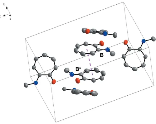
structure (Fig. 2). These interactions clearly contribute to the array of the molecules in the asymmetric unit (Fig. 2). The molecules show an interesting packing format in the unit cell. ‘Column’-like structures are formed by moleculeBpacking in a head-to-tail pattern with the aromatic rings overlapping (Fig. 3). A -interaction is observed, with a perpendicular distance of 3.4462 (19) A˚ between the overlapping aromatic rings of two inversion-relatedBmolecules (Fig. 4). These -interactions could not only possibly contribute to the packing
1130
Jansen van Vuurenet al. C8H9NO Acta Cryst.(2019). E75, 1128–1132
research communications
Figure 3
[image:3.610.314.565.72.272.2] [image:3.610.44.296.85.196.2]Packing of molecules viewed perpendicular to theacplane. Table 1
Hydrogen-bond geometry (A˚ ,).
D—H A D—H H A D A D—H A
N1A—HN1A O1A 0.884 (17) 2.099 (16) 2.5453 (16) 110.3 (13) N1A—HN1A O1B 0.884 (17) 2.248 (17) 2.9375 (17) 134.6 (14) N1B—HN1B O1B 0.893 (15) 2.085 (15) 2.5513 (16) 111.5 (12) N1B—HN1B O1C 0.893 (15) 2.385 (15) 3.1566 (18) 144.7 (13) N1C—HN1C O1C 0.890 (16) 2.130 (15) 2.5775 (16) 110.3 (12) N1C—HN1C O1Ai 0.890 (16) 2.313 (16) 2.9759 (17) 131.3 (13) C5C—H5C O1Bii 0.95 2.42 3.2914 (19) 153
C7B—H7B O1A 0.95 2.42 3.3446 (19) 165 C8C—H8C2 O1Ai 0.98 2.56 3.178 (2) 121
Symmetry codes: (i)x;yþ3
[image:3.610.111.511.446.720.2]2;z12; (ii)x;y1;z.
Figure 2
format of the molecules in the unit cell, but could also assist in the formation of one-dimensional infinite chains, as Wong et al.(2018) have found in water-soluble platinum (II) salts.
4. Database survey
A search of the Cambridge Structural Database (CSD, Version 5.40, update of February 2019; Groom et al., 2016) using a C7H5ONH fragment yielded four hits of
2-(alkyl-amino)tropones. These include 2-(isopropylamino)tropone (LIGVOM: Roesky & Bu¨rgstein, 1999), 2-(benzylamino)-tropone (NOPRUH: Barret et al., 2014),
2-(t-butylamino)-tropone (OZINUH: Siwatch et al., 2011) and 2-(cyclo
hexylamino)tropone (OTIMUB: Dwivediet al., 2016). Of the four structures, only the (isopropylamino)tropone and the 2-(benzylamino)tropone crystallize in theP21/cspace group.
5. Synthesis and crystallization
Tropolone (505 mg, 4.132 mmol) was dissolved in 20 mL of a 40% methylamine solution. The reaction mixture was stirred at room temperature for 7 d. The product was extracted three times with 30 mL of chloroform, and the organic layer was washed with 50 mL of water. The organic layer was dried with Na2SO4and the solvent removed under reduced pressure. A
46.03% yield (257.1 mg, 1.902 mmol) was obtained. Crystals suitable for single crystal X-ray diffraction data collection were obtained by recrystallization from hexane with slow evaporation. Yield: 0.2571 g, 46.03%. IR (cm1):NH= 3304,
CO = 1597. UV/Vis: max = 269 nm (" = 1.1885 105
Lmol1cm1).1H NMR (300 MHz, CDCl3):= 7.201 (m, 4H),
6.682 (t, 1H,J= 9.6 Hz), 6.501 (d, 1H,J= 10.5 Hz), 3.056 (d, 3H,J= 5.4 Hz).13C NMR (300MHz, CDCl3):= 177, 157, 137,
136, 129, 122, 108, 29.
6. Refinement
Crystal data, data collection and structure refinement details are summarized in Table 2. Methyl and aromatic hydrogen atoms were placed in geometrically idealized positions (C—H = 0.95–0.98 A˚ ) and constrained to ride on their parent atoms [Uiso(H) = 1.5Ueq(C) and 1.2Ueq(C)], while N—H
hydrogens were freely refined.
Acknowledgements
This work is based on the research supported in part by the National Research Foundation of South Africa. We would also like to thank the University of the Free State.
References
Barret, M., Bhatia, P., Kociok-Ko¨hn, G. & Molloy, K. (2014). Transition Met. Chem.39, 543–551.
Bhalla, G., Oxgaard, J., Goddard, W. & Periana, R. (2005). Organometallics,24, 3229–3232.
Boschi, A., Martini, P., Janevik-Ivanovska, E. & Duatti, A. (2018). Drug Discovery Today,23, 1489–1501.
Brandenburg, K. (2006).DIAMOND. Crystal Impact GbR, Bonn, Germany.
Bruker (2012). APEX2, SAINT, Bruker AXS Inc, Madison, Wisconsin, USA.
Crous, R., Datt, M., Foster, D., Bennie, L., Steenkamp, C., Huyser, J., Kirsten, L., Steyl, G. & Roodt, A. (2005).Dalton Trans.pp. 1108– 1116.
research communications
Acta Cryst.(2019). E75, 1128–1132 Jansen van Vuurenet al. C
[image:4.610.46.296.69.268.2]8H9NO
1131
Figure 4
–interaction (highlighted by the dashed line) between overlapping aromatic rings of molecule B, where B and B* are related through inversion.
Table 2
Experimental details.
Crystal data
Chemical formula C8H9NO
Mr 135.16
Crystal system, space group Monoclinic,P21/c
Temperature (K) 100
a,b,c(A˚ ) 17.635 (5), 7.817 (2), 16.718 (4)
() 110.639 (9)
V(A˚3) 2156.8 (10)
Z 12
Radiation type MoK
(mm1) 0.08
Crystal size (mm) 0.580.300.28
Data collection
Diffractometer Bruker X8 APEXII 4K Kappa
CCD
Absorption correction Multi-scanSADABS(Krauseet al., 2015)
Tmin,Tmax 0.970, 0.977
No. of measured, independent and observed [I> 2(I)] reflections
33879, 5192, 3575
Rint 0.046
(sin/)max(A˚
1
) 0.660
Refinement
R[F2> 2(F2)],wR(F2),S 0.039, 0.104, 1.03
No. of reflections 5192
No. of parameters 287
H-atom treatment H atoms treated by a mixture of independent and constrained refinement
max,min(e A˚
3
) 0.17,0.13
[image:4.610.313.563.92.385.2]Dias, H. V. R., Jin, W. & Ratcliff, R. E. (1995).Inorg. Chem.34, 6100– 6105.
Dwivedi, A., Binnani, C., Tyagi, D., Rawat, K., Li, P., Zhao, Y., Mobin, S. M., Pathak, B. & Singh, S. K. (2016).Inorg. Chem.55, 6739– 6749.
Farrugia, L. J. (2012).J. Appl. Cryst.45, 849–854.
Green, M. & Welch, M. (1989).Int. J. Radiat. Appl. Instrum. B,16, 435–448.
Groom, C. R., Bruno, I. J., Lightfoot, M. P. & Ward, S. C. (2016).Acta Cryst.B72, 171–179.
Guo, H., Roman, D. & Beemelmanns, C. (2019). Natural Product Reports.https://doi.org/10.1039/C8NP00078F.
Hsiao, C. J., Hsiao, S. H., Chen, W. L., Guh, J. H., Hsiao, G., Chan, Y. J., Lee, T. H. & Chung, C. L. (2012).Chem. Biol. Interact.197, 23– 30.
Krause, L., Herbst-Irmer, R., Sheldrick, G. M. & Stalke, D. (2015).J. Appl. Cryst.48, 3–10.
Liu, S. & Yamauchi, H. (2006).Biochem. Biophys. Res. Commun.351, 26–32.
Manicum, A., Schutte-Smith, M., Alexander, O., Twigge, L., Roodt, A. & Visser, H. (2019).Inorg. Chem. Commun.101, 93–98. Manicum, A., Schutte-Smith, M. & Visser, H. (2018). Polyhedron,
145, 80–87.
Markovitch, O. & Agmon, N. (2008).Mol. Phys.106, 485–495. Nepveu, F., Jasanada, F. & Walz, L. (1993).Inorg. Chim. Acta,211,
141–147.
Nishinaga, T., Aono, T., Isomura, E., Watanabe, S., Miyake, Y., Miyazaki, A., Enoki, T., Miyasaka, H., Otani, H. & Iyoda, M. (2010).Dalton Trans.39, 2293–2300.
Nozoe, T., Lin, L. C., Hsu, C., Tsay, S., Hakimelahib, G. H. & Hwu, J. R. (1997).J. Chem. Res. (S), pp. 362–363.
Ononye, S. N., VanHeyst, M. D., Oblak, E. Z., Zhou, W., Ammar, M., Anderson, A. C. & Wright, D. L. (2013).ACS Med. Chem. Lett.4, 757–761.
Roesky, P. & Bu¨rgstein, M. (1999).Inorg. Chem.38, 5629–5632. Roesky, P. W. (2000).Chem. Soc. Rev.29, 335–345.
Roodt, A., Otto, S. & Steyl, G. (2003).Coord. Chem. Rev.245, 121– 137.
Saleh, N. A., Zfiefak, A., Mordarski, M. & Pulverer, G. (1988). Zentralbl. Bakteriol. MikroBiol. Hyg. Med. Microbiol. Infect. Dis. Virol. Paras.270, 160–170.
Schutte, M., Kemp, G., Visser, H. & Roodt, A. (2011).Inorg. Chem.
50, 12486–12498.
Schutte, M., Roodt, A. & Visser, H. (2012).Inorg. Chem.51, 11996– 12006.
Schutte, M., Visser, H. G. & Roodt, A. (2010).Acta Cryst.E66, m859– m860.
Schutte-Smith, M., Roodt, A. & Visser, H. G. (2019).Dalton Trans. https://doi.org/10.1039/C9DT01528K.
Sheldrick, G. M. (2008).Acta Cryst.A64, 112–122. Sheldrick, G. M. (2015).Acta Cryst.C71, 3–8.
Shimanouchi, H. & Sasada, Y. (1973).Acta Cryst.B29, 81–90. Siwatch, R. K., Kundu, S., Kumar, S. & Nagendran, S. (2011).
Organometallics,30, 1998–2005.
Steyl, G., Muller, T. J. & Roodt, A. (2010).Acta Cryst.E66, m1508. Tavis, J. E. & Lomonosova, E. (2015).Antiviral Res.118, 132–138. Wong, V., Po, C., Leung, S., Chan, A., Yang, S., Zhu, B., Cui, X. &
Yam, V. W. (2018).J. Am. Chem. Soc.140, 657–666.
1132
Jansen van Vuurenet al. C8H9NO Acta Cryst.(2019). E75, 1128–1132
supporting information
sup-1
Acta Cryst. (2019). E75, 1128-1132
supporting information
Acta Cryst. (2019). E75, 1128-1132 [https://doi.org/10.1107/S2056989019009502]
Crystal structure of 2-(methylamino)tropone
Leandri Jansen van Vuuren, Hendrik G. Visser and Marietjie Schutte-Smith
Computing details
Data collection: APEX2 (Bruker, 2012); cell refinement: SAINT-Plus (Bruker, 2012); data reduction: SAINT-Plus (Bruker,
2012); program(s) used to solve structure: SHELXS97 (Sheldrick, 2008); program(s) used to refine structure:
SHELXL2018 (Sheldrick, 2015); molecular graphics: DIAMOND (Brandenburg, 2006); software used to prepare material
for publication: WinGX (Farrugia, 2012).
2-(Methylamino)cyclohepta-2,4,6-trien-1-one
Crystal data
C8H9NO
Mr = 135.16
Monoclinic, P21/c
Hall symbol: -P 2ybc
a = 17.635 (5) Å
b = 7.817 (2) Å
c = 16.718 (4) Å
β = 110.639 (9)°
V = 2156.8 (10) Å3
Z = 12
F(000) = 864
Dx = 1.249 Mg m−3
Mo Kα radiation, λ = 0.71073 Å
Cell parameters from 6617 reflections
θ = 3.5–23.7°
µ = 0.08 mm−1
T = 100 K
Cuboid, yellow 0.58 × 0.30 × 0.28 mm
Data collection
Bruker X8 APEXII 4K Kappa CCD diffractometer
Radiation source: fine-focus sealed tube Graphite monochromator
ω scans
Absorption correction: multi-scan
SADABS (Krause et al., 2015)
Tmin = 0.970, Tmax = 0.977
33879 measured reflections 5192 independent reflections 3575 reflections with I > 2σ(I)
Rint = 0.046
θmax = 28.0°, θmin = 3.7°
h = −23→23
k = −10→10
l = −20→22
Refinement
Refinement on F2
Least-squares matrix: full R[F2 > 2σ(F2)] = 0.039
wR(F2) = 0.104
S = 1.03
5192 reflections 287 parameters 0 restraints 0 constraints
Primary atom site location: structure-invariant direct methods
Secondary atom site location: structure-invariant direct methods
Hydrogen site location: mixed
H atoms treated by a mixture of independent and constrained refinement
w = 1/[σ2(F
o2) + (0.0361P)2 + 0.437P]
where P = (Fo2 + 2Fc2)/3 (Δ/σ)max < 0.001
Δρmax = 0.17 e Å−3
supporting information
sup-2
Acta Cryst. (2019). E75, 1128-1132 Extinction correction: SHELXL2018
(Sheldrick, 2015)
Extinction coefficient: 0.0113 (9)
Special details
Experimental. The intensity data was collected on a Bruker X8 ApexII 4K Kappa CCD diffractometer using an exposure
time of 10 seconds/frame. A total of 1436 frames was collected with a frame width of 0.5° covering up to θ = 27.99° with
99.7% completeness accomplished.
Geometry. All esds (except the esd in the dihedral angle between two l.s. planes) are estimated using the full covariance matrix. The cell esds are taken into account individually in the estimation of esds in distances, angles and torsion angles; correlations between esds in cell parameters are only used when they are defined by crystal symmetry. An approximate (isotropic) treatment of cell esds is used for estimating esds involving l.s. planes.
Fractional atomic coordinates and isotropic or equivalent isotropic displacement parameters (Å2)
x y z Uiso*/Ueq
C8A 0.12323 (10) 0.5412 (2) 0.21119 (10) 0.0626 (4)
H8A1 0.113602 0.421239 0.221702 0.094*
H8A2 0.172571 0.549845 0.197169 0.094*
H8A3 0.077051 0.584932 0.163359 0.094*
C8B 0.49672 (10) 0.4930 (2) 0.26816 (10) 0.0586 (4)
H8B1 0.530729 0.588791 0.263364 0.088*
H8B2 0.47058 0.440852 0.2118 0.088*
H8B3 0.530411 0.407538 0.307747 0.088*
C8C 0.19718 (11) 0.2656 (2) −0.10383 (9) 0.0588 (4)
H8C1 0.234672 0.189118 −0.117954 0.088*
H8C2 0.187004 0.367098 −0.140513 0.088*
H8C3 0.145975 0.20569 −0.113027 0.088*
N1A 0.13282 (7) 0.64142 (16) 0.28726 (8) 0.0460 (3)
N1B 0.43536 (8) 0.55487 (16) 0.29999 (8) 0.0454 (3)
N1C 0.23250 (7) 0.31715 (16) −0.01509 (7) 0.0450 (3)
O1A 0.17457 (6) 0.83578 (13) 0.41777 (6) 0.0530 (3)
O1B 0.30974 (5) 0.61826 (14) 0.33715 (6) 0.0515 (3)
O1C 0.28810 (7) 0.46038 (12) 0.13314 (6) 0.0574 (3)
HN1A 0.1803 (10) 0.689 (2) 0.3149 (10) 0.064 (5)*
HN1B 0.3829 (9) 0.5397 (19) 0.2695 (10) 0.052 (4)*
HN1C 0.2345 (9) 0.428 (2) −0.0016 (10) 0.056 (5)*
C1B 0.37461 (8) 0.67009 (17) 0.39220 (8) 0.0379 (3)
C2B 0.44995 (7) 0.63079 (16) 0.37612 (8) 0.0379 (3)
C3B 0.52868 (8) 0.66224 (19) 0.43132 (9) 0.0476 (3)
H3B 0.569782 0.625712 0.410663 0.057*
C4B 0.55733 (9) 0.7385 (2) 0.51194 (10) 0.0545 (4)
H4B 0.614666 0.74095 0.538203 0.065*
C5B 0.51628 (9) 0.8101 (2) 0.55919 (10) 0.0553 (4)
H5B 0.548086 0.855301 0.613404 0.066*
C6B 0.43262 (9) 0.82372 (19) 0.53599 (9) 0.0510 (4)
H6B 0.414495 0.882838 0.575565 0.061*
C7B 0.37249 (8) 0.76418 (18) 0.46432 (8) 0.0437 (3)
H7B 0.319314 0.790761 0.462424 0.052*
supporting information
sup-3
Acta Cryst. (2019). E75, 1128-1132
C2C 0.25275 (7) 0.21032 (16) 0.05204 (8) 0.0346 (3)
C3C 0.24942 (8) 0.03409 (16) 0.03938 (9) 0.0406 (3)
H3C 0.236584 −0.000441 −0.018381 0.049*
C4C 0.26179 (8) −0.09981 (17) 0.09743 (9) 0.0460 (3)
H4C 0.257336 −0.210933 0.073189 0.055*
C5C 0.27941 (9) −0.09661 (19) 0.18414 (10) 0.0495 (4)
H5C 0.283417 −0.204059 0.211879 0.059*
C6C 0.29204 (8) 0.0487 (2) 0.23568 (9) 0.0491 (4)
H6C 0.301651 0.026655 0.294322 0.059*
C7C 0.29278 (8) 0.21802 (19) 0.21506 (9) 0.0458 (3)
H7C 0.304073 0.293672 0.262311 0.055*
C1A 0.10733 (8) 0.76484 (17) 0.40289 (8) 0.0411 (3)
C2A 0.07897 (8) 0.65224 (16) 0.32681 (8) 0.0395 (3)
C3A 0.00556 (8) 0.56527 (19) 0.29566 (10) 0.0538 (4)
H3A −0.003564 0.505768 0.243542 0.065*
C4A −0.05654 (9) 0.5510 (2) 0.32809 (13) 0.0647 (5)
H4A −0.100588 0.48099 0.295409 0.078*
C5A −0.06451 (10) 0.6213 (2) 0.39924 (13) 0.0702 (5)
H5A −0.112064 0.59234 0.410595 0.084*
C6A −0.01009 (11) 0.7312 (3) 0.45709 (12) 0.0707 (5)
H6A −0.026039 0.768486 0.502906 0.085*
C7A 0.06298 (10) 0.7941 (2) 0.45827 (10) 0.0584 (4)
H7A 0.088461 0.870606 0.504039 0.07*
Atomic displacement parameters (Å2)
U11 U22 U33 U12 U13 U23
C8A 0.0635 (10) 0.0735 (11) 0.0479 (9) −0.0028 (9) 0.0162 (8) −0.0172 (8)
C8B 0.0646 (10) 0.0627 (10) 0.0585 (10) 0.0095 (8) 0.0343 (8) 0.0034 (8)
C8C 0.0758 (11) 0.0571 (9) 0.0380 (8) 0.0003 (8) 0.0132 (7) 0.0016 (7)
N1A 0.0412 (7) 0.0501 (7) 0.0437 (7) −0.0052 (6) 0.0114 (5) −0.0085 (6)
N1B 0.0432 (7) 0.0527 (7) 0.0421 (7) 0.0019 (6) 0.0171 (6) 0.0013 (5)
N1C 0.0587 (7) 0.0373 (6) 0.0384 (6) −0.0010 (5) 0.0164 (5) −0.0014 (5)
O1A 0.0484 (6) 0.0480 (6) 0.0582 (6) −0.0073 (5) 0.0132 (5) −0.0137 (5)
O1B 0.0353 (5) 0.0712 (7) 0.0441 (6) −0.0060 (5) 0.0091 (4) −0.0063 (5)
O1C 0.0861 (8) 0.0361 (5) 0.0494 (6) −0.0083 (5) 0.0230 (6) −0.0073 (5)
C1B 0.0360 (7) 0.0408 (7) 0.0354 (7) −0.0023 (5) 0.0106 (5) 0.0074 (5)
C2B 0.0385 (7) 0.0371 (7) 0.0388 (7) −0.0007 (5) 0.0146 (6) 0.0094 (6)
C3B 0.0355 (7) 0.0566 (9) 0.0517 (8) 0.0001 (6) 0.0166 (6) 0.0091 (7)
C4B 0.0353 (7) 0.0669 (10) 0.0522 (9) −0.0082 (7) 0.0042 (6) 0.0085 (8)
C5B 0.0516 (9) 0.0634 (10) 0.0422 (8) −0.0135 (7) 0.0056 (7) −0.0022 (7)
C6B 0.0576 (9) 0.0530 (9) 0.0425 (8) −0.0058 (7) 0.0177 (7) −0.0046 (7)
C7B 0.0409 (7) 0.0497 (8) 0.0417 (7) −0.0007 (6) 0.0158 (6) 0.0021 (6)
C1C 0.0402 (7) 0.0381 (7) 0.0416 (7) −0.0022 (5) 0.0158 (6) −0.0058 (6)
C2C 0.0327 (6) 0.0361 (6) 0.0365 (7) 0.0003 (5) 0.0141 (5) −0.0009 (5)
C3C 0.0445 (7) 0.0367 (7) 0.0417 (7) −0.0003 (6) 0.0164 (6) −0.0054 (6)
C4C 0.0478 (8) 0.0337 (7) 0.0571 (9) 0.0026 (6) 0.0192 (7) −0.0010 (6)
supporting information
sup-4
Acta Cryst. (2019). E75, 1128-1132
C6C 0.0462 (8) 0.0590 (9) 0.0393 (8) −0.0020 (7) 0.0117 (6) 0.0074 (7)
C7C 0.0492 (8) 0.0502 (8) 0.0372 (7) −0.0054 (6) 0.0143 (6) −0.0050 (6)
C1A 0.0396 (7) 0.0367 (7) 0.0420 (7) 0.0057 (6) 0.0080 (6) 0.0033 (6)
C2A 0.0368 (7) 0.0347 (7) 0.0417 (7) 0.0039 (5) 0.0072 (6) 0.0032 (6)
C3A 0.0406 (8) 0.0510 (8) 0.0620 (10) −0.0038 (6) 0.0086 (7) −0.0062 (7)
C4A 0.0392 (8) 0.0596 (10) 0.0907 (13) −0.0038 (7) 0.0170 (8) 0.0064 (9)
C5A 0.0445 (9) 0.0794 (12) 0.0920 (14) 0.0088 (9) 0.0308 (9) 0.0247 (11)
C6A 0.0661 (11) 0.0884 (13) 0.0683 (11) 0.0241 (10) 0.0372 (10) 0.0169 (10)
C7A 0.0603 (10) 0.0643 (10) 0.0504 (9) 0.0113 (8) 0.0194 (8) −0.0021 (8)
Geometric parameters (Å, º)
C8A—N1A 1.4518 (19) C4B—H4B 0.95
C8A—H8A1 0.98 C5B—C6B 1.391 (2)
C8A—H8A2 0.98 C5B—H5B 0.95
C8A—H8A3 0.98 C6B—C7B 1.3716 (19)
C8B—N1B 1.4470 (18) C6B—H6B 0.95
C8B—H8B1 0.98 C7B—H7B 0.95
C8B—H8B2 0.98 C1C—C7C 1.4293 (19)
C8B—H8B3 0.98 C1C—C2C 1.4832 (18)
C8C—N1C 1.4492 (18) C2C—C3C 1.3919 (18)
C8C—H8C1 0.98 C3C—C4C 1.3913 (19)
C8C—H8C2 0.98 C3C—H3C 0.95
C8C—H8C3 0.98 C4C—C5C 1.372 (2)
N1A—C2A 1.3376 (18) C4C—H4C 0.95
N1A—HN1A 0.884 (17) C5C—C6C 1.395 (2)
N1B—C2B 1.3444 (17) C5C—H5C 0.95
N1B—HN1B 0.893 (15) C6C—C7C 1.369 (2)
N1C—C2C 1.3423 (16) C6C—H6C 0.95
N1C—HN1C 0.890 (16) C7C—H7C 0.95
O1A—O1A 0.000 (3) C1A—C7A 1.426 (2)
O1A—C1A 1.2519 (16) C1A—C2A 1.4812 (19)
O1B—O1B 0.0000 (19) C2A—C3A 1.3906 (19)
O1B—C1B 1.2561 (15) C3A—C4A 1.387 (2)
O1C—O1C 0.000 (3) C3A—H3A 0.95
O1C—C1C 1.2537 (16) C4A—C5A 1.362 (3)
C1B—C7B 1.4240 (19) C4A—H4A 0.95
C1B—C2B 1.4777 (18) C5A—C6A 1.393 (3)
C2B—C3B 1.3917 (19) C5A—H5A 0.95
C3B—C4B 1.395 (2) C6A—C7A 1.373 (2)
C3B—H3B 0.95 C6A—H6A 0.95
C4B—C5B 1.366 (2) C7A—H7A 0.95
N1A—C8A—H8A1 109.5 C6B—C7B—C1B 132.22 (14)
N1A—C8A—H8A2 109.5 C6B—C7B—H7B 113.9
H8A1—C8A—H8A2 109.5 C1B—C7B—H7B 113.9
N1A—C8A—H8A3 109.5 O1C—C1C—O1C 0.00 (10)
supporting information
sup-5
Acta Cryst. (2019). E75, 1128-1132
H8A2—C8A—H8A3 109.5 O1C—C1C—C7C 119.73 (12)
N1B—C8B—H8B1 109.5 O1C—C1C—C2C 116.86 (12)
N1B—C8B—H8B2 109.5 O1C—C1C—C2C 116.86 (12)
H8B1—C8B—H8B2 109.5 C7C—C1C—C2C 123.36 (12)
N1B—C8B—H8B3 109.5 N1C—C2C—C3C 120.28 (12)
H8B1—C8B—H8B3 109.5 N1C—C2C—C1C 112.83 (11)
H8B2—C8B—H8B3 109.5 C3C—C2C—C1C 126.87 (12)
N1C—C8C—H8C1 109.5 C4C—C3C—C2C 130.60 (13)
N1C—C8C—H8C2 109.5 C4C—C3C—H3C 114.7
H8C1—C8C—H8C2 109.5 C2C—C3C—H3C 114.7
N1C—C8C—H8C3 109.5 C5C—C4C—C3C 130.16 (13)
H8C1—C8C—H8C3 109.5 C5C—C4C—H4C 114.9
H8C2—C8C—H8C3 109.5 C3C—C4C—H4C 114.9
C2A—N1A—C8A 125.69 (13) C4C—C5C—C6C 126.51 (13)
C2A—N1A—HN1A 115.0 (11) C4C—C5C—H5C 116.7
C8A—N1A—HN1A 118.8 (11) C6C—C5C—H5C 116.7
C2B—N1B—C8B 125.27 (13) C7C—C6C—C5C 130.18 (14)
C2B—N1B—HN1B 114.4 (10) C7C—C6C—H6C 114.9
C8B—N1B—HN1B 120.3 (10) C5C—C6C—H6C 114.9
C2C—N1C—C8C 125.07 (12) C6C—C7C—C1C 131.55 (13)
C2C—N1C—HN1C 114.7 (10) C6C—C7C—H7C 114.2
C8C—N1C—HN1C 119.5 (10) C1C—C7C—H7C 114.2
O1A—O1A—C1A 0 (10) O1A—C1A—O1A 0.00 (9)
O1B—O1B—C1B 0 (10) O1A—C1A—C7A 119.86 (13)
O1C—O1C—C1C 0 (10) O1A—C1A—C7A 119.86 (13)
O1B—C1B—O1B 0.00 (15) O1A—C1A—C2A 116.32 (12)
O1B—C1B—C7B 119.87 (12) O1A—C1A—C2A 116.32 (12)
O1B—C1B—C7B 119.87 (12) C7A—C1A—C2A 123.82 (13)
O1B—C1B—C2B 116.40 (12) N1A—C2A—C3A 120.88 (13)
O1B—C1B—C2B 116.40 (12) N1A—C2A—C1A 112.23 (11)
C7B—C1B—C2B 123.72 (12) C3A—C2A—C1A 126.88 (13)
N1B—C2B—C3B 121.26 (12) C4A—C3A—C2A 130.67 (16)
N1B—C2B—C1B 112.32 (11) C4A—C3A—H3A 114.7
C3B—C2B—C1B 126.40 (13) C2A—C3A—H3A 114.7
C2B—C3B—C4B 130.76 (14) C5A—C4A—C3A 130.39 (16)
C2B—C3B—H3B 114.6 C5A—C4A—H4A 114.8
C4B—C3B—H3B 114.6 C3A—C4A—H4A 114.8
C5B—C4B—C3B 130.44 (14) C4A—C5A—C6A 126.67 (16)
C5B—C4B—H4B 114.8 C4A—C5A—H5A 116.7
C3B—C4B—H4B 114.8 C6A—C5A—H5A 116.7
C4B—C5B—C6B 126.62 (14) C7A—C6A—C5A 130.12 (17)
C4B—C5B—H5B 116.7 C7A—C6A—H6A 114.9
C6B—C5B—H5B 116.7 C5A—C6A—H6A 114.9
C7B—C6B—C5B 129.43 (15) C6A—C7A—C1A 131.32 (16)
C7B—C6B—H6B 115.3 C6A—C7A—H7A 114.3
C5B—C6B—H6B 115.3 C1A—C7A—H7A 114.3
supporting information
sup-6
Acta Cryst. (2019). E75, 1128-1132
O1B—O1B—C1B—C2B 0.00 (11) C1C—C2C—C3C—C4C 7.0 (2)
C8B—N1B—C2B—C3B 2.3 (2) C2C—C3C—C4C—C5C 1.4 (3)
C8B—N1B—C2B—C1B −176.57 (13) C3C—C4C—C5C—C6C −2.5 (3)
O1B—C1B—C2B—N1B 4.66 (16) C4C—C5C—C6C—C7C −2.3 (3)
O1B—C1B—C2B—N1B 4.66 (16) C5C—C6C—C7C—C1C 1.4 (3)
C7B—C1B—C2B—N1B −174.08 (12) O1C—C1C—C7C—C6C −175.92 (15)
O1B—C1B—C2B—C3B −174.13 (13) O1C—C1C—C7C—C6C −175.92 (15)
O1B—C1B—C2B—C3B −174.13 (13) C2C—C1C—C7C—C6C 6.6 (2)
C7B—C1B—C2B—C3B 7.1 (2) O1A—O1A—C1A—C7A 0.00 (3)
N1B—C2B—C3B—C4B 179.84 (15) O1A—O1A—C1A—C2A 0.00 (2)
C1B—C2B—C3B—C4B −1.5 (2) C8A—N1A—C2A—C3A −0.8 (2)
C2B—C3B—C4B—C5B −2.6 (3) C8A—N1A—C2A—C1A 179.80 (13)
C3B—C4B—C5B—C6B −0.1 (3) O1A—C1A—C2A—N1A 1.88 (17)
C4B—C5B—C6B—C7B 3.1 (3) O1A—C1A—C2A—N1A 1.88 (17)
C5B—C6B—C7B—C1B 0.8 (3) C7A—C1A—C2A—N1A −177.78 (13)
O1B—C1B—C7B—C6B 174.25 (15) O1A—C1A—C2A—C3A −177.44 (13)
O1B—C1B—C7B—C6B 174.25 (15) O1A—C1A—C2A—C3A −177.44 (13)
C2B—C1B—C7B—C6B −7.1 (2) C7A—C1A—C2A—C3A 2.9 (2)
O1C—O1C—C1C—C7C 0.0 (2) N1A—C2A—C3A—C4A 176.30 (16)
O1C—O1C—C1C—C2C 0.0 (2) C1A—C2A—C3A—C4A −4.4 (3)
C8C—N1C—C2C—C3C 7.7 (2) C2A—C3A—C4A—C5A 1.6 (3)
C8C—N1C—C2C—C1C −174.00 (13) C3A—C4A—C5A—C6A 1.5 (3)
O1C—C1C—C2C—N1C −7.16 (17) C4A—C5A—C6A—C7A −0.6 (3)
O1C—C1C—C2C—N1C −7.16 (17) C5A—C6A—C7A—C1A −1.8 (3)
C7C—C1C—C2C—N1C 170.43 (12) O1A—C1A—C7A—C6A −178.88 (16)
O1C—C1C—C2C—C3C 171.01 (13) O1A—C1A—C7A—C6A −178.88 (16)
O1C—C1C—C2C—C3C 171.01 (13) C2A—C1A—C7A—C6A 0.8 (3)
C7C—C1C—C2C—C3C −11.4 (2)
Hydrogen-bond geometry (Å, º)
D—H···A D—H H···A D···A D—H···A
N1A—HN1A···O1A 0.884 (17) 2.099 (16) 2.5453 (16) 110.3 (13)
N1A—HN1A···O1B 0.884 (17) 2.248 (17) 2.9375 (17) 134.6 (14)
N1B—HN1B···O1B 0.893 (15) 2.085 (15) 2.5513 (16) 111.5 (12)
N1B—HN1B···O1C 0.893 (15) 2.385 (15) 3.1566 (18) 144.7 (13)
N1C—HN1C···O1C 0.890 (16) 2.130 (15) 2.5775 (16) 110.3 (12)
N1C—HN1C···O1Ai 0.890 (16) 2.313 (16) 2.9759 (17) 131.3 (13)
C5C—H5C···O1Bii 0.95 2.42 3.2914 (19) 153
C7B—H7B···O1A 0.95 2.42 3.3446 (19) 165
C8C—H8C2···O1Ai 0.98 2.56 3.178 (2) 121