Received 4 December 2019 Accepted 7 December 2019
Edited by W. T. A. Harrison, University of Aberdeen, Scotland
Keywords:crystal structure; short N—H N hydrogen bond.
CCDC reference:1970639
Supporting information:this article has supporting information at journals.iucr.org/e
An unusually short intermolecular N—H
N
hydrogen bond in crystals of the
hemi-hydro-chloride salt of 1-
exo
-acetamidopyrrolizidine
Minakshi Bhardwaj, Qianxiang Ai, Sean R. Parkin and Robert B. Grossman*
University of Kentucky, Lexington, Kentucky, 40506-0055, USA. *Correspondence e-mail: robert.grossman@uky.edu
The title compound [systematic name: (1R*, 8S )-2-acetamidooctahydropyrrol-izin-4-ium chloride–N-[(1R, 8S)-hexahydro-1H-pyrrolizin-2-yl)acetamide (1/1)], 2(C9H16N2O)HCl or C9H17N2O
+
ClC9H16N2O, arose as an unexpected product when 1-exo-acetamidopyrrolizidine (AcAP; C9H16N2O) was dissolved in CHCl3. Within the AcAP pyrrolizidine group, the unsubstituted five-membered ring is disordered over two orientations in a 0.897 (5):0.103 (5) ratio. Two AcAP molecules related by a crystallographic twofold axis link to H+and Cl ions lying on the rotation axis, thereby forming N—H N and N— H Cl H—N hydrogen bonds. The first of these has an unusually short N N separation of 2.616 (2) A˚ : refinement of different models against the present data set could not distinguish between a symmetrical hydrogen bond (H atom lying on the twofold axis and equidistant from the N atoms) or static or dynamic disorder models (i.e.N—H N + N H—N). Computational studies suggest that the disorder model is slightly more stable, but the energy difference is very small.
1. Chemical context
In the course of our ongoing studies on the biosynthesis of loline alkaloids (Schardlet al., 2007; Panet al., 2018), we had occasion to prepare 1-exo-acetamidopyrrolizidine (C9H16N2O; AcAP) by reduction of the corresponding oxime (Fig. 1) (Pan
et al., 2014). As part of our effort to prove unambiguously that the major diastereomer obtained in this reaction was indeed the exo diastereomer, we attempted to obtain crystals of AcAP that were suitable for X-ray analysis. We obtained sufficiently high-quality crystals by recrystallization from CHCl3, but to our surprise, the analysis showed that the crystals were not AcAP, but 2AcAPHCl, with the HCl presumably originating from amine-promoted decomposition of CHCl3. This paper describes the structure of crystalline 2AcAPHCl, which features an unusually short +N—H N interaction.
2. Structural commentary
The molecular structure of AcAP recrystallized from CHCl3is a hemi-hydrochloride, 2AcAPHCl (Fig. 2). Within the pyrrolizidine group, the unsubstituted five-membered ring is disordered over two orientations (Fig. 3). Each five-membered ring has an envelope conformation, but since the minor component occupancy is small [i.e., 0.103 (5)versus0.897 (5)], we restrict further comment to the major component only. The ‘flap’ atoms, C2 and C6, for the substituted (C1–C3/N4/C8) and unsubstituted (C5–C8/N4) rings, respectively, are displaced from the mean plane of the four remaining atoms (C1/C3/N4/C8 and C7/C5/N4/C8) by 0.622(1) and 0.633(2) A˚ , respectively. The dihedral angle between these mean planes is 56.07 (9). The acetamide group is characteristically almost
planar (r.m.s. deviation = 0.0181 A˚ ), but twisted from the mean plane of C1/C8/N4/C3 by a dihedral angle of 70.56 (7).
Aside from an unusually short N—H N hydrogen bond, which will be discussed in subsequent sections, all geometrical parameters are within their expected ranges (e.g., Allenet al., 1987).
3. Supramolecular features
The primary structural motif within crystals of 2AcAPHCl consists of a pair of homochiral AcAP molecules hydrogen bonded to H+and Cl ions about a crystallographic twofold axis of space groupC2/c. Based on electron density alone, the H+ cation and Cl anion both appear to be located on the twofold axis. As a result of the twofold symmetry axis, the hydrogen bond N4—H4N N4i[symmetry code: (i)x+ 1,y,
z+1
2] appears to be symmetric, and sincedN N= 2.616 (2) A˚ (Table 1), it is exceptionally short (see Database survey
[image:2.610.361.506.321.476.2]section, below). Similarly, since the refined H+position lies on
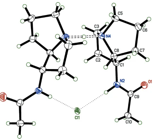
Figure 2
An ellipsoid plot (50% probability) showing the molecular structure of 2AcAPHCl. Unlabeled atoms are related to their labeled counterparts by a crystallographic twofold rotation (x+ 1,y,z+1
2). The unusually short N—H N hydrogen bond between the pyrrolizidine N atoms is highlighted by open dashed lines. Weaker N—H Cl hydrogen bonds between the acetamide NH group and the Clanion are shown as dotted
lines.
Figure 3
An ellipsoid plot (50% probability) showing disorder of the non-substituted half of the pyrrolizidine ring. The refined major:minor component occupancy factors are 0.897 (5):0.103 (5).
Table 1
Hydrogen-bond geometry (A˚ ,).
D—H A D—H H A D A D—H A
N4—H4N N4i 1.31 (1) 1.31 (1) 2.616 (2) 178 (3) N2—H2N Cl1 0.94 (2) 2.29 (2) 3.2263 (11) 176.4 (16)
Symmetry code: (i)xþ1;y;zþ1 2.
Figure 1
[image:2.610.47.297.436.665.2]Synthesis of AcAP.
Figure 4
[image:2.610.314.565.531.712.2]the twofold axis and is equidistant from N4 and N4i, the
refinedN H interatomic distance appears unusually long at 1.3080 (12) A˚ . The whole hydrogen-bonded ensemble makes an R3
3(12) supramolecular motif. For the short N4— H4N N4ihydrogen bond, a difference-Fourier map (Fig. 4) clearly shows an elongated region of electron density centered on the twofold axis corresponding to the position of the refined H4N hydrogen atom. The true location of H4N, however, remains ambiguous based on X-ray data alone. Possibilities include strictly symmetric (i.e., on the twofold axis exactly halfway between N4 and N4i), statically disordered (i.e., 50% on each of N4 and N4i) or dynamically disordered (i.e., exchanging rapidly between N4 and N4i). Alternative strategies for H4Ninclusion in the model refined equally well (see Refinement, below), so we settled on the simplest approach, following the recommendations of Fa´bry (2018). Nonetheless, the ambiguity prompted us to analyze the potential energy surface of the H4N position via computa-tional methods for singleversusdouble energy well character (seeComputational analysis, below). In addition to the strong N—H N hydrogen bond, weaker N2—H2—Cl1—H2i—N2i interactions link the twofold-related acetamide groups to the Clanion [N Cl = 3.2263 (11) A˚ ]. The twist of the
twofold-related hydrogen-bonded pyrrolizidine moieties relative to each other, as defined by the torsion angle C8—N4 N4i—C8i is 69.82 (16). The almost planar acetamide group forms a
dihedral angle with its twofold-related counterpart of 20.18 (3). The only other intramolecular interactions are van
der Waals contacts. Estimates of the relative fractions of intermolecular contacts between individual atom types derived from a Hirshfeld-surface analysis using Crystal-Explorer(Turneret al., 2017; Tanet al., 2019) are complicated by the ring disorder and by the N4—H4N N4i hydrogen bond. Nevertheless, all contacts appear to involve hydrogen atoms, with the overwhelming majority being H H (65.3%). The Cl anion and the acetamide O atom each reside in pockets surrounded by hydrogen atoms, giving H Cl/Cl H (16.2%) and H O/O H (12.4%), with the remainder being N H/H N and C H/H C contacts.
4. Database survey
A search of the Cambridge Structure Database (version 5.40, Nov. 2018; Groom et al., 2016) on the bicyclic pyrrolizidine core of AcAP yielded 584 hits. Of these, 41 are protonated at the ring N atom, but only three of those bear a substituent (other than H) at the 1-position of the pyrrolizidine ring system (assuming standard IUPAC ring numbering). CSD entry BRPYLZ (Wilson & Sawicki, 1979) is a bromide salt of a bromine derivative, and CPYRZD (Soderberg, 1971) is a zwitterion, with a carboxylate group at the 1-position and a bromo substituent at the 2-position. The relative stereo-chemistry of BRPYLZ and CPYRZD, however, are different from AcAP. CSD entry EDOTUP (Bhardwajet al., 2017) was a precursor to AcAP, and is the most closely related currently deposited structure.
The most striking feature of 2AcAPHCl is the unusually short N—H N hydrogen bond. A CSD search on the frag-ment ‘C-N(X)-H-N(X)-C0, where ‘X0 denotes ‘any group’,
gave 45 hits. Rejection of cases where apparent close N N distances were due to disorder (four entries), and those in which the N-bound H atom was missing from the model (two entries), left 39 structures, of which three were duplicates. In the remaining 36 structures the N—H N hydrogen bonds areintramolecular in 22 andintermolecular in 14. The closest N N separations occur in the intramolecular N—H N hydrogen-bonded structures, the shortest being 2.419 A˚ in EBOKOV (Wilkes et al., 2000). However, in these intra-molecular cases, the N N separation is largely dictated by the intramolecular geometry, which effectively forces the donor and acceptor N atoms into close proximity. Of the 14 CSD entries from the search that have intermolecular N— H N hydrogen bonding, the closest N N separation occurs in BECHOG (Glidewell & Holden, 1982), in which a bis(4-methylpyridine)hydrogen(I) cation sits on an inversion centre, giving an apparently symmetric N—H N hydrogen bond with N N distance of 2.610 (15) A˚ , and ROHTIR (Bocket al., 1997), for which the asymmetric unit contains two separate halves of a methylammonium-methylamine cation, [CH3NH2 -H-NH2CH3]+, each sitting on centres of inversion, giving N N distances of 2.620 and 2.641 A˚ . The N N separation in 2AcAPHCl is similar, at 2.616 (2) A˚ , although the difference is not significant, and well within the quoted precision estimate of BECHOG and the accuracy limits imposed by the sphe-rical-atom scattering-factor approximation (seee.g., Dawson, 1964).
5. Computational analysis
[image:3.610.315.565.527.713.2]A model consisting of the four 2AcAPHCl units present in one unit cell was relaxed at the DFT level (see below for details), both with and without symmetry constraints on
Figure 5
charge density. In the case where symmetry constraints were absent, a small displacement (0.06 A˚ ) was applied to the hydrogen atom in the N—H N hydrogen bond to break symmetry in the initial geometry. The relaxations led to two structures, one with constrained twofold symmetry (A), and one unconstrained (B). The volume difference between these theoretical models (calculations assumed absolute zero) was negligible (Avol= 1918.85 A˚3versus Bvol= 1919.63 A˚3). In the symmetric model, the N—H N hydrogen atom (corre-sponding to H4 in the crystallographic model) is equidistant between the two nitrogen atoms (N—H = 1.290 A˚ ), whereas in model B the N—H distances differ (N—H = 1.194 and 1.406 A˚ ). This is in agreement with the computed charge-density line profile of N—H N in structureB, as shown in Fig. 5. StructureBis calculated to be slightly more stable, but the energy difference (4.7 meV per unit cell) is vanishingly small (Fig. 6). The low energy barrier suggests dynamic disorder of the N—H N hydrogen atom.
Computational details: For this periodic system, density functional theory (DFT) calculations were carried out using the Viennaab initiosimulation Package (VASP) with Perdew– Burke–Ernzerhof (PBE) exchange-correlation functional (Kresse & Furthmu¨ller, 1996a,b; Kresseet al., 1994; Perdewet al., 1996). The electron-ion interactions were described with the projector augmented-wave (PAW) method (Blo¨chl, 1994; Kresse & Joubert, 1999). The valence electronic wavefunc-tions were expanded on a plane-wave basis with a kinetic energy cutoff at 520 eV, and Gaussian smearing with a width of 0.05 eV was employed. The convergence criterion of the total energy was set to 10 5 eV in the self-consistent field loop. The Brillouin zone was sampled with a 122 -centered grid. The experimental structures were relaxed until the Hellman–Feynman forces for each site were less than 0.005 eV A˚1, and Grimme’s DFT-D3 dispersion correction was applied with Becke–Johnson damping (Grimme et al., 2010, 2011).
6. Synthesis and crystallization
AcAP was synthesized and purified according to the published procedure (Pan et al., 2014). Crystals of 2AcAPHCl were obtained in the form of colorless plates by dissolving 20 mg of AcAP in 1 ml of CHCl3 in a 10 ml round-bottom flask and allowing the solution to stand in a refrigerator for about a month.
7. Refinement
Crystal data, data collection, and structure refinement details are given in Table 2. Non-disordered carbon-bound H atoms were found in difference-Fourier maps, but subsequently included in the refinement using riding models, with constrained distances set to 0.98 A˚ (RCH3), 0.99 A˚ (R2CH2) and 1.00 A˚ (R3CH). Following the advice of Fa´bry (2018), the hydrogen atom involved in the short N—H N hydrogen bond (H4N) was placed into difference-Fourier electron density and refined, albeit constrained to the twofold axis. An alternative model in which this H atom was allowed to ride at 50% occupancy on both N4 and N4i[symmetry code: (i)x+ 1,
y, z + 1
[image:4.610.312.565.88.380.2]2] refined equally well: the X-ray data alone being insufficient to establish a preference. The amide N—H hydrogen atom (H2N) was refined freely.Uiso(H) parameters for nitrogen-bound hydrogen atoms were refined, while for
Figure 6
Computed potential-energy surface as a function of N—H N hydrogen-atom displacement from the midpoint of the two nitrogen hydrogen-atoms.
Table 2
Experimental details.
Crystal data
Chemical formula C9H17N2O+Cl
C9H16N2O
Mr 372.93
Crystal system, space group Monoclinic,C2/c
Temperature (K) 90
a,b,c(A˚ ) 20.2125 (8), 9.6926 (4), 10.1458 (3)
() 100.445 (1)
V(A˚3) 1954.74 (13)
Z 4
Radiation type MoK
(mm1) 0.22
Crystal size (mm) 0.400.360.04
Data collection
Diffractometer Bruker D8 Venture dual source
Absorption correction Multi-scan (SADABS; Krauseet al., 2015)
Tmin,Tmax 0.885, 0.988
No. of measured, independent and observed [I> 2(I)] reflections
31903, 2235, 2096
Rint 0.032
(sin/)max(A˚1) 0.649
Refinement
R[F2> 2(F2)],wR(F2),S 0.037, 0.096, 1.12
No. of reflections 2235
No. of parameters 144
No. of restraints 82
H-atom treatment H atoms treated by a mixture of
independent and constrained refinement
max,min(e A˚3) 0.67,0.22
Computer programs: APEX3 (Bruker, 2016), SHELXT (Sheldrick, 2015a),
SHELXL2018/3(Sheldrick, 2015b),XPinSHELXTLandSHELX(Sheldrick, 2008),
[image:4.610.45.296.522.709.2]carbon-bound H atoms, Uiso(H) were set to values of either 1.2Ueq(R3CH,R2H2) or 1.5Ueq(RCH3) of the attached atom. The refined displacement parameters for the Cl anion (e.g., Figs. 2 and 4) appear a little small compared to the rest of the structure. In addition, the largest residual difference map peaks (0.67 and 0.65 e A˚3) were close (0.37 and 0.47 A˚ respectively) to Cl1. Refinement of the anion as mixed Cl and Br gave an occupancy ratio of 0.934 (2):0.066 (2), a lowerR -value (3.02%), and a flatter difference map ( = 0.29/-0.19 e A˚3). However, the reaction included no known source of Br, so the mixed anion model was not retained.
To ensure satisfactory refinement for disordered groups in the structure, a combination of constraints and restraints were employed. The constraints (SHELXLcommands EXYZ and EADP) were used to fix overlapping fragments. Restraints were used to ensure the integrity of ill-defined or disordered groups (SHELXLcommands SAME, SIMU, and RIGU). An alternative model using space group Cc (50:50 inversion twinned) was considered but rejected as it required hefty restraints and did not resolve the H4Natom ambiguity.
Acknowledgements
Supercomputing resources on the Lipscomb High-Perfor-mance Computing Cluster were provided by the University of Kentucky Information Technology Department and Center for Computational Sciences (CCS).
Funding information
Funding for this research was provided by: The National Science Foundation (MRI-CHE1625732 to SRP), and by the University of Kentucky. QA acknowledges the NSF under Award DMR-1627428.
References
Allen, F. H., Kennard, O., Watson, D. G., Brammer, L., Orpen, A. G. & Taylor, R. (1987).J. Chem. Soc., Perkin Trans. 2, pp. S1–S19.
Bhardwaj, M., Parkin, S. & Grossman, R. B. (2017). Private communication (refcode EDOTUP). CCDC, Cambridge, England. Blo¨chl, P. E. (1994).Phys. Rev. B,50, 17953–17979.
Bock, H., Vaupel, T. & Scho¨del, H. (1997).J. Prakt. Chem.339, 26–37. Bruker (2016).APEX3. Bruker AXS Inc., Madison, Wisconsin, USA. Dawson, B. (1964).Acta Cryst.17, 990–996.
Fa´bry, J. (2018).Acta Cryst.E74, 1344–1357.
Glidewell, C. & Holden, H. D. (1982).Acta Cryst.B38, 667–669. Grimme, S., Antony, J., Ehrlich, S. & Krieg, H. (2010).J. Chem. Phys.
132, 154104.
Grimme, S., Ehrlich, S. & Goerigk, L. (2011).J. Comput. Chem.32, 1456–1465.
Groom, C. R., Bruno, I. J., Lightfoot, M. P. & Ward, S. C. (2016).Acta Cryst.B72, 171–179.
Krause, L., Herbst-Irmer, R., Sheldrick, G. M. & Stalke, D. (2015).J. Appl. Cryst.48, 3–10.
Kresse, G. & Furthmu¨ller, J. (1996a).Comput. Mater. Sci.6, 15–50. Kresse, G. & Furthmu¨ller, J. (1996b).Phys. Rev. B,54, 11169–11186. Kresse, G., Furthmu¨ller, J. & Hafner, J. (1994).Phys. Rev. B, 50,
13181–13185.
Kresse, G. & Joubert, D. (1999).Phys. Rev. B,59, 1758–1775. Pan, J., Bhardwaj, M., Faulkner, J. R., Nagabhyru, P., Charlton, N. D.,
Higashi, R. M., Miller, A.-F., Young, C. A., Grossman, R. B. & Schardl, C. L. (2014).Phytochemistry,98, 60–68.
Pan, J., Bhardwaj, M., Zhang, B., Chang, W.-C., Schardl, C. L., Krebs, C., Grossman, R. B. & Bollinger, J. M. (2018).Biochemistry, 57, 2074–2083.
Parkin, S. (2013). CIFFIX. https://xray.uky.edu/Resources/scripts/ ciffix
Perdew, J. P., Burke, K. & Ernzerhof, M. (1996).Phys. Rev. Lett.77, 3865–3868.
Schardl, C. L., Grossman, R. B., Nagabhyru, P., Faulkner, J. R. & Mallik, U. P. (2007).Phytochemistry,68, 980–996.
Sheldrick, G. M. (2008).Acta Cryst.A64, 112–122. Sheldrick, G. M. (2015a).Acta Cryst.A71, 3–8. Sheldrick, G. M. (2015b).Acta Cryst.C71, 3–8. Soderberg, E. (1971).Acta Chem. Scand.,25, 615–624. Spek, A. L. (2009).Acta Cryst.D65, 148–155.
Tan, S. L., Jotani, M. M. & Tiekink, E. R. T. (2019).Acta Cryst.E75, 308–318.
Turner, M. J., Mckinnon, J. J., Wolff, S. K., Grimwood, D. J., Spackman, P. R., Jayatilaka, D. & Spackman, M. A. (2017).Crystal Explorer 17.The University of Western Australia.
Wilkes, E. N., Hambley, T. W., Lawrance, G. A. & Maeder, M. (2000). Aust. J. Chem.53, 517–521.
sup-1
Acta Cryst. (2020). E76, 77-81
supporting information
Acta Cryst. (2020). E76, 77-81 [https://doi.org/10.1107/S2056989019016517]
An unusually short intermolecular N
—
H
···
N hydrogen bond in crystals of the
hemi-hydrochloride salt of 1-
exo
-acetamidopyrrolizidine
Minakshi Bhardwaj, Qianxiang Ai, Sean R. Parkin and Robert B. Grossman
Computing details
Data collection: APEX3 (Bruker, 2016); cell refinement: APEX3 (Bruker, 2016); data reduction: APEX3 (Bruker, 2016);
program(s) used to solve structure: SHELXT (Sheldrick, 2015a); program(s) used to refine structure: SHELXL2018/3
(Sheldrick, 2015b); molecular graphics: XP in SHELXTL (Sheldrick, 2008); software used to prepare material for
publication: SHELX (Sheldrick, 2008), CIFFIX (Parkin, 2013) and PLATON (Spek, 2009).
(1R*, 8S)-2-acetamidooctahydropyrrolizin-4-ium chloride–N-[(1R, 8S)hexahydro-1H-pyrrolizin-2-yl)acetamide
(1/1)
Crystal data
C9H17N2O+·Cl−·C9H16N2O
Mr = 372.93
Monoclinic, C2/c a = 20.2125 (8) Å b = 9.6926 (4) Å c = 10.1458 (3) Å β = 100.445 (1)° V = 1954.74 (13) Å3
Z = 4
F(000) = 808 Dx = 1.267 Mg m−3
Mo Kα radiation, λ = 0.71073 Å Cell parameters from 9978 reflections θ = 2.3–27.5°
µ = 0.22 mm−1
T = 90 K Plate, colourless 0.40 × 0.36 × 0.04 mm
Data collection
Bruker D8 Venture dual source diffractometer
Radiation source: microsource Detector resolution: 7.41 pixels mm-1
φ and ω scans
Absorption correction: multi-scan
(SADABS; Krause et al., 2015)
Tmin = 0.885, Tmax = 0.988
31903 measured reflections 2235 independent reflections 2096 reflections with I > 2σ(I) Rint = 0.032
θmax = 27.5°, θmin = 2.3°
h = −26→26
k = −12→12
l = −11→13
Refinement
Refinement on F2 Least-squares matrix: full R[F2 > 2σ(F2)] = 0.037
wR(F2) = 0.096
S = 1.12 2235 reflections 144 parameters 82 restraints
Primary atom site location: structure-invariant direct methods
Secondary atom site location: difference Fourier map
Hydrogen site location: mixed
H atoms treated by a mixture of independent and constrained refinement
w = 1/[σ2(F
sup-2
Acta Cryst. (2020). E76, 77-81 (Δ/σ)max < 0.001
Δρmax = 0.67 e Å−3 Δρmin = −0.22 e Å−3
Extinction correction: SHELXL-2018/3 (Sheldrick 2018),
Fc*=kFc[1+0.001xFc2λ3/sin(2θ)]-1/4 Extinction coefficient: 0.005 (1)
Special details
Experimental. The crystal was mounted using polyisobutene oil on the tip of a fine glass fibre, which was fastened in a copper mounting pin with electrical solder. It was placed directly into the cold gas stream of a liquid-nitrogen based cryostat (Hope, 1994; Parkin & Hope, 1998).
Diffraction data were collected with the crystal at 90K, which is standard practice in this laboratory for the majority of flash-cooled crystals.
Geometry. All esds (except the esd in the dihedral angle between two l.s. planes) are estimated using the full covariance matrix. The cell esds are taken into account individually in the estimation of esds in distances, angles and torsion angles; correlations between esds in cell parameters are only used when they are defined by crystal symmetry. An approximate (isotropic) treatment of cell esds is used for estimating esds involving l.s. planes.
Refinement. Refinement progress was checked using PLATON (Spek, 2009) and by an R-tensor (Parkin, 2000). The final model was further checked with the IUCr utility checkCIF.
Fractional atomic coordinates and isotropic or equivalent isotropic displacement parameters (Å2)
x y z Uiso*/Ueq Occ. (<1)
C1 0.63783 (6) 0.58377 (14) 0.26083 (13) 0.0182 (3)
H1 0.684523 0.622089 0.275591 0.022*
C2 0.60256 (7) 0.61745 (15) 0.11816 (14) 0.0229 (3)
H2A 0.632627 0.601397 0.052759 0.027*
H2B 0.561093 0.562062 0.092144 0.027*
C3 0.58652 (7) 0.76915 (15) 0.12835 (14) 0.0232 (3)
H3A 0.626574 0.826636 0.124182 0.028*
H3B 0.549608 0.797166 0.055229 0.028*
N4 0.56555 (6) 0.78256 (11) 0.26254 (11) 0.0184 (3)
H4N 0.500000 0.781 (3) 0.250000 0.056 (9)*
C5 0.5885 (2) 0.9121 (2) 0.3373 (3) 0.0249 (7) 0.897 (5)
H5A 0.554374 0.945176 0.388570 0.030* 0.897 (5)
H5B 0.597261 0.985720 0.275018 0.030* 0.897 (5)
C6 0.65299 (9) 0.87111 (17) 0.43106 (16) 0.0237 (4) 0.897 (5)
H6A 0.663640 0.935711 0.507555 0.028* 0.897 (5)
H6B 0.691530 0.867102 0.383296 0.028* 0.897 (5)
C7 0.63499 (12) 0.7276 (2) 0.4771 (2) 0.0209 (5) 0.897 (5)
H7A 0.675941 0.673734 0.512861 0.025* 0.897 (5)
H7B 0.606530 0.733983 0.546696 0.025* 0.897 (5)
C8 0.59630 (6) 0.66296 (13) 0.34850 (13) 0.0178 (3) 0.897 (5)
H8 0.559801 0.602145 0.370299 0.021* 0.897 (5)
C5′ 0.595 (2) 0.9165 (19) 0.322 (2) 0.0249 (7) 0.103 (5)
H5′A 0.561158 0.991410 0.308580 0.030* 0.103 (5)
H5′B 0.634307 0.944647 0.282171 0.030* 0.103 (5)
C6′ 0.6157 (9) 0.8793 (15) 0.4690 (15) 0.032 (3) 0.103 (5)
H6′A 0.576222 0.871718 0.513823 0.039* 0.103 (5)
H6′B 0.647840 0.947113 0.517395 0.039* 0.103 (5)
sup-3
Acta Cryst. (2020). E76, 77-81
H7′A 0.655595 0.689122 0.544614 0.036* 0.103 (5)
H7′B 0.692818 0.749314 0.428958 0.036* 0.103 (5)
C8′ 0.59630 (6) 0.66296 (13) 0.34850 (13) 0.0178 (3) 0.103 (5)
H8′ 0.563026 0.605013 0.385455 0.021* 0.103 (5)
N2 0.64091 (6) 0.43798 (12) 0.29157 (11) 0.0184 (3)
H2N 0.6002 (10) 0.390 (2) 0.2837 (19) 0.035 (5)*
C9 0.69841 (6) 0.36514 (14) 0.31606 (12) 0.0178 (3)
O1 0.75448 (5) 0.41694 (11) 0.32142 (11) 0.0260 (2)
C10 0.68897 (7) 0.21311 (14) 0.33748 (14) 0.0224 (3)
H10A 0.715908 0.185291 0.423611 0.034*
H10B 0.703494 0.160954 0.265024 0.034*
H10C 0.641361 0.194212 0.337727 0.034*
Cl1 0.500000 0.27309 (5) 0.250000 0.02141 (15)
Atomic displacement parameters (Å2)
U11 U22 U33 U12 U13 U23
C1 0.0169 (6) 0.0170 (6) 0.0210 (6) −0.0016 (5) 0.0044 (5) 0.0007 (5)
C2 0.0266 (7) 0.0247 (7) 0.0185 (6) −0.0007 (5) 0.0071 (5) 0.0009 (5)
C3 0.0240 (7) 0.0244 (7) 0.0214 (7) −0.0021 (5) 0.0041 (5) 0.0056 (5)
N4 0.0201 (5) 0.0139 (5) 0.0201 (6) −0.0016 (4) 0.0006 (4) 0.0003 (4)
C5 0.0284 (13) 0.0147 (7) 0.0291 (10) −0.0044 (6) −0.0014 (9) −0.0013 (6)
C6 0.0240 (9) 0.0220 (8) 0.0234 (8) −0.0059 (6) 0.0001 (6) −0.0025 (6)
C7 0.0237 (10) 0.0206 (8) 0.0174 (9) 0.0000 (7) 0.0015 (7) −0.0004 (7)
C8 0.0189 (6) 0.0154 (6) 0.0187 (6) 0.0002 (5) 0.0020 (5) 0.0012 (5)
C5′ 0.0284 (13) 0.0147 (7) 0.0291 (10) −0.0044 (6) −0.0014 (9) −0.0013 (6)
C6′ 0.033 (7) 0.028 (5) 0.033 (5) −0.003 (5) −0.001 (5) −0.008 (4)
C7′ 0.034 (8) 0.034 (6) 0.017 (7) 0.002 (6) −0.003 (6) −0.006 (5)
C8′ 0.0189 (6) 0.0154 (6) 0.0187 (6) 0.0002 (5) 0.0020 (5) 0.0012 (5)
N2 0.0175 (5) 0.0155 (6) 0.0228 (6) −0.0015 (4) 0.0049 (4) −0.0005 (4)
C9 0.0200 (6) 0.0191 (6) 0.0142 (6) −0.0007 (5) 0.0028 (5) −0.0010 (5)
O1 0.0179 (5) 0.0247 (5) 0.0349 (6) −0.0010 (4) 0.0029 (4) 0.0037 (4)
C10 0.0246 (7) 0.0178 (7) 0.0239 (7) 0.0004 (5) 0.0022 (5) −0.0015 (5)
Cl1 0.0099 (2) 0.0177 (2) 0.0354 (3) 0.000 0.00096 (16) 0.000
Geometric parameters (Å, º)
C1—N2 1.4460 (17) C6—H6B 0.9900
C1—C2 1.5284 (19) C7—C8 1.529 (2)
C1—C8 1.5347 (18) C7—H7A 0.9900
C1—C8′ 1.5347 (18) C7—H7B 0.9900
C1—H1 1.0000 C8—H8 1.0000
C2—C3 1.513 (2) C5′—C6′ 1.521 (18)
C2—H2A 0.9900 C5′—H5′A 0.9900
C2—H2B 0.9900 C5′—H5′B 0.9900
C3—N4 1.5032 (18) C6′—C7′ 1.528 (17)
C3—H3A 0.9900 C6′—H6′A 0.9900
sup-4
Acta Cryst. (2020). E76, 77-81
N4—H4N 1.3080 (12) C7′—C8′ 1.576 (16)
N4—C5 1.497 (2) C7′—H7′A 0.9900
N4—C5′ 1.507 (17) C7′—H7′B 0.9900
N4—C8 1.5146 (16) C8′—H8′ 1.0000
N4—C8′ 1.5146 (16) N2—C9 1.3439 (17)
N4—H4N 1.3080 (12) N2—H2N 0.94 (2)
C5—C6 1.520 (4) C9—O1 1.2317 (16)
C5—H5A 0.9900 C9—C10 1.5066 (19)
C5—H5B 0.9900 C10—H10A 0.9800
C6—C7 1.532 (3) C10—H10B 0.9800
C6—H6A 0.9900 C10—H10C 0.9800
N2—C1—C2 114.05 (11) C8—C7—C6 102.97 (16)
N2—C1—C8 111.75 (11) C8—C7—H7A 111.2
C2—C1—C8 103.41 (11) C6—C7—H7A 111.2
N2—C1—C8′ 111.75 (11) C8—C7—H7B 111.2
C2—C1—C8′ 103.41 (11) C6—C7—H7B 111.2
N2—C1—H1 109.1 H7A—C7—H7B 109.1
C2—C1—H1 109.1 N4—C8—C7 105.73 (12)
C8—C1—H1 109.1 N4—C8—C1 105.05 (10)
C3—C2—C1 102.22 (11) C7—C8—C1 116.80 (13)
C3—C2—H2A 111.3 N4—C8—H8 109.7
C1—C2—H2A 111.3 C7—C8—H8 109.7
C3—C2—H2B 111.3 C1—C8—H8 109.7
C1—C2—H2B 111.3 N4—C5′—C6′ 101.8 (12)
H2A—C2—H2B 109.2 N4—C5′—H5′A 111.4
N4—C3—C2 104.18 (11) C6′—C5′—H5′A 111.4
N4—C3—H3A 110.9 N4—C5′—H5′B 111.4
C2—C3—H3A 110.9 C6′—C5′—H5′B 111.4
N4—C3—H3B 110.9 H5′A—C5′—H5′B 109.3
C2—C3—H3B 110.9 C5′—C6′—C7′ 100.7 (16)
H3A—C3—H3B 108.9 C5′—C6′—H6′A 111.6
H4N—N4—C5 106.1 (12) C7′—C6′—H6′A 111.6
H4N—N4—C3 110.96 (17) C5′—C6′—H6′B 111.6
C5—N4—C3 114.78 (16) C7′—C6′—H6′B 111.6
H4N—N4—C5′ 112 (2) H6′A—C6′—H6′B 109.4
C3—N4—C5′ 106.2 (15) C6′—C7′—C8′ 102.6 (11)
H4N—N4—C8 110.4 (12) C6′—C7′—H7′A 111.2
C5—N4—C8 107.04 (14) C8′—C7′—H7′A 111.2
C3—N4—C8 107.55 (10) C6′—C7′—H7′B 111.2
H4N—N4—C8′ 110.4 (12) C8′—C7′—H7′B 111.2
C3—N4—C8′ 107.55 (10) H7′A—C7′—H7′B 109.2
C5′—N4—C8′ 109.8 (9) N4—C8′—C1 105.05 (10)
H4N—N4—H4N 0 (3) N4—C8′—C7′ 101.6 (8)
C5—N4—H4N 106.1 (12) C1—C8′—C7′ 105.9 (9)
C3—N4—H4N 110.96 (17) N4—C8′—H8′ 114.3
C5′—N4—H4N 112 (2) C1—C8′—H8′ 114.3
sup-5
Acta Cryst. (2020). E76, 77-81
C8′—N4—H4N 110.4 (12) C9—N2—C1 123.73 (11)
N4—C5—C6 104.5 (2) C9—N2—H2N 118.0 (12)
N4—C5—H5A 110.9 C1—N2—H2N 117.9 (12)
C6—C5—H5A 110.9 O1—C9—N2 123.50 (13)
N4—C5—H5B 110.9 O1—C9—C10 122.14 (13)
C6—C5—H5B 110.9 N2—C9—C10 114.37 (12)
H5A—C5—H5B 108.9 C9—C10—H10A 109.5
C5—C6—C7 101.78 (16) C9—C10—H10B 109.5
C5—C6—H6A 111.4 H10A—C10—H10B 109.5
C7—C6—H6A 111.4 C9—C10—H10C 109.5
C5—C6—H6B 111.4 H10A—C10—H10C 109.5
C7—C6—H6B 111.4 H10B—C10—H10C 109.5
H6A—C6—H6B 109.3
N2—C1—C2—C3 162.24 (11) N2—C1—C8—C7 93.71 (16)
C8—C1—C2—C3 40.67 (13) C2—C1—C8—C7 −143.19 (14)
C8′—C1—C2—C3 40.67 (13) H4N—N4—C5′—C6′ 97 (2)
C1—C2—C3—N4 −39.46 (13) C3—N4—C5′—C6′ −142 (2)
C2—C3—N4—H4N −97.5 (15) C8′—N4—C5′—C6′ −26 (3)
C2—C3—N4—C5 142.30 (19) N4—C5′—C6′—C7′ 45 (3)
C2—C3—N4—C5′ 140.9 (15) C5′—C6′—C7′—C8′ −47 (2)
C2—C3—N4—C8 23.32 (13) H4N—N4—C8′—C1 123.3 (9)
C2—C3—N4—C8′ 23.32 (13) C3—N4—C8′—C1 2.12 (13)
H4N—N4—C5—C6 141.3 (8) C5′—N4—C8′—C1 −113 (2)
C3—N4—C5—C6 −95.81 (19) H4N—N4—C8′—C7′ −126.5 (13)
C8—N4—C5—C6 23.5 (3) C3—N4—C8′—C7′ 112.3 (10)
N4—C5—C6—C7 −39.9 (3) C5′—N4—C8′—C7′ −3 (2)
C5—C6—C7—C8 40.9 (2) N2—C1—C8′—N4 −149.53 (11)
H4N—N4—C8—C7 −112.6 (9) C2—C1—C8′—N4 −26.43 (13)
C5—N4—C8—C7 2.4 (2) N2—C1—C8′—C7′ 103.4 (9)
C3—N4—C8—C7 126.23 (14) C2—C1—C8′—C7′ −133.5 (9)
H4N—N4—C8—C1 123.3 (9) C6′—C7′—C8′—N4 30.6 (18)
C5—N4—C8—C1 −121.70 (19) C6′—C7′—C8′—C1 140.1 (14)
C3—N4—C8—C1 2.12 (13) C2—C1—N2—C9 113.29 (14)
C6—C7—C8—N4 −26.97 (19) C8—C1—N2—C9 −129.88 (13)
C6—C7—C8—C1 89.41 (17) C8′—C1—N2—C9 −129.88 (13)
N2—C1—C8—N4 −149.53 (11) C1—N2—C9—O1 3.8 (2)
C2—C1—C8—N4 −26.43 (13) C1—N2—C9—C10 −176.47 (12)
Hydrogen-bond geometry (Å, º)
D—H···A D—H H···A D···A D—H···A
N4—H4N···N4i 1.31 (1) 1.31 (1) 2.616 (2) 178 (3)
N2—H2N···Cl1 0.94 (2) 2.29 (2) 3.2263 (11) 176.4 (16)