ISSN Online: 2165-7416 ISSN Print: 2165-7408
DOI: 10.4236/ojoph.2019.92009 Apr. 15, 2019 70 Open Journal of Ophthalmology
Treatment Options of Macular Edema
Secondary to Retinal Vein Occlusion (RVO):
A Review
Aradhana Pokharel, Jie Luan
Department of Ophthalmology, Zhongda Hospital, Southeast University, Nanjing, China
Abstract
Retinal Vein Occlusion (RVO) is a common retinal vascular disease second-ary to diabetic retinopathy resulting in permanent loss of vision despite available treatment options. Main vision impending complication of retinal vein occlusion is macular edema. Laser photocoagulation has been an estab-lished method for treating macular edema for many years but nowadays intravitreal injection of Anti-Vascular endothelial growth factors (Ranibizu-mab, Aflibercept Bevacizumab and Pegaptanib sodium) is the treatment of choice for macular edema from retinal vein occlusion. Intra-vitreal corticos-teroids Triamcinolone Acetonide and Dexamethasone implant, are also being used to treat in some macular edema cases but with higher rates of side ef-fects. Numerous surgical methods have been attempted for treating RVO and preventing macular edema; they include pars plana vitrectomy, radial optic neurotomy, laser induced chorioretinal anastomosis, and arteriovenous sheathotomy. Surgical methods supposedly relieve compression of the central retinal vein, altering the pathophysiology of vein occlusion at the level of the lamina cribrosa thus improving blood flow and oxygenation. But limitations result from its complications.
Keywords
Retinal Vein Occlusion, Macular Edema, Branch Retinal Vein Occlusion, Central Retinal Vein Occlusion, Vascular Endothelial Growth Factor
1. Introduction
Retinal Vein Occlusion (RVO) is the cause of the second most common retinal vascular disease after diabetic retinopathy resulting in potential irreversible loss of vision. Main vision impending complication of RVO is macular edema (ME). How to cite this paper: Pokharel, A. and
Luan, J. (2019) Treatment Options of Ma-cular Edema Secondary to Retinal Vein Occlusion (RVO): A Review. Open Journal of Ophthalmology, 9, 70-83.
https://doi.org/10.4236/ojoph.2019.92009
Received: February 25, 2019 Accepted: April 12, 2019 Published: April 15, 2019
Copyright © 2019 by author(s) and Scientific Research Publishing Inc. This work is licensed under the Creative Commons Attribution International License (CC BY 4.0).
http://creativecommons.org/licenses/by/4.0/
DOI: 10.4236/ojoph.2019.92009 71 Open Journal of Ophthalmology Types of RVO are: branch retinal vein occlusion (BRVO) and central retinal vein occlusion (CRVO). CRVO is occlusion of central retinal vein, and BRVO occlu-sion in branch retinal vein [1]. Hayreh divided RVO into 3 types: 1) BRVO di-vided into major BRVO and macular BRVO; 2) CRVO didi-vided into ischemic (I-CRVO) and nonischemic (NI_CRVO); and 3) hemi-CRVO involving half of the retina and further divided into ischemic and nonischemic types [2]. Accord-ing to a pooled analysis of population-based data, RVO prevalence in the USA, Europe, Asia, and Australia containing 68,751 individuals from 31 to 101 years of age was 5.20 per 1000 for any RVO, 4.42 per 1000 for BRVO, and 0.80 per 1000 for CRVO. It is suggested around 16.4 million people are affected by RVO (BRVO = 13.9 million and CRVO = 2.5 million). BRVO is common than CRVO. Prevalence of RVO varied by ethnicity being highest in Hispanics and Asians and increased with age, but did not differ by gender [3]. Individuals with BRVO in 1 eye have a 10% risk of any RVO in the contra-lateral eye within 3 years. The estimated risk of contralateral involvement in people with CRVO is ~1% per year, which increases to 7% at 5 years [1].
RVO pathogenesis (Figure 1) is multifactorial. RVO may be due to Virchow’s triad: 1) hemodynamic changes (venous stasis), 2) degenerative changes of the vessel wall, and 3) blood hypercoagulability [5]. RVO may lead to fluid leak from capillaries draining into the obstructed vein, caused by secretion of VEGF and interleukin-6, and resulting in ME. [6]. Atherosclerosis is associated with RVO [7]. RVO can also be secondary to inflammation, vasospasm, or compression of retinal vessels [8]. RVO risk factors can be systemic and ophthalmic. Systemic risk includes DM, HTN, arteriosclerosis, vascular cerebral stroke, thrombophilia and blood hyperviscosity. Ophthalmic risk factors include glaucoma, ocular hypertension, decreased ocular perfusion pressure, with acquired and congenital retinal arteries changes. Hyperhomocysteinemia, factor V Leiden mutation and anti-cardiolipin antibodies also increase RVO risk. Vasculitis and Bechet disease may increase RVO risk [9].
[image:2.595.219.532.547.714.2]CRVO patient may present with dense central scotoma or sudden painless monocular vision loss. Patient with BRVO may be asymptomatic, or complain of
DOI: 10.4236/ojoph.2019.92009 72 Open Journal of Ophthalmology relative scotoma, or metamorphopsia. I-CRVO presentation is acute [10]. Neo-vascularization, secondary to RVO, can develop in the iris (NVI), angle (NVA), optic nerve head, or retina. (NVE). NVA can compromise the aqueous humor outflow, resulting in neovascular glaucoma. Visual field defects and RAPD can be found. Studies by Hayreh 2015 [11] [12] showed fundus changes in RVO as retinal and sub-internal limiting membrane hemorrhages, and papilledema in I-CRVO, initially more marked in NI-CRVO. ME was more marked in I-CRVO and major BRVO than NI-CRVO and macular BRVO. I-CRVO had more retinal venous engorgement than NI-CRVO. FFA showed significant fluorescein leak, dilatation and obliteration of retinal capillary, and capillary foveal arcade brea-kage in I-CRVO than NI-CRVO. Neovascularization in retina and disc was seen only in major BRVO.
When RVO is suspected, proper history taking and clinical examinations should be done to find disease severity and start early treatment. Assessing Visu-al Acuity, VisuVisu-al Field examination, Pupil examination, IOP, Color Doppler im-aging of ocular blood vessels, slit lamp exam with undilated gonioscopy, and di-lated fundoscopy to detect glaucoma, ocular neovascularization, and ME is neces-sary. OCT provides image of vitreomacular traction, retinal hole/detachments, ME and its thickness. FFA helps reveal delayed retinal venous filling, severity of retinal ischemia, retinal vascular leakage from retinal new vessels, or intraretinal pooling of the dye associated with ME [13]. Complete blood count, electrolytes, Lipid Profile, Blood Glucose level, ECG, Carotid Artery Doppler, BMI calcula-tion, measuring Blood Pressure may help diagnosing systemic diseases. Man-agement of RVO includes managing ME, treating associated risk factors and preventing neovascular complications.
2. Mechanism of Macular Edema from RVO (Figure 2)
[image:3.595.294.454.507.694.2]Macular edema is characterized by fluid collection within the retina, due to the
Figure 2. Mechanism of macular edema
DOI: 10.4236/ojoph.2019.92009 73 Open Journal of Ophthalmology downregulation of the blood-retinal barrier and fluid leak from the vasculature. VEGF is proven to play crucial role in pathological process. VEGF expression is up-regulated by hypoxia and other stimuli. Intravitreal VEGF levels were noted to be elevated in patients with RVO [14] [15] [16]. Main complication of retinal vein occlusion is vision loss due to macular edema (Table 1).
3. Treatment Options for ME Secondary to RVO
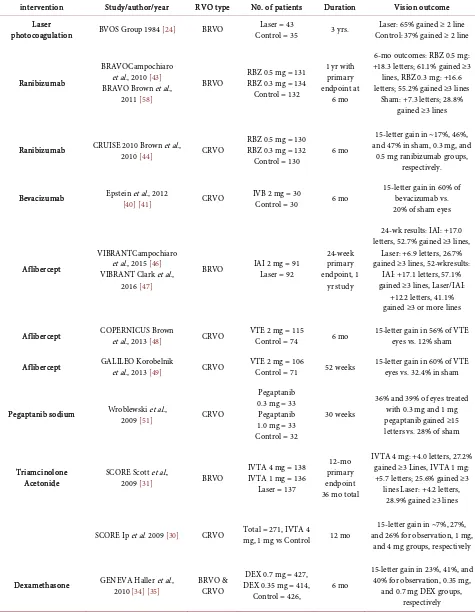
Treatment of RVO focuses on treating its outcome rather than restoring normal venous circulation. Management of RVO includes managing ME, treating risk factors and preventing occurrence of NVI, NVA, NVE. Multiple clinical trials have been done in the past (Table 2) and more ongoing in present to find most suitable treatment options for macular edema. Nowadays intravitreal injection of anti-VEGF, Triamcinolone Acetonide and dexamethasone implant, are in prior-ity to treat ME and other RVO’s complications. Surgical anastomoses [18] and laser have been tried [19] to relieve the obstruction by using thrombolytic ad-ministration [20] and by passing the congestion via optic nerve sheathotomy [21] but, limitations results from its complications.
3.1. Laser for ME
[image:4.595.209.538.554.700.2]Laser has been established method for treating RVO [22] [23]. Laser technique for resolving ME is macular grid photocoagulation while pan retinal (scatter) la-ser photocoagulation (PRPC) is used in treating retinal or disc neovasculariza-tion. Laser works by preventing secretion of chemical mediators originating from disturbed hemodynamics of retinal capillary and damaged tissue after RVO. The BVOS trial explored use of laser treatment for ME from BRVO re-porting vision improvement in about 1/3 of cases in first 3 months. Grid laser was considered after an FFA after 3 months in eyes with persistent ME and VA below 20/40. At 3 years endpoint, average of 1.33 lines was gained by treated eyes compared to 0.23 lines in control group. Since then, macular grid laser
Table 1. Vision loss mechanism in eyes with macular edema following retinal vein occlu-sion [17].
Anatomical Disorder Pathological mechanism reducing visual acuity Reversibility
Impairment of the
photoreceptors in the fovea Disruption of the inner and/or outer segments of photoreceptors irreversible* Probably
Impairment of the neurons in the inner retina
Neuronal loss in either the inner nuclear layer (INL) or the ganglion cell layer (GCL) due to
ischemia in the inner retina Irreversible
Neuro-retinal swelling due to macular edema
Light scattering resulting in a reduced amount of light received by photoreceptors at the fovea Compromised neural transduction from
photoreceptors to inner retinal neurons
Reversible
DOI: 10.4236/ojoph.2019.92009 74 Open Journal of Ophthalmology Table 2. Clinical trials in treating ME from RVO.
intervention Study/author/year RVO type N0. of patients Duration Vision outcome
Laser
photocoagulation BVOS Group 1984 [24] BRVO Control = 35 Laser = 43 3 yrs. Control: 37% gained ≥ 2 line Laser: 65% gained ≥ 2 line
Ranibizumab
BRAVOCampochiaro et al., 2010 [43] BRAVO Brown et al.,
2011 [58]
BRVO RBZ 0.5 mg = 131 RBZ 0.3 mg = 134 Control = 132
1 yr with primary endpoint at
6 mo
6-mo outcomes: RBZ 0.5 mg: +18.3 letters; 61.1% gained ≥3 lines, RBZ 0.3 mg: +16.6 letters; 55.2% gained ≥3 lines
Sham: +7.3 letters; 28.8% gained ≥3 lines
Ranibizumab CRUISE 2010 Brown et al., 2010 [44] CRVO RBZ 0.5 mg = 130 RBZ 0.3 mg = 132
Control = 130 6 mo
15-letter gain in ~17%, 46%, and 47% in sham, 0.3 mg, and
0.5 mg ranibizumab groups, respectively.
Bevacizumab Epstein et al., 2012 [40][41] CRVO IVB 2 mg = 30 Control = 30 6 mo 15-letter gain in 60% of bevacizumab vs. 20% of sham eyes
Aflibercept
VIBRANTCampochiaro et al., 2015 [46] VIBRANT Clark et al.,
2016 [47]
BRVO IAI 2 mg = 91 Laser = 92
24-week primary endpoint, 1
yr study
24-wk results: IAI: +17.0 letters, 52.7% gained ≥3 lines,
Laser: +6.9 letters, 26.7% gained ≥3 lines, 52-wkresults:
IAI: +17.1 letters, 57.1% gained ≥3 lines, Laser/IAI:
+12.2 letters, 41.1% gained ≥3 or more lines
Aflibercept COPERNICUS Brown et al., 2013 [48] CRVO VTE 2 mg = 115 Control = 74 6 mo 15-letter gain in 56% of VTE eyes vs. 12% sham
Aflibercept GALILEO Korobelnik et al., 2013 [49] CRVO VTE 2 mg = 106 Control = 71 52 weeks 15-letter gain in 60% of VTE eyes vs. 32.4% in sham
Pegaptanib sodium Wroblewski et al., 2009 [51] CRVO
Pegaptanib 0.3 mg = 33
Pegaptanib 1.0 mg = 33 Control = 32
30 weeks
36% and 39% of eyes treated with 0.3 mg and 1 mg pegaptanib gained ≥15 letters vs. 28% of sham
Triamcinolone
Acetonide SCORE Scott et al., 2009 [31] BRVO
IVTA 4 mg = 138 IVTA 1 mg = 136
Laser = 137
12-mo primary endpoint 36 mo total
IVTA 4 mg: +4.0 letters, 27.2% gained ≥3 Lines, IVTA 1 mg: +5.7 letters; 25.6% gained ≥3 lines Laser: +4.2 letters,
28.9% gained ≥3 lines
SCORE Ip et al. 2009 [30] CRVO Total = 271, IVTA 4 mg, 1 mg vs Control 12 mo and 26% for observation, 1 mg, 15-letter gain in ~7%, 27%, and 4 mg groups, respectively
Dexamethasone GENEVA Haller et al., 2010 [34][35] BRVO & CRVO DEX 0.35 mg = 414, DEX 0.7 mg = 427,
Control = 426, 6 mo
15-letter gain in 23%, 41%, and 40% for observation, 0.35 mg,
and 0.7 mg DEX groups, respectively
DOI: 10.4236/ojoph.2019.92009 75 Open Journal of Ophthalmology photocoagulation became gold standard for BRVO [24]. The central Vein occlu-sion group (CVOS) was planned to use laser for CRVO induced ME. It resulted in ME reduction but, no upgrade in visual acuity and final visual outcome was found compared to control group. Thus, observation became standard care for ME from CRVO [25]. Campochiaro in The RELATE Trial concluded that Scat-ter Photocoagulation does not reduce ME or treatment Burden in RVO eye [26]. Other study compared ranibizumab with standard grid laser for ME due to BRVO but found benefits in visual acuity gain and anatomic improvement with laser [27]. Some study say PRPC is not needed in NI-CRVO while a review [28] of 17 RCT concluded that though prophylactic PRPC did not prevent NVA/NVI or eliminated NVG risk for patient I-CRVO, it resulted in regression NVA/NVI and reduced progression to NVG. Today, intravitreal treatment has largely re-placed laser as choice of intervention for RVO.
3.2. Intravitreal Steroid for ME
1) Triamcinolone Acetonide: is intermediate acting powerful steroid. It dea-dens the humoral and cell-mediated component of inflammatory process and decreases vessel permeability, resolving ME. It blocks breakdown of the blood ocular barrier by modulating effector proteins downstream of VEGF receptor [29]. The SCORE Study (Standard Care versus Corticosteroid for Retinal Vein Occlusion) compared patients with both BRVO and CRVO using 4 and 1 mg of triamcinolone acetonide vs observation for 12 months [30] [31]. BRVO patient treated with 1 or 4 mg of triamcinolone versus focal laser showed that all groups achieved visual acuity score of ≥15 letters from baseline. In CRVO patient 7%, 27%, and 26% of participants achieved visual acuity score of ≥15 letters in the observation, 1-mg, and 4-mg groups. The complication rates of cataracts and increased IOP were greater in the 4 mg than 1 mg group. Despite SCORE’s result anti-VEGF has replaced corticosteroid as treatment choice for RVO because of side effects. High dose 20 mg IVTA has been found to be safe and cost effective alternative to Anti-VEGF in treating ME from RVO in lower-middle-income countries despite high risk of cataract and IOP rise when weighed against the vi-sion loss risk due to unaffordable anti-VEGF treatment [32].
DOI: 10.4236/ojoph.2019.92009 76 Open Journal of Ophthalmology significant increase in VA from baseline in both DEX implant groups than sham from day 30 - 180 with greatest response at day 60. With no statistical difference in two DEX groups at any follow-up visit. Mean decrease in CST was signifi-cantly high with DEX implant 0.7 mg (208 ± 201 µm) and 0.35 mg (177_ ± 197 µm) than with sham (85 ± 173 µm) at day 90 but not at day 180. An upgrade of ≥15-letter in BCVA from baseline by 30% and 32% of patients after 60 days of first and second DEX implant. After second 0.7 mg DEX implant the reduction in CST was similar to the first injection. Safety profile was also similar to the first treatment, except more cataract progression. The IOP increases were short-term and controlled by medication or observation. Cobalt study evaluated the effect of 0.7 mg IVD (n = 71) with ME < 3 month from BRVO. Retreatment was allowed ≥4 months from the last injection.At 6 and 12 months, mean BCVA improve-ment was 18.6 ± 12.9 and 15.3 ± 15.0 letters, respectively. with maximum sponse (70%) after 1 week. Over 12-month period, 32% and 49% of patients re-ceived 1 and 3 injections, respectively, with a mean ± SD interval of 20.0 ± 5.0 weeks. Patients requiring 3 injections had higher CRT and larger macular non-perfusion at baseline. Adverse events were increased IOP (35%) and newly di-agnosed cataract (16%). Study concluded DEX implant with interval of ≥4 months provides rapid and significantly better improvement in BCVA and CRT in BRVO-associated ME [36]. A study compared 4 mg of IVTA (n = 20) and 0.7 mg IVD (n = 20) for treating ME from NI-CRVO over 6 months. Study resulted that even patient achieving ≥15 letters was 40% in both groups, IOP rise and cataract progression was 2.4 and 3.5 times higher respectively in IVTA than IVD. Thus concluding Intravitreal steroids to be effective in managing ME from RVO, while IVD being significantly safer than IVTA [37].
3.3. Anti-VEGF
Currently four anti-VEGF drugs are available to treat CRVO and BRVO. Rani-bizumab and aflibercept are US FDA approved, while bevacizumab and pegap-tanib sodium is an off-label drug. Anti-VEGF therapy is now the treatment of choice for RVO disease.
DOI: 10.4236/ojoph.2019.92009 77 Open Journal of Ophthalmology letters compared to 33.3% in the (sh/bz) group. Mean decrease CRT was 435 μm and 404 μm in the bz/bz and sh/bz group respectively. Study concluded IVB given every 6 weeks for 12 months improves VA and reduce ME significantly, while delayed treatment may limit visual improvement. [40] [41]. A study eva-luated 1-year efficacy and safety of low-frequency IVB in treating ME due RVO. Patients with CRVO (n = 33) and BRVO (n = 55) were included. After 1 month, BCVA and OCT were recorded. If <30% improvement in BCVA and CMT, two injections were added at 1.5-month intervals. others were injected as per need. After a year 74% had clinically significant improvement of BCVA (>0.3 log MAR) with 1.98 average number of injections. Both groups had clinically signif-icant mean BCVA improvement from baseline vision and decrease in CMT. Pan retinal laser photocoagulation was done in 75.8% of all eyes with CRVO and sectoral photocoagulation in 49.1% of eyes with BRVO [42].
2) Ranibizumab: (Lucentis) is a monoclonal antibody fragment which binds all forms of VEGF. The BRAVO Study (Ranibizumab for the treatment of ma-cular edema following BRVO) studied the Efficacy and Safety of Ranibizumab Injection in Patients with ME Secondary to BRVO. Study showed, 55% patient receiving 0.3 mg and 61% of patients receiving 0.5 mg ranibizumab, achieved 3-line vision improvement compared with 29% in the control group. Continued PRN treatment showed stabilizing vision, although laser was added in almost 50% of patients [43]. The CRUISE Trial (Ranibizumab for the treatment of ma-cular edema after Central Retinal Vein Occlusion Study) studied Ranibizumab for treating ME from CRVO. 392 patients with ME secondary to CRVO were randomized to 0.3 mg or 0.5 mg of ranibizumab, or sham injection. At 6 months, 46% in 0.3 mg group and 48% in 0.5 mg group, showed improvement of 3 lines vision versus 17% in the control group [44]. Recently a study concluded no notable difference in outcome between IVR and IVB when treating ME from RVO for a year in routine clinical practice. This information might be of use in uplifting treatment burden of patient in the developing nations [45].
ef-DOI: 10.4236/ojoph.2019.92009 78 Open Journal of Ophthalmology ficacy of IAI in treating ME from CRVO. After one year Over 50% of treated eyes gained ≥15 ETDRS letters from baseline in both studies [48] [49]. A study Compared Efficacy of Intravitreal Aflibercept and Bevacizumab for ME due to BRVO. For 12 months patients (n = 27) received PRN IVB or (n = 25) PRN IAI with monthly follow-ups. Around 2/3 of the patients in both groups had final BCVA ≥ 20/40 and visual gains > 3 lines from baseline to month 12. Mean number of aflibercept and bevacizumab injection was 2.12 and 2.22 during period of 12-month. Both IAI and IVB were effective in reducing macular thickness and improving visual acuity [50].
4) Pegaptanib sodium: (Macugen) is a selective antagonist binding to 165 iso-forms of VEGF. For ME from CRVO Wroblewski et al. reported, double-masked phase two trial. Patients with CRVO for ≤6 months were randomly assigned (1:1:1) to injection of pegaptanib (n = 33) 1 mg and 0.3 mg, or sham (n = 32) every 6 weeks for 24 weeks. At 30 weeks eyes gaining≥15 letters among treat-ment groups was not significantly different. Study concluded pegaptanib appears to provide both visual and anatomical benefits in the treating ME from CRVO. Wroblewski et al. also (level II) assessed use of pegaptanib sodium for ME from BRVO. Subjects received pegaptanib 0.3 mg (n = 15) and 1 mg (n = 5) at base-line and at weeks 6 and 12 with subsequent injections at 6-week intervals up to 48 weeks. Both groups had similar outcomes from baseline to week 54 in mean BCVA (+14 ± 13 letters) and central subfield thickness (−201 ± 153 µm) [51] [52].
4. Surgical and Other Treatments
Numerous surgical methods have been attempted for treating RVO. Treatments include pars plana vitrectomy (PPV) with or without internal limiting mem-brane (ILM) peeling, radial optic neurotomy (RON), and laser induced chori-oretinal anastomosis (LCA), tissue plasminogen activator (tPA) and arterioven-ous sheathotomy. These methods supposedly relieve compression of the central retinal vein, altering the pathophysiology of RVO at the level of the lamina cribrosa (RON) or by improving blood flow and oxygenation (PPV, LCA) [53] [54]. Some studies [55] show LMWH role in the acute treatment of RVO while other study [56] shows Parnaparin to be more effective than aspirin in prevent-ing functional worsenprevent-ing in RVO. Study by Hayreh [57] showed use of aspirin, other anti-platelet aggregating agents, or anticoagulants in patients with CRVO and hemi-CRVO was associated with a worse visual outcome and no credible benefit. Case reports of patients treated with clopidogrel, tPA (both intravitreal and into a vein), heparin, aspirin, LMWH, or hemodilution, have shown variable results. But none of the treatment methods has shown to prevent the vision loss from RVO and larger clinical trial allowing a guidance is required.
5. Summary
retino-DOI: 10.4236/ojoph.2019.92009 79 Open Journal of Ophthalmology pathy. The main complication of retinal vein occlusion is vision loss due to ma-cular edema. Anti-VEGF therapy is the current standard of care for treating ME due to less side effect profile than intravitreal steroids which has a higher inci-dence of increased intraocular pressure and cataract formation. Future awaits more effective single or combined treatments for macular edema while mini-mizing health care cost.
Conflicts of Interest
The authors declare no conflicts of interest regarding the publication of this pa-per.
References
[1] Hayreh, S.S., Zimmerman, M.B. and Podhajsky, P. (1994) Incidence of Various Types of Retinal Vein Occlusion and Their Recurrence and Demographic Characte-ristics. American Journal of Ophthalmology, 117, 429-441.
https://doi.org/10.1016/S0002-9394(14)70001-7
[2] Hayreh, S.S. (2005) Prevalent Misconceptions about Acute Retinal Vascular Occlu-sive Disorders. Progress in Retinal and Eye Research, 24, 493-519.
https://doi.org/10.1016/j.preteyeres.2004.12.001
[3] Rogers, S., McIntosh, R.L., Cheung, N., Lim, L., Wang, J.J., Mitchell, P., et al. (2010) The Prevalence of Retinal Vein Occlusion: Pooled Data from Population Studies from the United States, Europe, Asia, and Australia. Ophthalmology, 117, 313-319e1.
[4] Marcucci, R., Sofi, F., Grifoni, E., Sodi, A. and Prisco, D. (2011) Retinal Vein Occlu-sions: A Review for the Internist. Internal and Emergency Medicine, 6, 307-314.
https://doi.org/10.1007/s11739-010-0478-2
[5] Zhou, J.Q., Xu, L., Wang, S., Wang, Y.X., You, Q.S., Tu, Y., et al. (2013) The 10-Year Incidence and Risk Factors of Retinal Vein Occlusion: The Beijing Eye Study. Ophthalmology, 120, 803-808. https://doi.org/10.1016/j.ophtha.2012.09.033
[6] Glanville, J., Patterson, J., McCool, R., Ferreira, A., Gairy, K. and Pearce, I. (2014) Efficacy and Safety of Widely Used Treatments for Macular Oedema Secondary to Retinal Vein Occlusion: A Systematic Review. BMC Ophthalmology, 14, 7.
https://doi.org/10.1186/1471-2415-14-7
[7] O’Mahoney, P.R., Wong, D.T. and Ray, J.G. (2008) Retinal Vein Occlusion and Traditional Risk Factors for Atherosclerosis. Archives of Ophthalmology, 126, 692-699. https://doi.org/10.1001/archopht.126.5.692
[8] Yen, Y.C., Weng, S.F., Chen, H.A. and Lin, Y.S. (2013) Risk of Retinal Vein Occlu-sion in Patients with Systemic Lupus Erythematosus: A Population-Based Cohort Study. The British Journal of Ophthalmology, 97, 1192-1196.
https://doi.org/10.1136/bjophthalmol-2013-303265
[9] Kolar, P. (2014) Risk Factors for Central and Branch Retinal Vein Occlusion: A Meta-Analysis of Published Clinical Data. Journal of Ophthalmology, 2014, Article ID: 724780. https://doi.org/10.1155/2014/724780
[10] Khayat, M., Williams, M. and Lois, N. (2018) Ischemic Retinal Vein Occlusion: Characterizing the More Severe Spectrum of Retinal Vein Occlusion. Survey of Ophthalmology, 63, 816-850. https://doi.org/10.1016/j.survophthal.2018.04.005
DOI: 10.4236/ojoph.2019.92009 80 Open Journal of Ophthalmology
Occlusion. Retina, 35, 1016-1027. https://doi.org/10.1097/IAE.0000000000000418
[12] Hayreh, S.S. and Zimmerman, M.B. (2015) Fundus Changes in Central Retinal Vein Occlusion. Retina, 35, 29-42. https://doi.org/10.1097/IAE.0000000000000256
[13] Sivaprasad, S., Amoaku, W.M., Hykin, P. and RVO Guideline Group (2015) The Royal College of Ophthalmologists Guidelines on Retinal Vein Occlusions: Execu-tive Summary. Eye, 29, 1633-1638. https://doi.org/10.1038/eye.2015.164
[14] Vinores, S.A., Derevjanik, N.L., Ozaki, H., Okamoto, N. and Campochiaro, P.A. (1999) Cellular Mechanisms of Blood-Retinal Barrier Dysfunction in Macular Ede-ma. Documenta Ophthalmologica, 97, 217-228.
https://doi.org/10.1023/A:1002136712070
[15] Aiello, L.P., Avery, R.L., Arrigg, P.G., Keyt, B.A., Jampel, H.D., Shah, S.T., et al.
(1994) Vascular Endothelial Growth Factor in Ocular Fluid of Patients with Diabet-ic Retinopathy and Other Retinal Disorders. The New England Journal of Medicine, 331, 1480-1487. https://doi.org/10.1056/NEJM199412013312203
[16] Boyd, S.R., Zachary, I., Chakravarthy, U., Allen, G.J., Wisdom, G.B., Cree, I.A., et al.
(2002) Correlation of Increased Vascular Endothelial Growth Factor with Neovas-cularization and Permeability in Ischemic Central Vein Occlusion. Archives of Ophthalmology, 120, 1644-1650. https://doi.org/10.1001/archopht.120.12.1644
[17] Iijima, H. (2018) Mechanisms of Vision Loss in Eyes with Macular Edema Asso-ciated with Retinal Vein Occlusion. Japanese Journal of Ophthalmology, 62, 265-273. https://doi.org/10.1007/s10384-018-0586-5
[18] Fekrat, S. and de Juan Jr., E. (1999) Chorioretinal Venous Anastomosis for Central Retinal Vein Occlusion: Transvitreal Venipuncture. Ophthalmic Surgery and Las-ers, 30, 52-55.
[19] McAllister, I.L., Gillies, M.E., Smithies, L.A., Rochtchina, E., Harper, C.A., Daniell, M.D., et al. (2010) The Central Retinal Vein Bypass Study: A Trial of Laser-Induced Chorioretinal Venous Anastomosis for Central Retinal Vein Occlusion. Ophthal-mology, 117, 954-965. https://doi.org/10.1016/j.ophtha.2009.10.026
[20] Feltgen, N., Junker, B., Agostini, H. and Hansen, L.L. (2007) Retinal Endovascular Lysis in Ischemic Central Retinal Vein Occlusion: One-Year Results of a Pilot Study.
Ophthalmology, 114, 716-723.e1. https://doi.org/10.1016/j.ophtha.2006.06.064
[21] Arevalo, J.F., Garcia, R.A., Wu, L., Rodriguez, F.J., Dalma-Weiszhausz, J., Qui-roz-Mercado, H., et al. (2008) Radial Optic Neurotomy for Central Retinal Vein Occlusion: Results of the Pan-American Collaborative Retina Study Group (PACORES). Retina, 28, 1044-1052. https://doi.org/10.1097/IAE.0b013e3181744153
[22] McIntosh, R.L., Mohamed, Q., Saw, S.M. and Wong, T.Y. (2007) Interventions for Branch Retinal Vein Occlusion: An Evidence-Based Systematic Review. Ophthal-mology, 114, 835-854. https://doi.org/10.1016/j.ophtha.2007.01.010
[23] Mohamed, Q., McIntosh, R.L., Saw, S.M. and Wong, T.Y. (2007) Interventions for Central Retinal Vein Occlusion: An Evidence-Based Systematic Review. Ophthal-mology, 114, 507-519. https://doi.org/10.1016/j.ophtha.2006.11.011
[24] The Branch Vein Occlusion Study Group (1984) Argon Laser Photocoagulation for Macular Edema in Branch Vein Occlusion. American Journal of Ophthalmology, 98, 271-282. https://doi.org/10.1016/0002-9394(84)90316-7
[25] (1993) Central Vein Occlusion Study of Photocoagulation Therapy. Baseline Find-ings. Central Vein Occlusion Study Group. The Online Journal of Current Clinical Trials, No. 95.
DOI: 10.4236/ojoph.2019.92009 81 Open Journal of Ophthalmology
I., et al. (2015) Scatter Photocoagulation Does Not Reduce Macular Edema or Treatment Burden in Patients with Retinal Vein Occlusion: The RELATE Trial.
Ophthalmology, 122, 1426-1437. https://doi.org/10.1016/j.ophtha.2015.04.006
[27] Tan, M.H., McAllister, I.L., Gillies, M.E., Verma, N., Banerjee, G., Smithies, L.A., et al. (2014) Randomized Controlled Trial of Intravitreal Ranibizumab versus Stan-dard Grid Laser for Macular Edema Following Branch Retinal Vein Occlusion.
American Journal of Ophthalmology, 157, 237-247e1.
[28] Rehak, M. and Wiedemann, P. (2010) Retinal Vein Thrombosis: Pathogenesis and Management. Journal of Thrombosis and Haemostasis: JTH, 8, 1886-1894.
https://doi.org/10.1111/j.1538-7836.2010.03909.x
[29] Edelman, J.L., Lutz, D. and Castro, M.R. (2005) Corticosteroids Inhibit VEGF-Induced Vascular Leakage in a Rabbit Model of Blood-Retinal and Blood-Aqueous Barrier Breakdown. Experimental Eye Research, 80, 249-258.
https://doi.org/10.1016/j.exer.2004.09.013
[30] Ip, M.S., Scott, I.U., VanVeldhuisen, P.C., Oden, N.L., Blodi, B.A., Fisher, M., et al.
(2009) A Randomized Trial Comparing the Efficacy and Safety of Intravitreal Tri-amcinolone with Observation to Treat Vision Loss Associated with Macular Edema Secondary to Central Retinal Vein Occlusion: The Standard Care vs Corticosteroid for Retinal Vein Occlusion (SCORE) Study Report 5. Archives of Ophthalmology, 127, 1101-1114. https://doi.org/10.1001/archophthalmol.2009.234
[31] Scott, I.U., Ip, M.S., VanVeldhuisen, P.C., Oden, N.L., Blodi, B.A., Fisher, M., et al.
(2009) A Randomized Trial Comparing the Efficacy and Safety of Intravitreal Tri-amcinolone with Standard Care to Treat Vision Loss Associated with Macular Edema Secondary to Branch Retinal Vein Occlusion: The Standard Care vs Corti-costeroid for Retinal Vein Occlusion (SCORE) Study Report 6. Archives of Oph-thalmology, 127, 1115-1128. https://doi.org/10.1001/archophthalmol.2009.233
[32] Ghoraba, H.H., Leila, M., Elgouhary, S.M., Elgemai, E.E.M., Abdelfattah, H.M., Ghoraba, H.H., et al. (2018) Safety of High-Dose Intravitreal Triamcinolone Aceto-nide as Low-Cost Alternative to Anti-Vascular Endothelial Growth Factor Agents in Lower-Middle-Income Countries. Clinical Ophthalmology, 2018, 2383-2391.
https://doi.org/10.2147/OPTH.S185274
[33] Felinski, E.A. and Antonetti, D.A. (2005) Glucocorticoid Regulation of Endothelial Cell Tight Junction Gene Expression: Novel Treatments for Diabetic Retinopathy.
Current Eye Research, 30, 949-957. https://doi.org/10.1080/02713680500263598
[34] Haller, J.A., Bandello, F., Belfort Jr., R., Blumenkranz, M.S., Gillies, M., Heier, J., et al. (2010) Randomized, Sham-Controlled Trial of Dexamethasone Intravitreal Im-plant in Patients with Macular Edema Due to Retinal Vein Occlusion. Ophthalmol-ogy, 117, 1134-1146e3.
[35] Haller, J.A., Bandello, F., Belfort Jr., R., Blumenkranz, M.S., Gillies, M., Heier, J., et al. (2011) Dexamethasone Intravitreal Implant in Patients with Macular Edema Re-lated to Branch or Central Retinal Vein Occlusion Twelve-Month Study Results.
Ophthalmology, 118, 2453-2460. https://doi.org/10.1016/j.ophtha.2011.05.014
[36] Yoon, Y.H., Kim, J.W., Lee, J.Y., Kim, I.T., Kang, S.W., Yu, H.G., et al. (2018) Dexamethasone Intravitreal Implant for Early Treatment and Retreatment of Ma-cular Edema Related to Branch Retinal Vein Occlusion: The Multicenter COBALT Study. Ophthalmologica, 240, 81-89. https://doi.org/10.1159/000487547
DOI: 10.4236/ojoph.2019.92009 82 Open Journal of Ophthalmology
https://doi.org/10.1186/s40942-018-0114-2
[38] Yilmaz, T. and Cordero-Coma, M. (2012) Use of Bevacizumab for Macular Edema Secondary to Branch Retinal Vein Occlusion: A Systematic Review. Graefe’s Arc-hive for Clinical and Experimental Ophthalmology, 250, 787-793.
https://doi.org/10.1007/s00417-012-2016-6
[39] Hoeh, A.E., Ruppenstein, M., Ach, T. and Dithmar, S. (2010) OCT Patterns of Ma-cular Edema and Response to Bevacizumab Therapy in Retinal Vein Occlusion.
Graefe’s Archive for Clinical and Experimental Ophthalmology, 248, 1567-1572.
https://doi.org/10.1007/s00417-010-1419-5
[40] Epstein, D.L., Algvere, P.V., von Wendt, G., Seregard, S. and Kvanta, A. (2012) Be-vacizumab for Macular Edema in Central Retinal Vein Occlusion: A Prospective, Randomized, Double-Masked Clinical Study. Ophthalmology, 119, 1184-1189.
https://doi.org/10.1016/j.ophtha.2012.01.022
[41] Epstein, D.L., Algvere, P.V., von Wendt, G., Seregard, S. and Kvanta, A. (2012) Benefit from Bevacizumab for Macular Edema in Central Retinal Vein Occlusion: Twelve-Month Results of a Prospective, Randomized Study. Ophthalmology, 119, 2587-2591. https://doi.org/10.1016/j.ophtha.2012.06.037
[42] Ivanovska Adjievska, B., Boskurt, S., Orovcanec, N. and Dimovska-Jordanova, V. (2017) The Outcome of Low-Frequency Intravitreal Bevacizumab Therapy for Ma-cular Edema in Retinal Vein Occlusions. Clinical Ophthalmology, 2017, 1183-1190.
https://doi.org/10.2147/OPTH.S137380
[43] Campochiaro, P.A., Heier, J.S., Feiner, L., Gray, S., Saroj, N., Rundle, A.C., et al.
(2010) Ranibizumab for Macular Edema Following Branch Retinal Vein Occlusion: Six-Month Primary End Point Results of a Phase III Study. Ophthalmology, 117, 1102-1112e1.
[44] Brown, D.M., Campochiaro, P.A., Singh, R.P., Li, Z., Gray, S., Saroj, N., et al. (2010) Ranibizumab for Macular Edema Following Central Retinal Vein Occlusion: Six-Month Primary End Point Results of a Phase III Study. Ophthalmology, 117, 1124-1133e1.
[45] Khan, M., Wai, K.M., Silva, F., Srivastava, S., Ehlers, J., Rachitskaya, A., Babiuch, A., Deasy, R., Kaiser, P., Schachat, A., Yuan, A. and Singh, R. (2017) Comparison of Ranibizumab and Bevacizumab for Macular Edema Secondary to Retinal Vein Oc-clusions in Routine Clinical Practice. Ophthalmic Surgery, Lasers and Imaging Re-tina, 48, 465-472. https://doi.org/10.3928/23258160-20170601-04
[46] Campochiaro, P.A., Clark, W.L., Boyer, D.S., Heier, J.S., Brown, D.M., Vitti, R., et al. (2015) Intravitreal Aflibercept for Macular Edema Following Branch Retinal Vein Occlusion: The 24-Week Results of the VIBRANT Study. Ophthalmology, 122, 538-544. https://doi.org/10.1016/j.ophtha.2014.08.031
[47] Clark, W.L., Boyer, D.S., Heier, J.S., Brown, D.M., Haller, J.A., Vitti, R., et al. (2016) Intravitreal Aflibercept for Macular Edema Following Branch Retinal Vein Occlu-sion: 52-Week Results of the VIBRANT Study. Ophthalmology, 123, 330-336.
https://doi.org/10.1016/j.ophtha.2015.09.035
[48] Brown, D.M., Heier, J.S., Clark, W.L., Boyer, D.S., Vitti, R., Berliner, A.J., et al.
(2013) Intravitreal Aflibercept Injection for Macular Edema Secondary to Central Retinal Vein Occlusion: 1-Year Results from the Phase 3 COPERNICUS Study.
American Journal of Ophthalmology, 155, 429-437e7.
[49] Korobelnik, J.F., Holz, F.G., Roider, J., Ogura, Y., Simader, C., Schmidt-Erfurth, U.,
DOI: 10.4236/ojoph.2019.92009 83 Open Journal of Ophthalmology Ophthalmology, 121, 202-208. https://doi.org/10.1016/j.ophtha.2013.08.012
[50] Wang, J.K., Su, P.Y., Hsu, Y.R., Chen, Y.J., Chen, F.T. and Tseng, Y.Y. (2016) Comparison of the Efficacy of Intravitreal Aflibercept and Bevacizumab for Macular Edema Secondary to Branch Retinal Vein Occlusion. Journal of Ophthalmology, 2016, Article ID: 8421940. https://doi.org/10.1155/2016/8421940
[51] Wroblewski, J.J., Wells 3rd, J.A., Adamis, A.P., Buggage, R.R., Cunningham Jr., E.T., Goldbaum, M., et al. (2009) Pegaptanib Sodium for Macular Edema Secondary to Central Retinal Vein Occlusion. Archives of Ophthalmology, 127, 374-380.
https://doi.org/10.1001/archophthalmol.2009.14
[52] Wroblewski, J.J., Wells III, J.A. and Gonzales, C.R. (2010) Pegaptanib Sodium for Macular Edema Secondary to Branch Retinal Vein Occlusion. American Journal of Ophthalmology, 149, 147-154. https://doi.org/10.1016/j.ajo.2009.08.005
[53] Yeh, S., Kim, S.J., Ho, A.C., Schoenberger, S.D., Bakri, S.J., Ehlers, J.P., et al. (2015) Therapies for Macular Edema Associated with Central Retinal Vein Occlusion: A Report by the American Academy of Ophthalmology. Ophthalmology, 122, 769-778. https://doi.org/10.1016/j.ophtha.2014.10.013
[54] Ehlers, J.P., Kim, S.J., Yeh, S., Thorne, J.E., Mruthyunjaya, P., Schoenberger, S.D., et al. (2017) Therapies for Macular Edema Associated with Branch Retinal Vein Oc-clusion: A Report by the American Academy of Ophthalmology. Ophthalmology, 124, 1412-1123. https://doi.org/10.1016/j.ophtha.2017.03.060
[55] Squizzato, A., Manfredi, E., Bozzato, S., Dentali, F. and Ageno, W. (2010) Antith-rombotic and Fibrinolytic Drugs for Retinal Vein Occlusion: A Systematic Review and a Call for Action. Thrombosis and Haemostasis, 103, 271-276.
https://doi.org/10.1160/TH09-09-0626
[56] Ageno, W., Cattaneo, R., Manfredi, E., Chelazzi, P., Venco, L., Ghirarduzzi, A., et al.
(2010) Parnaparin versus Aspirin in the Treatment of Retinal Vein Occlusion. A Randomized, Double Blind, Controlled Study. Thrombosis Research, 125, 137-141.
https://doi.org/10.1016/j.thromres.2009.05.007
[57] Hayreh, S.S., Podhajsky, P.A. and Zimmerman, M.B. (2011) Central and hemiCentral Retinal Vein Occlusion: Role of Anti-Platelet Aggregation Agents and Anticoagulants. Ophthalmology, 118, 1603-1611.
https://doi.org/10.1016/j.ophtha.2011.04.036
[58] Brown, D.M., Campochiaro, P.A., Bhisitkul, R.B., Ho, A.C., Gray, S., Saroj, N., et al.
![Figure 1. Pathogenesis of RVO [4].](https://thumb-us.123doks.com/thumbv2/123dok_us/9076033.404210/2.595.219.532.547.714/figure-pathogenesis-of-rvo.webp)
![Figure 2. Mechanism of macular edema from retinal vein occlusion [14] [15] [16].](https://thumb-us.123doks.com/thumbv2/123dok_us/9076033.404210/3.595.294.454.507.694/figure-mechanism-macular-edema-from-retinal-vein-occlusion.webp)
![Table 1. Vision loss mechanism in eyes with macular edema following retinal vein occlu-sion [17]](https://thumb-us.123doks.com/thumbv2/123dok_us/9076033.404210/4.595.209.538.554.700/table-vision-mechanism-macular-edema-following-retinal-occlu.webp)