ISSN Online: 2161-6809 ISSN Print: 2161-6795
DOI: 10.4236/wjnst.2019.94011 Sep. 9, 2019 147 World Journal of Nuclear Science and Technology
Diagnostic Reference Level in Frontal Chest
X-Ray in Western Côte d’Ivoire
Issa Konate1, George A. Monnehan1,2*, Douo B. L. H. Gogon1,2, Tekpo P. A. Dali1, Aka A. Koua1,2, Koudou Djagouri3
1Laboratory of Nuclear Physics and Radiation Protection, Training and Research Unit, Sciences of Matter and Technology,
Félix Houphouet-Boigny University, Abidjan, Côte d’Ivoire
2Radiation Protection and Nuclear Security Authority, Abidjan, Côte d’Ivoire 3High Normal School, Abidjan, Côte d’Ivoire
Abstract
Our study aims to determine diagnostic reference levels (DRL) for chest front examination in postero anterior (PA) for optimizing patient entrance surface dose (ESD) and dose-area product (DAP) of patients in west of Côte d’Ivoire. 90 patients from three hospitals undergoing conventional radiology were considered. The ESD and DAP for each patient were obtained during chest radiography (PA) examination. The measurements were performed with the device call Dose-Area Product-meter (DAP-meter) with brand Diamentor M4-KDK, type 11017. The DRL were obtained in applying the 75th percentile statistical method to the obtained ESD and DAP. The obtained DRL in ESD for chest radiography for all rooms is 0.40 mGy and in DAP is 54.85 cGy∙cm2.
Our DRL for ESD is higher than those obtained in Abidjan District and in other countries like UK and Cameroon. Our DRL for DAP is higher than those from Abidjan and all other countries for which a similar study was made. The comparison of these values to those from Abidjan and other countries, shows us that radiology technicians can make efforts to choose ra-diological parameters to reduce ESD. They must use convenable the X-rays tube to reduce DAP by reducing the patient exposure surface.
Keywords
Conventional Radiology, Entrance Surface-Dose, Dose-Area-Product, Dose-Area Product-Meter, Diagnostic Reference Levels
1. Introduction
The excessive variability of doses delivered to patients of the same body size,
How to cite this paper: Konate, I., Mon-nehan, G.A., Gogon, D.B.L.H., Dali, T.P.A., Koua, A.A. and Djagouri, K. (2019) Diag-nostic Reference Level in Frontal Chest X-Ray in Western Côte d’Ivoire. World Journal of Nuclear Science and Technology, 9, 147-158.
https://doi.org/10.4236/wjnst.2019.94011
Received: August 8, 2019 Accepted: September 6, 2019 Published: September 9, 2019
Copyright © 2019 by author(s) and Scientific Research Publishing Inc. This work is licensed under the Creative Commons Attribution International License (CC BY 4.0).
DOI: 10.4236/wjnst.2019.94011 148 World Journal of Nuclear Science and Technology during the same examination for the same medical purpose led the International Commission for Radiological Protection (ICRP) to make recommendations in 1996 [1]. The goal is to determine the Diagnostic Reference Levels (DRLs) for the most-performed and most-irradiating examinations to optimize the dose and dose-area product in conventional radiology received by the patient. From then on, many states have transposed the ICRP recommendations into their laws. Today the determination of the DRL is part of the technical cooperation project of the International Atomic Energy Agency (IAEA), n˚ RAF 9059 [2]. It is within this framework that we are inscribing our work to determine the DRL in western Côte d'Ivoire for three (3) radiology rooms for frontal chest examina-tion for 90 patients. The final objective is to provide convenexamina-tional radiology practicians with some reference dose values in order to ensure the management of the doses delivered and the efficient control of the exposure of patients in Côte d’Ivoire. Our work was conducted on patients with a measuring device called DAP-meter. In Côte d’Ivoire, Issa Konaté et al. have published in the same conditions two DRLs studies, one on Abidjan frontal chest (PA) examinations [3] and the other on Abidjan lumbar spine examinations [4]. It is worth noting that a preliminary work was conducted by Monnehan et al. [5] using a phantom (water-filled can) and TLD dosimeters. The reading of the dose at the entrance was made from a Harshaw 4500 reader.
For this work, we described the material, then the results were presented, then the discussions and conclusion closed our presentation.
2. Materials and Methods
2.1. Sampling MethodsThree (3) conventional radiology rooms corresponding to 3 hospitals, all located in three (3) cities of western Côte d'Ivoire, were selected. These are the Regional Hospital Center (CHR) in Daloa, the Regional Hospital Center (CHR) in San Pedro, and the General Hospital (HG) in Bangolo. All these rooms comply with the Ivorian standards of at least 25 m2 of base area and a ceiling height of at least
3.5 m [6] and have been inspected by the competent body. In each of the rooms, we took into account in our measurements, 30 patients [7], all equipped with a bulletin for the examination of the frontal chest and all, 18 years old at least. It should be noted that the examination of the frontal chest is the examination most practiced in these rooms. We excluded from our study patients on bed or chair. In total for our study we had 90 patients.
2.2. Data Collection
DOI: 10.4236/wjnst.2019.94011 149 World Journal of Nuclear Science and Technology mass, size, and thickness of the patient portion to be examined. We also noted technical parameters such as source-skin distance, voltage and electrical charge.
2.3. Materials
In a conventional radiology room, we have equipment such as the X-ray genera-tor, X-ray tube, desk, wall stand and X-ray viewer. For our study, we brought with us in each room, a DAP-meter brand Diamentor M4-KDK and type 11,017 manufactured by the German company PTW. This device has been previously calibrated by the PTW-Freiburg calibration laboratory. It consists of an ioniza-tion chamber and an electrometer connected by two cords. The ionizaioniza-tion chamber is placed at the exit of the tube at the collimator and the electrometer is placed at the desk behind the screen. When the beam passes through the ioniza-tion chamber, it deposits energy that is transferred by the leads to the electro-meter. At this level, this energy is transformed into to dose in air (Dair) and DAP [8].
2.4. Methods for Determining DRLs
We obtained the Entrance Surface Dose (ESD) for each patient, from the Dair by the following equation:
ESD Dair BSF= × (1)
where Dair is the dose in air
(BSF = backscattering factor) with BSF = 1.35 for voltage values included be-tween 60 and 80 kV or 1.5 above 80 kV [9].
We determined the Diagnostic Reference Levels (DRLs) for each examination according to the 75th percentile statistical method in accordance with the rec-ommendations of the European Commission. This is to take as DRL the value of the 75th percentile of ESD or DAP for a given exam on a large number of pa-tients and on a large number of ESD and DAP values. This is a method used in statistics to remove the limit values from the sample. The 75th percentile of n values in ascending order is the value of rank k, given by the following mathe-matical equation:
75 100
n
k= (2)
[10]; where n is the number of patient.
3. Results
3.1. Determination of the DRL for the ESD per Center for the Examination of the Frontal Chest (PA)
DOI: 10.4236/wjnst.2019.94011 150 World Journal of Nuclear Science and Technology
Figure 1. Comparison of DRLs values in mGy.
3.2. Comparison of Voltage Values in kV and Electrical Charge in mAs of the Rooms of Our Study for the Examination of the Frontal Thorax (AP)
It is observed on Figure 2, that the highest mean value of 117.2 kV of the voltage is obtained at HG Bangolo and the lowest average value 81.66 kV is obtained at the CHR of Daloa.
On Figure 3, the higher mean value of the electrical charge is obtained at the CHR of Daloa and the lowest is obtained at HG Bangolo.
3.3. Determination of the DRL for the DAP per Center for the Examination of the Frontal Chest (PA)
On Figure 4, DAP have their highest value obtained at CHR of Daloa (74.02 cGy∙cm2) and the lowest value obtained at Bangolo HG (15.2 cGy∙cm2).
3.4. Characteristics of the X-Ray Tubes in Each of the Radiology Rooms of Our Study
In Table 1, the higher filtration of the tube is observed at the CHR San-Pédro (2 mm Al) and the lowest is obtained at CHR Daloa and HG Bangolo (1 mm Al). The tube at CHR Daloa is almost the same age as that of HG Bangolo. However that of CHR San-Pédro is older than four (4) years. All these devices arrived new in the corresponding radiology rooms.
3.5. Voltage in kV and Electrical Charge in mAs, DRLs in mGy and in cGycm2 for All the Centers of Our Study for the Examination of the Frontal Thorax
We calculated the mean tension and the average electrical charge (arithmetic mean) [11]. We have adopted the following notations: average (minimum, maximum) in kV and mAs. Finally, we determined the DRL (75th percentile) in ESD and DAP. The values are shown in Table 2.
In Table 2, we have reported the mean values of voltages and mAs with their corresponding intervals. We also reported the DRLs values in ESD (0.40 mGy) and in DAP (54.85 cGy∙cm2) of all 3 rooms in our study.
0 0.05 0.1 0.15 0.2 0.25 0.3 0.35 0.4 0.45
CHR Daloa CHR San-Pédro HG Bangolo
DOI: 10.4236/wjnst.2019.94011 151 World Journal of Nuclear Science and Technology
[image:5.595.209.427.216.321.2]Figure 2. Comparison of voltage mean [11] values.
[image:5.595.211.527.356.511.2]Figure 3. Comparison of electrical charge mean [11] values.
[image:5.595.204.540.561.626.2]Figure 4. Comparison of DRL values in cGy∙cm2.
Table 1. Comparison of age and total filtration of X-ray tubes in our radiology rooms.
Hospital centers X-ray tubes model X-ray tubes year of installation X-ray tubes total filtration
CHR DALOA Dongmum 2011 1 mm Al
CHR SAN-PEDRO Toshiba 2015 2 mm Al
HG BANGOLO Dongmum 2012 1 mm Al
Table 2. Exposure parameters with mean values and range (in bracket) and DRL of ESD and DAP for all the hospital centers of our study.
Examination Voltage (kV) mAs DRL
ESD (mGy) DAP (cGy∙cm2)
Frontal chest (PA) (80 - 122) 101.6 (1.3 - 16) 6 0.40 54.85
0 20 40 60 80 100 120 140
CHR Daloa CHR San-Pédro HG Bangolo
Voltage (mean) in kV
0 2 4 6 8 10 12 14
CHR Daloa CHR San-Pédro HG Bangolo
Electrical charge (mean) in mAs
0 10 20 30 40 50 60 70 80
CHR DALOA CHR San Pédro HG Bangolo
DOI: 10.4236/wjnst.2019.94011 152 World Journal of Nuclear Science and Technology 3.6. DRL of Our Study and DRL Obtained in the District of Abidjan
for the Examination of the Frontal Thorax (PA) [3]
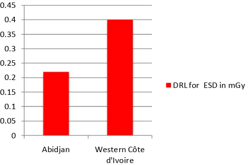
On Figure 5, we observe that the DRL for ESD in our study in the west of Côte d’Ivoire is higher than the one of Abidjan.
On Figure 6, the voltage use in Abidjan for the frontal chest exam is higher than the one of our study.
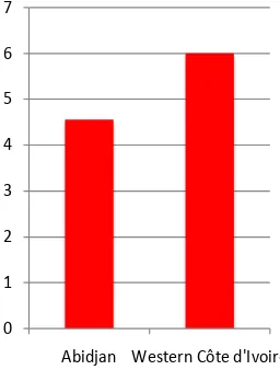
On Figure 7, the electrical charge use in Abidjan for the frontal chest exam is smaller than the one use in our study in western Côte d’Ivoire.
We observe on Figure 8, that the DRL for DAP in our study in the west of Côte d’Ivoire is higher than the one of Abidjan.
3.7. DRL from Our Study and DRL Obtained in Other Countries On Figure 9, we compare the DRL in ESD of our study with those obtained in other countries and institutions outside Africa. Our value 0.40 mGy is close to that of Iran and IAEA [12][13] but larger than all other values in the figure [14].
On Figure 10, we can compare our value of DRL for ESD with those obtained in other African countries. Our DRL value in ESD is equal to that of Nigeria [15], close to that of Cameroon [16] and higher than all other values [17][18].
On Figure 11, which presents the comparison of the DRL for DAP obtained in our study with those of IRNS and other countries outside Africa, the smallest value is obtained in the United Kingdom (UK), 10 cGy∙cm2[14], and the most
large, 54.85 cGy∙cm2 in our study [19][20].
4. Discussion
[image:6.595.251.503.536.703.2]According to the results of our study, for the examination of the frontal chest, we note that the highest DRL value in ESD of these radiology centers is obtained at CHR Daloa, it is 0.425 mGy. The lowest DRL value is obtained at CHR San-Pédro, 0.141 mGy (Figure 1). We explain this situation by the fact that the average value of the voltage used at the CHR Daloa, 81.66 kV is the smallest
Figure 5. Comparison of the DRL for ESD of our study to the one of Abidjan.
0 0.05 0.1 0.15 0.2 0.25 0.3 0.35 0.4 0.45
Abidjan Western Côte d'Ivoire
DOI: 10.4236/wjnst.2019.94011 153 World Journal of Nuclear Science and Technology
[image:7.595.233.361.301.470.2] [image:7.595.239.511.505.702.2]Figure 6. Comparison of the voltage to the one of Abidjan.
Figure 7. Comparison of the electrical charge to the one of Abidjan.
Figure 8. Comparison of the DRL for DAP of our study to the one of Abidjan.
100 100.5 101 101.5 102 102.5 103 103.5 104 104.5
Abidjan Western Côte
d'Ivoire
Voltage(mean) in kV
0 1 2 3 4 5 6 7
Abidjan Western Côte d'Ivoire
Electrical charge (mean) in mAs
52 52.5 53 53.5 54 54.5 55
Abidjan Western Côte
d'Ivoire
DOI: 10.4236/wjnst.2019.94011 154 World Journal of Nuclear Science and Technology
[image:8.595.230.520.291.474.2]Figure 9. Comparison of DRL for ESD obtained in our study with those ob-tained in other countries and institutions.
Figure 10. Comparison of DRL for ESD obtained in our study with those ob-tained in other African countries.
Figure 11. Comparison of DRL for DAP obtained in our study with those ob-tained in other countries.
0 0.05 0.1 0.15 0.2 0.25 0.3 0.35 0.4 0.45
Western Côte d'Ivoire
Iran [12] IAEA [13] UK [14] IRNS [1]
DRL for ESD in mGy
0 0.05 0.1 0.15 0.2 0.25 0.3 0.35 0.4 0.45
DRL for ESD in mGy
0 10 20 30 40 50 60
[image:8.595.230.522.525.688.2]DOI: 10.4236/wjnst.2019.94011 155 World Journal of Nuclear Science and Technology value compared to other centers (Figure 2) and the average value of the electric-al charge used in this center, 12.8 mAs, is the largest (Figure 3). Indeed, the lower the voltage and the electrical charge, the dose received by the patient is high. According to the French Society of Radiology (FSR) [21], the recom-mended average voltage is 125 kV in a voltage range (115 - 140) kV and the recommended charging interval is (1.5 - 3) mAs for the examination of the frontal chest with a tendency to increase the tension and decrease the electrical charge. But in our work, the electrical charge is 4 times the maximum value of the recommended interval (1.5 - 3) mAs. The DRL of ESD at CHR San-Pédro is 0.141 mGy. This value is slightly lower than that obtained at Bangolo HG which is 0.156 mGy. It is noted that the average value of the voltage at San Pedro is slightly lower than that of Bangolo’s general hospital and that the average elec-trical charge at San Pedro is slightly larger than at Bangolo’s General Hospital, which can be explained by through filtration (Table 1). Indeed the filtration of the X-ray tube lowers the dose at the entrance of a patient. The greater the filtra-tion, the lower the dose at entry [22]. FSR recommends total filtration greater than or equal to 3 mm Al. The tube of the CHR of San-Pédro has a total filtra-tion of 2 mm Al while that of HG of Bangolo has a total filtrafiltra-tion of 1 mm Al. For DRL in PDS, the highest value is obtained at the CHR of Daloa (74.02 cGy∙cm2) (Figure 4), which is explained by the DRL in ESD which is also the
largest in this radiology center. In addition we know that the DAP is propor-tional to the dose and the surface. Note that the DRL in ESD is lower in the CHR of San-Pédro than the one in HG Bangolo, but the DAP in CHR San-Pédro (16.23 cGy∙cm2) is higher than the one obtained at HG Bangolo (15.2 cGy∙cm2).
It is therefore clear that the area of exposure of patients is greater at the CHR of San-Pédro than HG of Bangolo. Operators should properly manipulate the tube diaphragm at the CHR San-Pédro to avoid unnecessary patient exposure to the X-ray beam. We obtained, for all the centers of our study, for the examination of the frontal thorax (PA), the DRL in ESD equal to 0.40 mGy (Table 2). The comparison of this value with that obtained in Abidjan for five (5) centers (0.22 mGy) [3], indicates that the DRL in ESD in west of Côte d’Ivoire is larger than that obtained in Abidjan in the south of the same country (Figure 5). An expla-nation for this result comes from the radiological parameters. The average vol-tage used in Abidjan (104 kV) is greater than the average volvol-tage used in western Côte d'Ivoire (101.6 kV) (Figure 6) and the average electrical charge used in Abidjan (4.55 mAs) is smaller than that used in western Côte d'Ivoire (6 mAs) (Figure 7). The higher the voltage and the lower the electrical charge, the dose at the entrance is low. We also observe for DRL in DAP that the value obtained in Abidjan (53.26 cGy∙cm2) (Table 2 and Figure 8) is lower than that obtained in
our study in western Côte d'Ivoire (54.85 cGy∙cm2). The explanation comes from
DOI: 10.4236/wjnst.2019.94011 156 World Journal of Nuclear Science and Technology that our value is equal to that obtained in Nigeria and by the IAEA, close to those obtained in Iran and Cameroon. However it is larger than those obtained by the IRNS and the other countries. We can reduce the DRL in ESD of our cen-ters of study, if the operators of medical imagery increase the tension more and reduce the charge. The FSR recommends a voltage range of (115 - 140) kV with a tendency to increase the voltage and electrical charge range (1.5 - 3) mAs with a tendency to reduce the charge [21]. The comparison of DRL in DAP of our study (54.85 cGy∙cm2) with DRL in DAP obtained by the IRNS and other
coun-tries, (Figure 11), shows us that our value is greater. It is therefore necessary to take corrective measures by reducing the ESD and the area of exposure of the patients in the rooms of our study for the examination of the frontal chest.
5. Conclusions
We were able to achieve our goal of determining the DRL for ESD and DAP in western Côte d’Ivoire, for the postero-anterior frontal chest examination. The values that we obtained are for the DRL, in ESD 0.40 mGy and for the DRL in DAP, 54.85 cGy∙cm2. The DRL values obtained for each of the sites in our study
are different, which again justifies the need for the establishment of regional and national DRL. This disparity of values is justified by the choice of the radiologi-cal parameters by the technicians in the different rooms: voltage and electriradiologi-cal charges but also by the poor focusing of the beam. We also noted the importance of filtration in reducing the dose at the patient's entrance. Our DRL value in De is equal to that of the IAEA but greater than that of Abidjan and those of many countries such as the United Kingdom, Ghana and France. So there are efforts to be made in the rooms of our study to optimize the dose to patients. This involves the appropriate choice of voltage and electrical charge in accordance with IRNS recommendations and also by the equipment and a total equivalent filtration of 3 mm Al.
The value of DRL of DAP obtained in our study in western Côte d’Ivoire for the examination of the frontal chest (54.85 cG∙cm2) is greater than those
ob-tained in Abidjan and in several countries. It is therefore necessary not only to reduce the ESD but also to use the diaphragm of the tube to expose just the part of the patient’s body to examine.
Acknowledgements
The authors would like to express their gratitude to the Director of the National Public Health Laboratory (NPHL) and the staff of the (NPHL) for their availa-bility and their DAPmeter available to us. The authors send their thanks to the Directors General of the three health establishments for having accepted to par-ticipate in this campaign.
Conflicts of interest
DOI: 10.4236/wjnst.2019.94011 157 World Journal of Nuclear Science and Technology
References
[1] IRSN PRP-HOM (2014) Analyse des données relatives à la mise à jour des niveaux de référence diagnostiques en radiologie et en médecine nucléaire. Bilan 2011-2012. Rapport parut en 2014.
[2] Autorité de Sûreté Nucléaire (ASN) France (2013) Recueil de textes réglementaires relatifs à la radioprotection. Publié le 22/08/2013.
[3] Konaté, I., Monnehan, G., Gogon, D., Koua, A. and Dali, T. (2017) Détermination des Niveaux de Référence Diagnostiques en radiologie thoracique à Abidjan. Jour-nal Africain d’Imagerie Médicale, 9, 1-6.
[4] Konaté, I., Monnehan, G., Gogon, D., Kezo, C., Dali, T., Koua, A. and Koudou, D. (2017) Diagnostic Reference Level in Lumbar Radiography in Abidjan, Côte d’Ivoire. The International Journal of Engineering and Science, 6, 31-35.
https://doi.org/10.9790/1813-0602023135
[5] Monnehan, G., Anouan, K., Onoma, D., Yao, K., Kouadio, L., Koua, A. and Dali, T. (2009) Détermination des Niveaux de Référence Diagnostiques en Côte d’Ivoire: Cas de la radiographie du thorax de face et de l’abdomen sans preparation (ASP) de face chez l’adulte dans le district d’Abidjan et dans la region du sud comoé. Revue Internationale des Sciences et Technologie, 14, 45-53.
[6] Journal Officiel (1967) Article 4D 497 du Décret n 67-321 du 21 juillet 1967, portant codification des dispositions règlementaires prises pour application du titre VI “hy-giène et sécurité service medical” de la loi n 64-290 du 1er Aout 1964, portant code du travail 9 juillet 1968, Côte d’Ivoire.
[7] Institut de Radioprotection et de Sûreté Nucléaire (IRSN) (2016) Analyse des données relatives à la mise à jour des niveaux de référence diagnostiques en radi-ologie et en nucléaire. Bilan 2013-2015. Rapport de mission 2016.
[8] PTW. User manual diasoft (2014) Version 5.2. D154.131.O/7. Manuel d’instructions 2014.
[9] Leclet, H. (2016) La métrologie des niveaux de dose dans les pratiques radiodiag-nostiques. http://www.bivi.metrologie.afnor.org
[10] Glossaire-Statistica, Centile (2016).
http://www.statsoft.fr/concepts6statistiques/glossaire/C/centile [11] Mazerolle. Moyenne arithmétique (2012) Notes de cours.
[12] Asadinezhad, M. and Toosi, M. (2008) Doses to Patients in Some Routine Diagnos-tic X-Ray Examinations in Iran/Proposed the First Iranian DiagnosDiagnos-tic Reference Levels. Radiation Protection Dosimetry, 132, 409-414.
https://doi.org/10.1093/rpd/ncn308
[13] Homolka, P. (2009) Center for Biomedical Engineering and Physics Medical Uni-versity of Vienna, Austria, IAEA. Diagnostic Reference Levels (Guidance Levels). IAEA Training Course on Medical Physics in Diagnostic Radiology. Trieste, Italy May 11-15 2009.
[14] Hart, D., Hillier, M. and Shrimpton, P. (2012) Doses to Patients from, Radiographic and Fluoroscopic X-Ray Imaging Procedures in the UK-2010. Review HPA-CRCE-034, R June 2012, 81 p.
[15] Obed, R., Ademola, A., Adewoyin, K. and Akunade, O. (2007) Doses to Patients in Routine X-Ray Examinations of Chest, Skull, Abdomen and Pelvis in Nine Selected Hospitals in Nigeria. Research Journal of Medical Sciences, 1, 209-214.
DOI: 10.4236/wjnst.2019.94011 158 World Journal of Nuclear Science and Technology
Médicale, 3, 152-162.
[17] Suliman, I.I., Abbas, N. and Habbani, F. (2007) Entrance Surface Doses to Patients Undergoing Selected Diagnostic X-Ray Examinations in Sudan. Radiation Protec-tion Dosimetry, 123, 209-214.https://doi.org/10.1093/rpd/ncl137
[18] Muhogora, W., Ahmed, N., Almosabihi, A., Alsuwaidi, J., Beganovic, A., Ci-raj-Bjélac, O., Shandorf, C., Rehani, M., Ramanazndraibe, M., Mukwadag, M. and Rouzitalab, J. (2008) Patient Doses in Radiographic Examinations in 12 Countries in Asia, Africa and Eastern Europe: Initial Results from IAEA Projects. American Journal of Roentgenology, 190, 1453-1461.https://doi.org/10.2214/AJR.07.3039
[19] Bundesamt für Strahlenschutz (2018) The Updated Diagnostic and Interventional X-Ray Examinations. Germany Federal Gazette.
http://www.bfs.de/EN/topics
[20] Office fédéral de la santé publique, Berne, Suisse Directive R-06-04 (2011) Niveaux de référence diagnostiques (NRD) en radiologie par projection 2011. Division radi-oprotection. http://www.sfr-rad.ch
[21] Société Française de radiologie (SFR) (2014) Guide des procédures radiologiques. Critère de qualité et d’optimisation. 26/03/2014. Rapport SFR/OPRI.

![Figure 2. Comparison of voltage mean [11] values.](https://thumb-us.123doks.com/thumbv2/123dok_us/8983514.394868/5.595.207.434.77.185/figure-comparison-of-voltage-mean-values.webp)