Original Article
Differential loss of natural killer cell activity in patients
with acute myocardial infarction and stable
angina pectoris
Wenwen Yan, Lin Zhou, Siwan Wen, Qianglin Duan, Feifei Huang, Yu Tang, Xiaohong Liu, Yongyan Chai, Lemin Wang
Department of Cardiology, Tongji Hospital, Tongji University School of Medicine, 389 Xincun Rd, Putuo District, Shanghai 200065, China
Received September 9, 2015; Accepted October 22, 2015; Epub November 1, 2015; Published November 15, 2015
Abstract: Background: To evaluate the activity of natural killer cells through their inhibitory and activating receptors and quantity in peripheral blood mononuclear cells extracted from patients with acute myocardial infarction, stable angina pectoris and the controls. Methods: 100 patients with myocardial infarction, 100 with stable angina, and 20 healthy volunteers were recruited into the study. 20 randomly chosen people per group were examined for the whole human genome microarray analysis to detect the gene expressions of all 40 inhibitory and activating natural killer cell receptors. Flow cytometry analysis was applied to all 200 patients to measure the quantity of natural killer cells. Results: In myocardial infarction group, the mRNA expressions of six inhibitory receptors KIR2DL2, KIR3DL3, CD94, NKG2A, KLRB1, KLRG1, and eight activating receptors KIR2DS3, KIR2DS5, NKp30, NTB-A, CRACC, CD2, CD7 and CD96 were significantly down-regulated (P<0.05) compared with both angina patients and the controls. There was no statistical difference in receptor expressions between angina patients and control group. The quan-tity of natural killer cells was significantly decreased in both infarction and angina patients compared with normal range (P<0.001). Conclusions: The significant mRNAs down-regulation of several receptors in myocardial infarction group and reduction in the quantity of natural killer cells in both myocardial infarction and angina patients showed a quantitative loss and dysfunction of natural killer cells in myocardial infarction patients.
Keywords: Myocardial infarction, stable angina pectoris, natural killer cells, natural killer cell inhibitory receptors, natural killer cell activating receptors, gene expression
Introduction
Cardiovascular diseases, with high morbidity and mortality worldwide, are caused mainly by atherosclerosis, a multifactorial and highly complex disease in which innate and adaptive immunity operates together in the progression of lesion [1]. Natural killer (NK) cells are a key cellular component of innate immune response characterized by strong cytolytic activity against susceptible target cells and the ability to release several cytokines. NK cells provide the first-line defense against infecting microbes, tumors and autoimmune diseases [2]. Although NK cells do not express classical antigen recep-tors of the immunoglobulin-gene family, they use an array of innate receptors to sense the environment and respond to alterations caused
by infections, cellular stress, and transforma-tion. They recognize the absence of self major histocompatibility complex class I (MHC-I) as a way to discriminate normal cells from cells in distress. In humans, this “missing-self” recogni-tion is ensured by inhibitory receptors, which dampen NK cell activation upon interaction with their MHC-I ligands [3, 4]. The array of acti-vating receptors expressed by NK cells can trig-ger cytolytic process, as well as cytokine or che-mokine secretion [5, 6]. Synergistic signals from combinations of inhibitory and activating receptors are integrated to activate NK cells [7].
loss of NK cell functions occurred in patients with coronary atherosclerosis diseases (CAD) [9-12]. Jonasson et al. suggested that the impaired function of NK cells in CAD patients might be a mainly quantitative defect [9]. Backteman et al. described the sustained reduction of NK cells was associated with low-grade inflammation [10]. Li et al. found that the rate of spontaneous NK cell apoptosis was increased in CAD patients [11]. However, the mechanisms of controlling the suppression of NK cells in CAD patients have not been fully investigated. Only a couple of studies were per-formed towards all the NK cell receptors in dif-ferent stage of CAD patients. We designed this
in vitro study to investigate the activity of NK cells through the expression of their receptors and quantity in patients with AMI and SA. Human microarray analysis was used to sys-tematically examine the mRNA expressions of both inhibitory and activating NK cell receptors in peripheral blood mononuclear cells (PBMCs) extracted from AMI patients, SA patients and the control group. Flow cytometry analysis was applied to test the difference in NK cell propor-tion in PBMCs between AMI and SA patients.
Materials and methods
Patient information
The study recruited 100 patients with myocar-dial infarction, 100 with stable angina, and 20 healthy volunteers. Human microarray analysis was performed for 20 randomly selected AMI patients, 20 randomly selected SA patients,
[image:2.629.102.531.96.255.2]and 20 healthy volunteers. The sample sizes, the number of subjects per group, were based on an assumed within-group variance of 0.50 and the targeted nominal power of 0.95 [13].
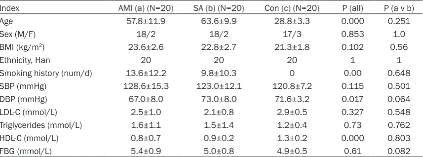
Table 1 showed the baseline demographic data. The AMI patients were admitted no more than 12 hours from the onset of symptoms to our Coronary Care Unit between January and June 2013, included 18 male and two female, with an age of 58±12 (mean ± s.d.) years. The SA group has 20 patients (18 male, two female, age 64±10). 20 volunteers (17 male, three female, age 29±3) were enrolled as the control group during the same period with similar male/female ratio. Histories, physical examina-tion, ECG, chest radiography and routine chemi-cal analyses showed the controls had no evi-dence of coronary heart diseases.
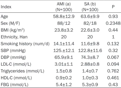
For the flow cytometry analysis, 100 AMI (88 male, 12 female, age 59±13) and 100 SA patients (82 male, 18 female, age 63±10) from Coronary Care Unit and Department of Cardiology were recruited between January and December 2013 (Table 2).
All AMI patients were diagnosed on the basis of following criteria [14]: detection of a rise of car-diac biomarker values [preferably carcar-diac tro-ponin (cTn)] with at least one value above the 99th percentile upper reference limit (URL) and with at least one of the following: 1) Symptoms of ischemia. 2) New or presumed new signifi -cant ST-segment-T wave (ST-T) changes or new left bundle branch block (LBBB). 3) Development of pathological Q waves in the ECG. 4) Imaging
Table 1. Baseline demographic data in three groups (_x±s.d.)
Index AMI (a) (N=20) SA (b) (N=20) Con (c) (N=20) P (all) P (a v b)
Age 57.8±11.9 63.6±9.9 28.8±3.3 0.000 0.251
Sex (M/F) 18/2 18/2 17/3 0.853 1.0
BMI (kg/m2) 23.6±2.6 22.8±2.7 21.3±1.8 0.102 0.56
Ethnicity, Han 20 20 20 1 1
Smoking history (num/d) 13.6±12.2 9.8±10.3 0 0.00 0.648
SBP (mmHg) 128.6±15.3 123.0±12.1 120.8±7.2 0.115 0.501
DBP (mmHg) 67.0±8.0 73.0±8.0 71.6±3.2 0.017 0.064
LDL-C (mmol/L) 2.5±1.0 2.1±0.8 2.9±0.5 0.327 0.548
Triglycerides (mmol/L) 1.6±1.1 1.5±1.4 1.2±0.4 0.73 0.762
HDL-C (mmol/L) 0.8±0.7 0.9±0.2 1.3±0.2 0.000 0.803
FBG (mmol/L) 5.4±0.9 5.0±0.8 4.9±0.5 0.61 0.082
evidence of new loss of viable myocardium or new regional wall motion abnormality. 5) Identification of an intracoronary thrombus by angiography.
All SA patients had exclusively effort-related angina with a positive exercise stress test and at least one coronary stenosis detected at angi-ography (>70% reduction of lumen diameter). There were no significant differences between AMI and SA patients in age, sex, smoking sta-tus, BMI, systolic blood pressure, diastolic blood pressure, LDL-C, HDL-C, triglycerides and fasting plasma glucose(FBG) (Tables 1 and 2). The exclusion criteria for three groups were as follows: venous thrombosis, history of severe renal or hepatic diseases, haematological dis-orders, acute or chronic inflammatory diseases and malignancy.
The study protocol was approved by the ethics committee of Tongji University and informed consent form was obtained.
Gene expression chips
Agilent G4112F Whole Human Genome Oligo Microarrays purchased from Agilent (USA) were used in the chip analysis. A microarray is com-posed of more than 41,000 genes or tran-scripts, including targeted 19,596 entrez gene RNAs. Sequence information used in the micro-arrays was derived from the latest databases of RefSeq, Goldenpath, Ensembl and Unigene
cked for a RIN number to inspect RNA integra-tion by an Agilent Bioanalyzer 2100 (Agilent technologies, Santa Clara, CA, US). The sample was considered qualified when both 2100 RIN and 28S/18S are no less than 0.7.
RNA amplification and labeling
Total RNA was amplified and labeled by Low Input Quick Amp Labeling Kit, One-Color (Cat#5190-2305, Agilent technologies, Santa Clara, CA, US), following the manufacturer’s instructions. Labeled cRNA was purified by RNeasy mini kit (Cat#74106, QIAGEN, GmBH, Germany).
Microarray hybridization
Each slide was hybridized with 1.65 μg Cy3-labeled cRNA using Gene Expression Hybri- dization Kit (Cat#5188-5242, Agilent technolo-gies, Santa Clara, CA, US) in Hybridization Oven (Cat#G2545A, Agilent technologies, Santa Clara, CA, US), following the manufacturer’s instructions. After 17 hours of hybridization, slides were washed in staining dishes (Cat#121, Thermo Shandon, Waltham, MA, US) with Gene Expression Wash Buffer Kit (Cat#5188-5327, Agilent technologies, Santa Clara, CA, US), according to the manufacturer’s operation manual.
Chip scan and data acquisition
[image:3.629.100.334.105.275.2]Slides were scanned using Agilent Microarray Scanner (Cat#G2565CA, Agilent technologies,
Table 2. Baseline demographic data for flow cytometric analysis (_x±s.d.)
Index (N=100)AMI (a) (N=100)SA (b) P
Age 58.8±12.9 63.6±9.9 0.93
Sex (M/F) 88/12 82/18 0.2348
BMI (kg/m2) 23.8±3.2 22.6±3.0 0.44
Ethnicity, Han 20 20 1
Smoking history (num/d) 14.1±11.4 11.6±9.8 0.132
SBP (mmHg) 125.±12.1 122.8±11.6 0.32
DBP (mmHg) 65.9±9.1 74.3±8.7 0.067
LDL-C (mmol/L) 3.01±1.1 2.88±0.8 0.094
Triglycerides (mmol/L) 1.5±0.8 1.4±0.7 0.762
HDL-C (mmol/L) 0.9±0.2 1.0±0.3 0.461
FBG (mmol/L) 5.4±1.2 5.3±0.9 0.43
Footnotes: LDL-C = low-density lipoprotein cholesterol; HDL-C: high-density lipoprotein cholesterol; FBG: Fasting Plasma Glucose.
[15]. The functions of more than 70% of the genes in the microarray are already known. All patients were subjected to the chip analysis.
Total RNA isolation
Santa Clara, CA, US) with default settings, Dye channel: Green, Scan resolution =3 μm, 20 bit. Data were extracted with Feature Extraction software 10.7 (Agilent technologies, Santa Clara, CA, US). Raw data were normalized using Quantile algorithm, Gene Spring Software 11.0 (Agilent technologies, Santa Clara, CA, US).
RT-PCR
The spots in the microarray were randomly selected and their expressions were confirmed by RT-PCR. Among all the genes with differen-tial expressions, three genes were randomly selected and subjected to RT-PCR, along with the house keeping genes (GAPDH). The relative expressions were indicated as the expression of the target genes normalized to the expres-sion of GAPDH (2-ΔΔCt). The melting curve and
the 2-ΔΔCt-method were used to detect the
dif-ferences in the expressions among the three groups. The results from RT-PCR were consis-tent with the microarray analysis.
Flow cytometry
Five milliliter peripheral bloods from median cubital vein were drawn immediately from 100
per 100 peripheral blood mononuclear cells. Isotype-identical antibodies served as controls. Double color flow cytometry analyses were per -formed using aEBeckman Coulter (Coulter, USA) flow cytometer and performed in duplicate.
Statistical analysis
Descriptive statistics were expressed as mean ± s.d. Differences between groups were exam-ined by one-way analysis of variance (ANOVA). After ANOVA the test of all pairwise group mean comparison was performed using the Tukey’s method. Density curve for NK cells was delin-eated using R software. Data were analyzed using SPSS 17.0, and P-values <0.05 were con -sidered statistically significant.
Results
Gene expression of inhibitory NK cell receptors
[image:4.629.98.389.79.356.2]The results showed mRNA expressions of 15 inhibitory NK cell receptors. The inhibitory receptors are generally divided into MHC-I spe-cific (Figure 1A) and non-MHC specific (Figure
Figure 1. mRNA expression of specific MHC inhibitory NK cell receptors (A) and non-MHC inhibitory NK cell receptors (B) in PBMCs from three groups. Three groups *: P<0.05, **: P<0.01. a v c #: P<0.05, ##: P<0.01; b v c &: P<0.05, &&: P<0.01; a v b +: P<0.05, ++: P<0.01.
1B). MHC-I specific receptors include killer cell immunoglobulin-like receptors (KIRs), killer lec-tin-like receptors (KLRs) and LIR-1. Non-MHC specific receptors contain KLRB1, KLRG1, Siglec-7 (sialic acid binding Ig-like lectin 7), CEACAM1 (carcinoembryonic antigen-related cell adhesion molecule 1, Siglec-9) and IRp60. The KIRs consist of KIR2DL1, KIR2DL2, KIR2DL3, KIR2DL5, KIR3DL1, KIR3DL2 and KIR3DL3. The KLRs comprise CD94 (KLRD1), NKG2A, KLRG1, and KLRB1. The last two belong to non-MHC receptors. In sum the gene expressions of 15 NK cell inhibitory receptors in PBMCs from three groups of patients were detected. In PBMCs from the three groups, expressions of the genes encoding KIR2DL2, KIR3DL3, CD94, NKG2A, KLRB1 and KLRG1 were significantly different (P<0.05). In AMI group, all the 11 gene expressions of MHC spe-cific receptors were the lowest among three groups, and mRNA expressions of KIR2DL2, KIR3DL3, CD94 and NKG2A were significantly lower (P<0.05) than in SA patients and the con -trols. The mRNA expression of non-MHC recep-tors, KLRB1 and KLRG1 in AMI patients was
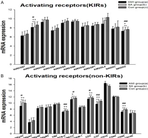
44, NKp46 and NKP80. 2B4 (CD244), NTB-A (CD352) and CRACC (CD319) are members of the recently defined family of SRR. In PBMCs from the three groups, expressions of the gene encoding KIR2DS2, NKG2D, NKp30, NTB-A, CRACC, CD2, CD7 and CD96 were significantly different (P<0.01). In AMI group 23 gene expressions of activating receptors were lowest among the three groups. KIR2DS2, NKG2D, NKp30, NTB-A, CRACC, CD2, CD7 and CD96 mRNAs in AMI patients were significantly down-regulated when compared with in SA patients and controls (P<0.05). Between the SA and controls there is no statistically significant dif -ference in NK cell activating receptors.
NK cells counting
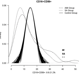
[image:5.629.99.395.80.355.2]Density curves for three groups were delineat-ed using R software and the normal range for NK cells is from 8.6-21.1% (Figure 3) [16]. The two density curves for AMI and SA patients were substantially left shift. The number of NK cells was significantly decreased in both AMI and SA patients in PBMCs when compared with Figure 2. mRNA expression of activating NK cell receptors in PBMCs from three
groups: KIRs NK cell activating receptors (A) and other NK cell activating recep-tors (B). Three groups *: P<0.05, **: P<0.01. a v c #: P<0.05, ##: P<0.01; b v c &: P<0.05, &&: P<0.01; a v b +: P<0.05, ++: P<0.01.
significantly down-regulat -ed (P<0.05) in comparison with the SA patients and the controls. There was no statistical difference in NK cell inhibitory receptor mR- NA expressions between SA and the controls.
Gene expression of activat-ing NK cell receptors
the normal range (all P<0.01). However, there was no significant difference between AMI and SA patients in the quantity of NK cells.
Discussion
The NK cells express an array of inhibitory and activating receptors, and the inhibitory recep-tors are responsible for self-tolerance and adjusting NK-cell activity based on the expres-sion level of self-MHC I [17, 18]. Considerable evidences confirmed that the signal transmit -ted by NK inhibitory receptors was dominant when they are present together with the acti-vating receptors for the normal cells protection [7, 19]. KIRs are the most important NK cell receptors, which recognize classical MHC class I. KIRs with short cytoplasmic domains are acti-vating receptors, whereas ligation of the KIR with long cytoplasmic domains to their cognate HLA class I ligands transmits a cascade of inhibitory signals that mediates NK cell toler-ance to self cells [20, 21]. In our present study, the gene expressions of KIR2DL2, KIR3DL3, CD94 and NKG2A were significantly lower than in SA patients and the controls, suggesting the impaired ability to protect the normal cells. Recent work revealed that there was another
As to SA patients, the gene expressions of inhibitory NK receptors were not statistically different from those in the controls, indicating that no initiation of any inhibitory NK receptors was activated in SA group.
The activating receptors are widely expressed on the surface of NK cells. NKG2D is a central activating NK cell receptor, which can be bound to many ligands that are induced on cells under stress due to infection, transformation, or DNA damage. Therefore, NKG2D played an impor-tant role in targeting NK cell responses toward abnormal cells and eventually the lysis [26, 27]. NCRs including NKp30, NKp44, NKp46 and NKp80 are one of the most important activat-ing receptors that mediate the NK-cell cytotox-icity and cytokine production [7]. NCRs have been shown to recognize a broad spectrum of ligands, ranging from viral-, parasite- and bac-terial-derived ligands to cellular ligands, and play a central role in eliminating infected or transformed cells, particularly malignant cells [28-30]. The 2B4, NTB-A and CRACC receptors belong to the family of SRR, and transmit acti-vating signals through the SLAM-associated protein (SAP) [31]. Like NKG2D ligands, DNAM-Figure 3. The comparison of NK cells counting among the AMI, SA patients
and the normal range. *: P<0.05, **: P<0.01. a v c #: P<0.05, ##: P<0.01; b v c &&: P<0.01; +: P<0.05, ++: P<0.01; a v b &: P<0.05.
[image:6.629.103.376.80.339.2]1 ligands are frequently expressed on stressed cells, and DNAM-1 receptor participates in a cytotoxic immune response, when bound to the ligands [32-34]. Many other receptors, includ-ing CD2, CD7, CD16, CD44, CD96 and CD160 contribute to NK cell activation, but much work remains to be done in determining how and when the various NK cell receptors deliver sig-nals for activation [7, 35]. Among the activating receptors, the most dominant NKG2D and NCRs receptors, however, do not activate the NK cells by their own. Activation of NK cells by any of the tested receptors requires comple-mentation with another activating receptor, such as 2B4, NTB-A, CRACC or DNMA-1 to obtain synergistic activation signals, and then releases the perforin and granzyme, leading to the target cells lysis directly [36]. In the mean-while the requirement for combination may serve as a safeguard to prevent unrestrained activation of NK cells [37]. In our study, 23 gene expressions of activating receptors in AMI patients were lowest among the three groups, and KIR2DS2, NKG2D, NKp30, NTB-A, CRACC, CD2, CD7 and CD96 mRNAs were significantly decreased in comparison with SA patients and controls respectively. The result showed the transduction of activating signal was inhibited in patients with AMI. As a result, the NK cells immune activity to the targeted cells was inhib-ited. Just as the NK cell inhibitory receptors, there was no statistical difference in mRNA expressions between the SA patients and con-trols in activating receptors, indicating the NK receptors in SA patients was in a nearly inactive state.
In the present study the significant down-regu -lation of NK cell inhibitory receptors in AMI patients suggested the decreased protection of normal cells and the hyporesponsive level of NK cells. Meanwhile, the significant down-regu -lated activating NK receptors in AMI group pre-sented the declined cytotoxicity and cytokine production activity of NK cells. However, there was no statistical difference in gene expres-sions of inhibitory and activating receptors between the SA patients and controls. Previous studies found the reduced proportions of NK cells in peripheral blood of CAD [5-7], but the reason is still controversial [9-12]. In line with this, the similar loss of NK cell numbers in both AMI and SA patients were also demonstrated in our study (Figure 3). We may conclude that the impaired NK cell function in different stages of
CAD patients is not just a quantitative defect. In AMI patients both numbers and receptor activ-ity decreased, while only reduction of quantactiv-ity was found in SA patients. As a result, we specu-lated that the pathogenesis of AMI may be associated with NK cell receptor deficient or the progress of AMI restrained the NK cell innate immunity. Both sides showed the close relationship between AMI pathogenesis and the NK cell receptor activity.
In conclusion, the loss of NK cell activity in AMI patients is quantitative and involves the dys-function of NK cells receptor activity. Consequently, improving NK cell immunity may be considered as a potential target for medical interventions in the patients with AMI.
Limitation
A limitation of our study is the statistically sig-nificant difference in age between AMI and SA patients and the controls as aging may affect the immune status. However, our study showed no statistically significant difference in the activity of NK cell receptors between the older SA patients and the younger controls. This may indicate that the aging effect is only marginal for the activity of NK cell receptors.
Acknowledgements
The present study was supported by the National Natural Science Foundation of China (No. 30570809).
Disclosure of conflict of interest
None.
Address correspondence to: Dr. Lemin Wang, De- partment of Cardiology, Tongji Hospital, Tongji University School of Medicine, 389 Xincun Rd, Putuo District, Shanghai 200065, China. Tel: 86-21-66111329; Fax: 86-21-86-21-66111329; E-mail: [email protected]
References
[1] Hansson GK, Hermansson A. The immune sys-tem in atherosclerosis. Nat Immunol 2011; 12: 204-12.
[2] Martín-Fontecha A, Carbone E. The social life of NK cells. Arch Immunol Ther Exp (Warsz) 2001; 49 Suppl 1: S33-9.
Middleton D, Romagné F, Ugolini S, Vivier E. Human NK cell education by inhibitory recep-tors for MHC class I. Immunity 2006; 25: 331-42.
[4] Joncker NT, Fernandez NC, Treiner E, Vivier E, Raulet DH. NK cell responsiveness is tuned commensurate with the number of inhibitory receptors for self-MHC class I: the rheostat model. J Immunol 2009; 182: 4572-80. [5] Vivier E, Nunès JA, Vély F. Natural killer cell
sig-naling pathways. Science 2004; 306: 1517-9. [6] André P, Castriconi R, Espéli M, Anfossi N,
Juarez T, Hue S, Conway H, Romagné F, Don-dero A, Nanni M, Caillat-Zucman S, Raulet DH, Bottino C, Vivier E, Moretta A, Paul P. Compara-tive analysis of human NK cell activation in-duced by NKG2D and natural cytotoxicity re-ceptors. Eur J Immunol 2004; 34: 961-71. [7] Whitman SC, Rateri DL, Szilvassy SJ,
Yokoya-ma W, Daugherty A. Depletion of natural killer cell function decreases atherosclerosis in low-density lipoprotein receptor null mice. Arterio-scler Thromb Vasc Biol 2004; 24: 1049-54. [8] Long EO, Kim HS, Liu D, Peterson ME,
Rajago-palan S. Controlling natural killer cell respons-es: integration of signals for activation and in-hibition. Annu Rev Immunol 2013; 31: 227-58. [9] Jonasson L, Backteman K, Ernerudh J. Loss of natural killer cell activity in patients with coro-nary artery disease. Atherosclerosis 2005; 183: 316-21.
[10] Backteman K, Ernerudh J, Jonasson L. Natural killer (NK) cell deficit in coronary artery dis -ease: no aberrations in phenotype but sus-tained reduction of NK cells is associated with low-grade inflammation. Clin Exp Immunol 2014; 175: 104-12.
[11] Li W, Lidebjer C, Yuan XM, Szymanowski A, Backteman K, Ernerudh J, Leanderson P, Nils-son L, Swahn E, JonasNils-son L. NK cell apoptosis in coronary artery disease: relation to oxidative stress. Atherosclerosis 2008; 199: 65-72. [12] Hak Ł, Myśliwska J, Wieckiewicz J, Szyndler K,
Trzonkowski P, Siebert J, Myśliwski A. NK cell compartment in patients with coronary heart disease. Immun Ageing 2007; 4: 3.
[13] Dobbin K, Simon R. Sample size determination in microarray experiments for class compari-son and prognostic classification. Biostatistics 2005; 6: 27-38.
[14] Thygesen K, Alpert JS, Jaffe AS, Simoons ML, Chaitman BR, White HD; Joint ESC/ACCF/AHA/ WHF Task Force for Universal Definition of Myo -cardial Infarction; Authors/Task Force Mem-bers Chairpersons, Thygesen K, Alpert JS, White HD; Biomarker Subcommittee, Jaffe AS, Katus HA, Apple FS, Lindahl B, Morrow DA; ECG Subcommittee, Chaitman BR, Clem-mensen PM, Johanson P, Hod H; Imaging
Sub-committee, Underwood R, Bax JJ, Bonow JJ, Pinto F, Gibbons RJ; Classification Subcommit -tee, Fox KA, Atar D, Newby LK, Galvani M, Hamm CW; Intervention Subcommittee, Uretsky BF, Steg PG, Wijns W, Bassand JP, Me-nasche P, Ravkilde J; Trials & Registries Sub -committee, Ohman EM, Antman EM, Wallentin LC, Armstrong PW, Simoons ML; Trials & Regis -tries Subcommittee, Januzzi JL, Nieminen MS, Gheorghiade M, Filippatos G; Trials & Regis -tries Subcommittee, Luepker RV, Fortmann SP, Rosamond WD, Levy D, Wood D; Trials & Regis -tries Subcommittee, Smith SC, Hu D, Lopez-Sendon JL, Robertson RM, Weaver D, Tendera M, Bove AA, Parkhomenko AN, Vasilieva EJ, Mendis S; ESC Committee for Practice Guide-lines (CPG), Bax JJ, Baumgartner H, Ceconi C, Dean V, Deaton C, Fagard R, Funck-Brentano C, Hasdai D, Hoes A, Kirchhof P, Knuuti J, Kolh P, McDonagh T, Moulin C, Popescu BA, Reiner Z, Sechtem U, Sirnes PA, Tendera M, Torbicki A, Vahanian A, Windecker S; Document Review-ers, Morais J, Aguiar C, Almahmeed W, Arnar DO, Barili F, Bloch KD, Bolger AF, Botker HE, Bozkurt B, Bugiardini R, Cannon C, de Lemos J, Eberli FR, Escobar E, Hlatky M, James S, Kern KB, Moliterno DJ, Mueller C, Neskovic AN, Pieske BM, Schulman SP, Storey RF, Taubert KA, Vranckx P, Wagner DR. Third universal defi -nition of myocardial infarction. J Am Coll Cardi-ol 2012; 60: 1581-98.
[15] Wiltgen M, Tilz GP. DNA microarray analysis: principles and clinical impact. Hematology 2007; 12: 271-287.
[16] Cooper MA, Colonna M, Yokoyama WM. Hid-den talents of natural killers: NK cells in innate and adaptive immunity. EMBO Rep 2009; 10: 1103-10.
[17] Backström E, Kristensson K, Ljunggren HG. Ac-tivation of natural killer cells: underlying mo-lecular mechanisms revealed. Scand J Immu-nol 2004; 60: 14-22.
[18] Middleton D, Curran M, Maxwell L. Natural killer cells and their receptors. Transpl Immu-nol 2002; 10: 147-64.
[19] Moretta L, Bottino C, Pende D, Vitale M, Min-gari MC, Moretta A. Different checkpoints in human NK-cell activation. Trends Immunol 2004; 25: 670-6.
[20] Campbell KS, Purdy AK. Structure/function of human killer cell immunoglobulin-like recep-tors: lessons from polymorphisms, evolution, crystal structures and mutations. Immunology 2011; 132: 315-25.
prolong animal survival. J Immunol 2014; 193: 6192-206.
[22] Kumar V, McNerney ME. A new self: MHC-class-I-independent natural-killer-cell self-tol-erance. Nat Rev Immunol 2005; 5: 363-74. [23] Lebbink RJ, Meyaard L. Non-MHC ligands for
inhibitory immune receptors: novel insights and implications for immune regulation. Mol Immunol 2007; 44: 2153-64.
[24] Ito M, Maruyama T, Saito N, Koganei S, Yama-moto K, MatsuYama-moto N. Killer cell lectin-like re-ceptor G1 binds three members of the classi-cal cadherin family to inhibit NK cell cytotoxicity. J Exp Med 2006; 203: 289-95.
[25] Li Y, Hofmann M, Wang Q, Teng L, Chlewicki LK, Pircher H, Mariuzza RA. Structure of natural killer cell receptor KLRG1 bound to E-cadherin reveals basis for MHC-independent missing self recognition. Immunity 2009; 31: 35-46. [26] Raulet DH, Gasser S, Gowen BG, Deng W, Jung
H. Regulation of ligands for the NKG2D activat-ing receptor. Annu Rev Immunol 2013; 31: 413-41.
[27] Zafirova B, Wensveen FM, Gulin M, Polić B. Regulation of immune cell function and differ-entiation by the NKG2D receptor. Cell Mol Life Sci 2011; 68: 3519-29.
[28] Kruse PH, Matta J, Ugolini S, Vivier E. Natural cytotoxicity receptors and their ligands. Immu-nol Cell Biol 2014; 92: 221-9.
[29] Koch J, Steinle A, Watzl C, Mandelboim O. Acti-vating natural cytotoxicity receptors of natural killer cells in cancer and infection. Trends Im-munol 2013; 34: 182-91.
[30] Bhat R, Rommelaere J. NK-cell-dependent kill-ing of colon carcinoma cells is mediated by natural cytotoxicity receptors (NCRs) and stim-ulated by parvovirus infection of target cells. BMC Cancer 2013; 13: 367.
[31] Claus M, Meinke S, Bhat R, Watzl C. Regulation of NK cell activity by 2B4, NTB-A and CRACC. Front Biosci 2008; 13: 956-65.
[32] de Andrade LF, Smyth MJ, Martinet L. DNAM-1 control of natural killer cells functions through nectin and nectin-like proteins. Immunol Cell Biol 2014; 92: 237-44.
[33] Tahara-Hanaoka S, Miyamoto A, Hara A, Honda S, Shibuya K, Shibuya A. Identification and characterization of murine DNAM-1 (CD226) and its poliovirus receptor family ligands. Bio-chem Biophys Res Commun 2005; 329: 996-1000.
[34] El-Sherbiny YM, Meade JL, Holmes TD, McG-onagle D, Mackie SL, Morgan AW, Cook G, Fey-ler S, Richards SJ, Davies FE, Morgan GJ, Cook GP. The requirement for DNAM-1, NKG2D, and NKp46 in the natural killer cell-mediated kill-ing of myeloma cells. Cancer Res 2007; 67: 8444-9.
[35] Chan CJ, Smyth MJ, Martinet L. Molecular mechanisms of natural killer cell activation in response to cellular stress. Cell Death Differ 2014; 21: 5-14.
[36] Bryceson YT, March ME, Ljunggren HG, Long EO. Activation, coactivation, and costimulation of resting human natural killer cells. Immunol Rev 2006; 214: 73-91.