www.impactjournals.com/oncotarget/ Oncotarget, Vol. 7, No. 23
AMPK-dependent and independent effects of AICAR and
compound C on T-cell responses
Enyu Rao1, Yuwen Zhang1, Qiang Li2, Jiaqing Hao1, Nejat K. Egilmez1, Jill Suttles1 and Bing Li1
1 Department of Microbiology and Immunology, University of Louisville, Louisville, KY, USA
2 Department of Burn and Plastic Surgery, Shandong Provincial Hospital Affiliated to Shandong, University, Jinan, Shandong,
China
Correspondence to: Bing Li, email: b.li@louisville.edu
Keywords: AMPK, T cell responses, Immunology and Microbiology Section, Immune response, Immunity Received: March 16, 2016 Accepted: April 27, 2016 Published: May 10, 2016
AbstrAct
As a master metabolic sensor, AMP-activated protein kinase (AMPK) is involved in different fundamental cellular processes. Regulation of AMPK activity either by agonists (e.g., AICAR) or by antagonists (e.g., Compound C) has been widely employed to study the physiological functions of AMPK. However, mounting evidence indicates AMPK-independent effects for these chemicals and how they regulate immune cell functions remains largely unknown. Herein, using T cells from AMPK conditional knockout mice and their wild type littermates, we demonstrate that AICAR and Compound C can, indeed, activate or inhibit AMPK activity in T cells,
respectively. Specifically, AICAR inhibits, but Compound C promotes, Ca2+-induced
T cell death in an AMPK-dependent manner. In contrast, our data also demonstrate that AICAR and Compound C inhibit T cell activation and cytokine production in an
AMPK-independent manner. Moreover, we find that the AMPK-independent activity
of AICAR and Compound Cis mediated via the mTOR signaling pathway in activated T cells. Our results not only reveal the critical role of AMPK in regulating T cell survival and function, but also demonstrate AMPK-dependent and independent rolesof AICAR/ Compound C in regulating T cell responses, thus suggesting a context-dependent effect of these “AMPK regulators”.
IntroductIon
As an evolutionarily conserved metabolic sensor, AMPK has been shown to regulate various aspects of cellular fundamental functions including cell proliferation, survival and metabolism [1-3]. The AMPK complex is composed of three subunits, one catalytic α subunit and two regulatory β and γ subunits. In different tissues of mammals, AMPK displays distinct expression pattern of subunits, which contain two α subunits (α1, α2), twoβ subunits (β1, β2), and three γ subunits (γ1, γ2 and γ3) [2, 4]. AMPK activity can be regulated by intracellular factors, such as the cellular AMP/ATP ratio, as well as auto-inhibitory features and phosphorylation status of its subunits. Full activation of AMPK requires specific phosphorylation of the α subunit at the conserved threonine residue (Thr172) by upstream kinases including LKB1, CAMKKs and TAK1. Protein phosphatases
2A and 2C also regulate the activation of AMPK by dephosphorylation of Thr172 [1, 5]. It is well-established that activation of AMPK is critical in restoring the intracellular energy balance to sustain cell survival and function under stress via turning off ATP-consuming anabolic pathways and stimulating ATP-producing catabolic processes [2, 4, 6].
injury and to improve myocardial protection in coronary artery bypass grafting [14-16]. Moreover, AICAR is also a promising drug for the treatment of B-cell neoplasms and chronic lymphocytic leukemia [17-19]. In contrast, Compound C (6-[4-(2-Piperidin-1-ylethoxy) phenyl]-3-pyridin-4-ylpyrazolo [1,5-a]pyrimidine) (CC), is well-known for its potent inhibitory effect on AMPK activation. In combination with AMPK agonists (e.g. AICAR), Compound C is often used as an AMPK antagonist to study AMPK-dependent cellular events [5, 20, 21]. However, mounting evidence indicates AICAR and Compound C are able to regulate cellular functions via AMPK-independent mechanisms [19, 22-30]. In addition, Compound C has been shown to inhibit activities of many other kinases, such as ERK8, ALK2, Src, Lck, etc, besides AMPK [31, 32]. Thus, pharmacological application of AICAR or Compound C may result in both AMPK-dependent and inAMPK-dependent effects in different types of cells.
In our previous studies using AMPK conditional knockout mice, we have demonstrated that expression of AMPK in T cells is indispensable for their activation, but critical to promoting their survival and anti-tumor functions in mouse tumor models [10]. Thus, clinical application of AMPK agonists/antagonists will most likely influence T cell survival and function. Although the effects of AICAR and Compound C on T cell activity has been studied [30, 33-37], whether these effects are dependent or independent of AMPK is still unclear. Here, using AMPK-deficient T cells, we investigated the effects of AICAR and Compound C on T cell survival and function. We found that AICAR promoted, but Compound C inhibited, Ca2+ signaling-induced T cell death in an AMPK-dependent manner. Importantly, both AICAR and Compound C exerted an inhibitory effect on T cell activation and function in an AMPK-independent manner.
rEsuLts
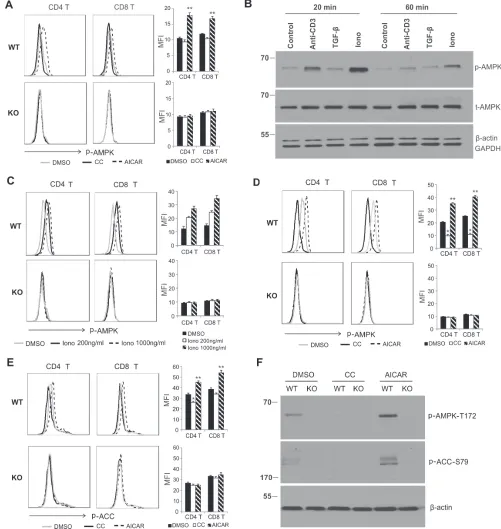
AIcAr promotes, but compound c inhibits, AMPK activation in t cells
Although AICAR/Compound C have been commonly used as an agonist/antagonist of AMPK, respectively, whether or not they are able to activate/ inhibit AMPK in T cells remains unclear [18, 30, 34]. Our previous data demonstrated that AMPK is specifically deleted in T cells from CD4-Cre+ AMPKα1fl/fl (KO) mice, but is intact in T cells from CD4-Cre- AMPKα1fl/ fl (WT) mice [10]. We thus continued to use this model to dissect the effects of AICAR/Compound C on AMPK in T cells. We first measured the AMPK activation using resting T cells from lymph nodes of WT and KO mice. Intracellular staining of phosphorylation of AMPK
Thr-172 (p-AMPK) showed that AMPK was not or only weakly activated in resting WT T cells as compared to KO T cells. Interestingly, treatment with AICAR significantly increased phosphorylation of AMPK in WT T cells, but not in KO T cells, suggesting a specific activation of AMPK with AICAR. We did not observe any obvious inhibition of p-AMPK with Compound C treatment (Figure 1A), which may be due to the non- or weak activation of AMPK in resting T cells. As Ionomycin (Iono) was able to induce much stronger AMPK activation than anti-CD3 antibody or TGF-β in LN cells (Figure 1B), and it increased the levels of p-AMPK in WT T cells in a dose-dependent manner (Figure 1C), we next measured the effects of AICAR/Compound C on AMPK activation using Iono-activated T cells. Importantly, pretreatment of T cells with AICAR enhanced, but Compound C suppressed, phosphorylation of AMPK in Iono-activated T cells from WT mice, but not from KO mice, further suggesting a specific effect of AICAR and Compound C on AMPK activity in activated T cells (Figure 1D). We also investigated the impact of AICAR/Compound C treatment on acetyl-CoA carboxylase (ACC), the downstream target of activated AMPK in T cells. Similarly, AICAR promoted, while Compound C inhibited, phosphorylation of ACC (Ser-79) in Iono-activated CD4+ and CD8+ T cells from WT mice (Figure 1E). Using Western blot analysis, we further confirmed that AICAR enhanced, but Compound C inhibited, the phosphorylation of AMPK and ACC in T cells from WT mice, but not from KO mice (Figure 1F). Altogether, using CD4-Cre-AMPKα1fl/fl mice, our data clearly indicate a specific AMPK activation/ inhibition effect of AICAR/Compound C in T cells.
AIcAr inhibits, but compound c promotes, ca2+-induced t cell death in an AMPK-dependent
manner
Figure 1: AIcAr promotes, but compound c inhibits, AMPK activation in t cells. A. Cells from lymph nodes of WT and
KO mice were treated with DMSO, Compound C (CC, 10µΜ) or AICAR (500µM) for 30 minutes and were analyzed for p-AMPKT172 levels in CD4+ and CD8+ T cellsby intracellular staining. The mean value of median fluorescence intensity (MFI) in DMSO, CC or AICAR group is shown in the right panel (**, p < 0.01 as compared to DMSO group). b. LN cells were treated with anti-CD3 (5µg/ml), TGF-β (5ng/ml) or ionomycin (1µg/ml), respectively. Cells were collected for western blot analysis at indicated time points. c.LN cells were treated with DMSO or indicated concentrations of ionomycin (200ng/ml or 1000ng/ml) for 20 minutes. p-AMPKT172 levels in CD4+ and CD8+ T cells were analyzed by intracellular staining. (D, E) Cells from lymph nodes of WT and KO mice were pretreated with DMSO, AICAR (500µM) or CC(10µM) for 30 minutes and then stimulated with PMA/Ionomycin (P/I) for another 20 minutes, p-AMPKT172d. and
p-ACCS79E.in CD4+ and CD8+ T cells were analyzed by intracellular staining. MFI in DMSO, CC or AICAR-treated group is shown in the right panel (*, p < 0.05; **, p < 0.01 as compared to DMSO group). F. Sorted CD4+ T cells were pretreated with DMSO, CCand AICAR for 30 minutes and then followed by Ionomycinstimulation for another 20 minutes. Cells were collected for analysis of p-AMPKT172 and
Figure 2: AIcAr inhibits, but compound c promotes, ca2+ signaling-induced t cell death in an AMPK-dependent
manner. Cells from lymph nodesof WT and KO mice were pretreated with indicated concentrations of AICAR or Compound C for 30 minutes, and then stimulated with PMA(10ng/ml)/Ionomycin (1000ng/ml) for 6 hours. The survival of CD8+ T cells and CD4+ Tcells was analyzed with Annexin V and 7-AAD staining. The representative dot blots showing the survival of CD8+ T cells treated with AICAR A. or
in WT mice (57.6%) as compared to those in KO mice (23.3%) under PMA/Ionomycin stimulation for 6 hours. Interestingly, pretreatment with AICAR further promoted the survival of WT CD8+ T cells, but not in CD8+ T cells from KO mice (Figure 2A). In contrast, pretreatment with Compound C significantly decreased the survival of WT CD8+ T cells, but not KO CD8+ T cells (Figure 2B). These data suggest an AMPK-specific effect of AICAR and Compound C on CD8+ T cell survival. When we further analyzed the impact of AICAR/Compound C treatment on the survival of CD4+ T cells, we observed the same effects of AICAR/Compound C on the survival of CD4+ T cells as what we observed in CD8+ T cells (Figure 2C, 2D). In addition, we also measured T cell survival after longer period of activation with PMA/Ionomycin (12 hours) and confirmed the similar effects of AICAR and Compound C on T cell survival (Figure S2). Thus, all data suggest that AICAR promotes, but Compound C inhibits, Ca2+-induced T cell death in an AMPK-dependent manner.
AIcAr and compound c suppress t cell activation in an AMPK-independent manner
After determining the effects of AICAR/Compound C on T cell survival, we next investigated whether these agents affect T cell activation. The early T cell activation marker CD69 was expressed at low levels on resting lymphocytes (Figure S3), but was rapidly upregulated following activation. Interestingly, AICAR inhibited CD69 expression on live CD4+ T cells (7-AAD-) in a dose-dependent manner in both WT and KO mice (Figure 3A). Moreover, CD69 was expressed at similar levels on live T cells from WT and KO mice with or without AICAR treatment, implying that AMPK deficiency has no impact on CD69 expression (Figure 3B). The low percentage of 7-ADD- CD69+ T cells in KO mice may be due to the elevated cell death in the absence of AMPK. Thus, AICAR inhibited CD69 expression regardless of AMPK expression. Unexpectedly, treatment of Compound C also inhibited CD69 expression on PMA/Ionomycin-activated CD4+ T cells in the same pattern as AICAR treatment (Figure 3C, 3D). To further confirm these observations, we measured CD69 expression on T cells activated with anti-CD3/CD28 antibodies. Both AICAR and Compound C inhibited CD69 expression on anti-CD3/CD28-activated T cells from WT and KO mice. In addition, other T cell activation markers, including CD25, CD71, were also inhibited by AICAR or Compound C treatment (Figure 3E, 3F). Taken together, these data suggest that T cell activation does not require AMPK expression and both AICAR and Compound C are able to inhibit T cell activation in an AMPK-independent manner.
AIcAr and compound c inhibit t cell cytokine production in an AMPK-independent manner
We next determined whether AICAR or Compound C affects T cell function by measurement of cytokine production in activated T cells. In line with our previous data [10], deletion of AMPK in T cells greatly impaired production of IL-2, IFNγ and TNFα in PMA/Ionomycin-activated CD8+ T cells (DMSO control group in Figure 4A, 4B, 4C), implying that AMPK activation contributes to cytokine production in T cells. Interestingly, when we pretreated T cells with either AICAR or Compound C before PMA/Ionomycin activation, the production of cytokines, including IL-2, IFNγ and TNFα, were inhibited substantially in CD8+ T cells from both WT and KO mice (Figure 4A, 4B, 4C), suggesting that AICAR- or Compound C-mediated cytokine inhibition is independent of AMPK expression/activation in CD8+ T cells. Of note, similar inhibitory effects were also observed in CD4+ T cells, regardless of AMPK expression (Figure 4D, 4E). As AMPK deficiency promoted Ca2+ overload-induced T cell death (e.g. 1000ng/ml Ionomycin), which may contribute to the reduced cytokine production in KO T cells, we further measured cytokine production in T cells stimulated with a low dose of Ionomycin (200ng/ml), which induced no discernible T cell death in the presence or absence of AICAR or Compound C between WT and KO mice (Figure S4). We found that AMPK expression was still able to promote cytokine production in both CD4+ and CD8+ T cells (left panel, Figure S5). However, treatment with either AICAR or Compound C inhibited cytokine production irrespective of AMPK expression in T cells (middle and right panels, Figure S5). Furthermore, we measured cytokine production in supernatants collected from anti-CD3/CD28-activated T cells and demonstrated that both Compound C and AICAR inhibited levels of IL-2, IFNγ and TNFα regardless of AMPK expression (Figure 5). Thus, our data indicate that, although AMPK is critical in promoting cytokine production in Ca2+- and TCR-activated T cells, the cytokine-inhibitory effects of AICAR/Compound C on T cells are independent of AMPK.
treatment of AIcAr and compound c inhibits the mtor signaling pathway in t cells
Figure 3: AIcAr and compound c suppress t cell activation in an AMPK-independent manner. Cells from lymph
Figure 4: AIcAr and compound c inhibit cytokine production in PMA/Ionomycin-activated t cells. Cells from lymph
nodes of WT and KO mice were pretreated with DMSO, Compound C (10µM) or AICAR (500µM) for 30 minutes, and then stimulated with PMA(10ng/ml)/Ionomycin (1000ng/ml) and Golgiplug for 5 hours. Cells were collected and stained for cytokines production. A. IL-2 production in CD8+ T cells. b. IFNγ production in CD8+ T cells.c. TNFα production in CD8+ T cells. d. IL-2 production in CD4+ T cells.
pathway involved in the AICAR/Compound C-mediated effects. Interestingly, phosphorylation of S6K, S6P and 4EBP1 was greatly inhibited by either AICAR or Compound C treatment in T cells, suggesting a critical role of mTOR in AICAR/Compound C-mediated effects. By taking advantage of the single cell intracellular staining techniques, we demonstrated that treatment with either AICAR or Compound C reduced phosphorylation of Serine 235/236 of S6P (p-S6S235/236) in both CD4+ and CD8+ T cells (Figure 6B). As S6P can be phosphorylated on other sites, such as Serine 240/244, we also measured the effect of AICAR and Compound C on the phosphorylation of Serine240/244 (p-S6S240/244), and found the same inhibitory effect (Figure 6C). In agreement with our Western blotting results, AICAR and Compound C exhibited no obvious effects on the phosphorylation of ERK in T cells from both WT and KO mice (Figure 6D). Taken together, our data suggest that AICAR and Compound C may inhibit T cell activation and function via suppression of mTOR pathway.
dIscussIon
Due to the critical role of AMPK in the regulation of fundamental cellular functions, pharmacological modulation of AMPK activity has been widely used in experimental and clinical studies. Targeting AMPK by different agonists, such as AICAR and metformin, has been shown to exhibit therapeutic benefits in different diseases, including type II diabetes, cardiac ischemic injury and tumor development [7, 8]. While much attention has been focused on how modulation of AMPK activity affects non-hematopoietic cell pathology, the effect of AMPK agonists/antagonists on immune cell
survival and function is yet to be determined. Using AMPK conditional knockout mice, we and others have previously demonstrated that AMPK is dispensable for T cell development and proliferation in normal physiological conditions, but indispensable for T cell survival and anti-tumor functions under tumor or other metabolic stresses [10, 42, 43]. In this follow-up study, we further investigated the effects of AICAR/Compound C treatment on T cell responses. We found that treatment with AICAR or Compound C results in pleiotropic effects, i.e. an dependent effect on T cell survival and an AMPK-independent activity on T cell activation and function.
AICAR/Compound C is commonly used as an agonist/antagonist to study AMPK-dependent cellular pathways. By specific deletion of AMPK in T cells using CD4-Cre-AMPK fl/fl mice, we confirmed that AICAR and Compound C can indeed activate or inhibit AMPK, respectively, in T cells from WT mice (Figure 1). Importantly, treatment with AICAR or Compound C has no impact on AMPK KO T cell death, but respectively promotes or inhibits the survival of AMPK WT T cells in response to high concentration Ionomycin-activated T cells death (Figure 2). These data further substantiate the pro-survival role of AMPK in T cells, and reveal an AMPK-dependent effect of AICAR/Compound C on T cell survival. It is worth noting that the AMPK-dependent effect on T cell survival is not prominent when T cells are activated with anti-CD3/CD28 signals, which may be attributed to the weak activation of AMPK under the anti-CD3 stimulation (Figure 1B).
[image:8.612.61.552.481.652.2]During our previous studies, we also noticed that AMPK deficiency has no influence on the expression of T cell activation markers, such as CD25, CD69, etc.,
Figure 5: AIcAr and compound c inhibit cytokines in anti-cd3/cd28 activated t cells. Cells from lymph nodes of WT
implying that AMPK is dispensable for T cell activation. In the present study, we found that these early activation markers are only expressed in 7-AAD- live T cells after
activation regardless of AMPK expression. Although the
[image:9.612.62.560.118.606.2]overall expression levels of these markers are higher in total T cells from WT mice than from KO mice, the MFI of these markers on live T cells is comparable in both strains (Figure 3). Thus, AMPK deficiency seems to impact cell
Figure 6: treatment of AIcAr and compound c inhibits mtor signaling in t cells. CD4+ T cells separated from lymph nodes of WT and KO mice with a flow sorter were pretreated with DMSO, Compound C (10µM) and AICAR (500µM) for 30 minutes, and then stimulated with PMA(10ng/ml)/Ionomycin (1000ng/ml) for 20 minutes. Phosphorylation of ERK, S6K, S6P, 4EBP1 in total cells was analyzed by western blotting A.. Intracellular staining was performed to analyze the phosphorylation of p-S6S235/236b., p-S6S240/244
c. and p-ERKT202/Y204d. in individual populations of CD4+ and CD8+ T cells. The values of MFI are shown above the histograms and representtreatments of no P/I control, P/I+DMSO, P/I+CC and P/I+AICAR from left to right, respectively. Data represent one of at least
survival, but not activation, of T cells. More interestingly, both AICAR and Compound C significantly inhibit T cell activation as determined by the decreased expression of activation markers. Thus, these results further indicate that AMPK is not involved in T cell activation. In line with other studies that reported AMPK-independent activity of AICAR/Compound on cellular physiology [19, 24-27, 44] , our data provide new evidence that these reagents inhibit T cell activation in an AMPK-independent manner.
In addition, AMPK-deficient T cells exhibit reduced cytokine production as compared to AMPK-sufficient controls (Figure 4). This could be explained by two possibilities: 1) AMPK deficiency may promote elevated cell death during T cell activation, thereby leading to reduced cytokine production. 2) AMPK signaling itself may regulate cytokine production pathways. To address this question, we activated T cells either with overload Ca2+ signals which mainly induce T cell death, or with low Ca2+ signals which mainly induce T activation, and measured intracellular cytokine production under both conditions (Figure 4 and Figure S5). Our results suggest that AMPK affects T cell cytokine production via both pathways. Intriguingly, addition of either AICAR or Compound C significantly suppresses cytokine production in PMA/Ionomycin-activated T cells from both WT and KO T cells. Moreover, similar results were observed when T cells were stimulated with anti-CD3/CD28 signaling (Figure 5). Of note, the cytokine inhibitory effects of AICAR/Compound C are not due to the non-specific toxicity of these compounds as none of the doses used induced cell death in our experimental settings (Figure 2, Figure S3). Thus, these data provide additional evidence that AICAR/Compound C inhibits T cell function independent of AMPK status. It has been shown that AICAR and Compound C can regulate T cell function and promote T cell anergy by inhibiting different pathways, such as PKC, NFAT, AP1 and GSK-3β [33-36]. During the investigation of possible mechanisms by which AICAR/ Compound C induces AMPK-independent inhibition of T cell activation and function, our results suggest that they may also exert inhibitory effects through suppressing mTOR activation in activated T cells (Figure 6).
So far, the exact role of AMPK in T cell function is not fully understood and sometimes controversial. Depending on the experimental setting, different studies demonstrated that AMPK is either dispensable, or can influence IFNγ production in T cells [33-35, 45-47]. While it is known that AMPK is dispensable for T cell development, AMPK deficiency has been shown to impair the generation of memory T cells [45]. By specific deletion of AMPK in T cells using a genetic approach, our studies reveal several important functions of AMPK in T cells. 1) AMPK protects T cells against Ca2+-induced cell death. 2) AMPK promotes T cell function by enhancing cytokine production. 3) AICAR and Compound C exert both AMPK-dependent and independent effects in T cells
depending on different functional context. In the context of T cell early activation and cytokine production, both AICAR and Compound C inhibit these events independent of AMPK. However, in the setting of Ca2+-induced T cell activation and death, AICAR and Compound C can either promote or inhibit T cell survival in an AMPK-dependent manner. Taken together, our data clarify the role of AMPK in T cells, as well as shed light on the implications of chemical intervention of AMPK activity in different diseases in vivo.
MAtErIALs And MEthods
Mice
Floxed Prkαα1 mice were purchased from Jackson Laboratory and CD4-Cre mice were from purchased from Taconic. Floxed Prkαα1 mice were crossed to CD4-cre mice to conditionally delete AMPKα1 expression in T cells during the double positive stage of T cell development. Genotyping of mice was performed by PCR with following primers: CCT GGA AAA TGC TTC TGT CCG TTT G and ACG AAC CTG GTC GAA ATC AGT GCG for Cre, CCC ACC ATC ACT CCA TCT CT and AGC CTG CTT GGC ACA CTT AT for floxed Prkaα1. Eight to twelve-week-old AMPKα1fl/fl CD4-Cre+ (KO) mice and AMPKa1fl/fl CD4-Cre- (WT) littermates were
used in experiments. All mice were bred and maintained in the animal facility in accordance with protocols approved by the Institutional Animal Care and Use Committee.
cell culture and treatment
Cells were prepared from lymph nodes (LN cells) and cultured with RPMI 1640 with L-glutamine (Corning Cellgro), 5% FBS and 50µg/ml gentamycin. PMA (10ng/ ml, Sigma-Aldrich) in combination with 200ng/ml or 1000ng/ml Ionomycin (sigma-Aldrich) were used to stimulate LN cells. The anit-CD3 (2µg/ml, clone 145-2C11, Biolegemd) antibody and anti-CD28 antibody (2µg/ ml, clone 37.51, Biolegend) were also used to activate T cells. The designated concentrations (in text and figures) of Compound C Aldrich) and AICAR (Sigma-Aldrich) were used in our experiments. Each experiment contained a group with equal concentrations of DMSO as a control.
Flow cytometric analyses
(Tree Star). The following antibodies were used for surface staining, which included CD4 (clone RM4-5), anti-CD8 (clone 53-6.7), anti-CD69 (clone H1.2F3), anti-CD25 (clone PC61), anti-CD71 (clone RI7217). For intracellular staining of cytokines, LN cells with different treatments were stimulated with PMA/Ionomycin and golgiplug (BD Bioscience) for 5 hours. The cells were collected and the cytokines were stained by anti-interferon-γ (IFNγ, clone XMG1.2), anti-IL-2 (clone JES6-5H4), or anti-TNFα (clone MP6-XT22) using the BD Cytofix/Cytoperm and Fixation/Permeabilization Solution kit.
Analysis of t cell survival
T cell survival was analyzed by flow cytometric staining for Annexin V (BD Bioscience) and 7-AAD (BD Bioscience) at different conditions. For in vitro activation, cells from lymph nodes were activated with PMA/Ionomycin at indicated time points and collected for analysis. Annexin V and 7-AAD double negative population was considered as live T cells.
Phosphorylation analysis by intracellular staining
For detection of phosphorylated proteins, cells from lymph nodes were cultured under different treatment conditions for the designated time points and were immediately fixed with Phosflow Lyse/Fix buffer (BD Biosciences) and permeabilized by Phosflow Perm buffer III (BD Biosciences). Cells were stained with the Alex488 conjugated antibody for p-S6S235/236 (clone D57.2.2E; Cell Signaling Technology), p-S6S240/244 (clone D68F8, Cell Signaling Technology) and p-ErkT202/Y204 (clone D13.14.4E Cell Signaling Technology). Cells were also stained with the antibody for p-AMPKα (Thr172) (clone 40H9, Cell Signaling Technology), p-ACC (Acetyl-CoA Carboxylase, Ser79) (clone D7D11, Cell Signaling Technology). FITC conjugated anti-Rabbit IgG (H+L) was used as a secondary antibody (Invitrogen, #65-6111).
Western blotting
CD4+ T cells were isolated from lymph nodes with CD4+ T cells isolation kit (Stem cell). After pretreatment with Compound C or AICAR for 30 minutes, CD4+ T cells were stimulated with PMA/Ionomycin for another 20 minutes. Cells were lysed in buffers with protease and phosphorylation inhibitors. Protein concentrations were determined by BCA assay (Thermo Scientific). The antibodies of p-AMPKT172 (#4188), p-ACCS79 (#3661), p-S6S235/236 (#4803), p-S6S240/244 (#4803), p-ERKT202/Y204 (#4370), p-4EBP1T37/46 (#7547), p-AKTS473 (#4060), p-S6KT389 (#9205) were purchased from Cell Signaling Technology. β-actin was used as a loading control.
ELIsA
Mouse IL-2, IFNγ and TNF-
α
ELISA kit (Biolegend) was used to measure the level of IL-2, IFNγ and TNF-α
in cultural supernatants according to manufacturer’s protocol.statistical analysis
Values shown in the figures represent means±SD. Unpaired, two-tailed Student’s t test was performed for the comparison of results from different treatments. A p value of less than 0.05 is considered statistically significant.
AcKnoWLEdgEMEnts
This work was supported by Career Transition Fellowship (NMSS, TA3047-A-1, B. Li), NIH R01-AI048850 (J. Suttles), and NIH R01-CA180986-01A1, CA177679-01A1 (B. Li).
conFLIcts oF IntErEsts
The authors declare that they have no conflict of interests.
Authors’ contribution
E.R., J.S., and B.L. conceived of and designed the experiments and interpreted the data. E.R., Y.Z., Q.L., J.H., performed experimentation. E.R., N.K.E., J.S., and B.L. wrote the paper.
rEFErEncEs
1. Hardie DG: AMP-activated/SNF1 protein kinases: conserved guardians of cellular energy. Nat Rev Mol Cell Biol. 2007;8:774-785.
2. Hardie DG, Ross FA, Hawley SA: AMPK: a nutrient and energy sensor that maintains energy homeostasis. Nat Rev Mol Cell Biol. 2012;13:251-262.
3. Hardie DG: AMP-activated protein kinase: an energy
sensor that regulates all aspects of cell function. Genes Dev.
2011;25:1895-1908.
4. Hardie DG, Carling D, Carlson M: The AMP-activated/ SNF1 protein kinase subfamily: metabolic sensors of the eukaryotic cell? Annu Rev Biochem. 1998;67:821-855. 5. Viollet B, Horman S, Leclerc J, Lantier L, Foretz M,
Billaud M, Giri S, Andreelli F: AMPK inhibition in health and disease. Crit Rev Biochem Mol Biol. 2010;45:276-295. 6. Liang J, Mills GB: AMPK: a contextual oncogene or tumor
7. Hardie DG: AMP-activated protein kinase as a drug target. Annu Rev Pharmacol Toxicol. 2007;47:185-210.
8. Hardie DG, Ross FA, Hawley SA: AMP-activated protein kinase: a target for drugs both ancient and modern. Chem Biol. 2012;19:1222-1236.
9. Hardie DG: AMPK: a target for drugs and natural products
with effects on both diabetes and cancer. Diabetes.
2013;62:2164-2172.
10. Rao E, Zhang Y, Zhu G, Hao J, Persson XM, Egilmez NK, Suttles J, Li B: Deficiency of AMPK in CD8+ T cells
suppresses their anti-tumor function by inducing protein
phosphatase-mediated cell death. Oncotarget. 2015;6:7944-7958. doi: 10.18632/oncotarget.3501.
11. Sullivan JE, Brocklehurst KJ, Marley AE, Carey F, Carling D, Beri RK: Inhibition of lipolysis and lipogenesis in isolated rat adipocytes with AICAR, a cell-permeable activator of AMP-activated protein kinase. FEBS Lett. 1994;353:33-36.
12. Guigas B, Sakamoto K, Taleux N, Reyna SM, Musi N, Viollet B, Hue L: Beyond AICA riboside: in search of new specific AMP-activated protein kinase activators. IUBMB Life. 2009;61:18-26.
13. Corton JM, Gillespie JG, Hawley SA, Hardie DG: 5-aminoimidazole-4-carboxamide ribonucleoside. A specific method for activating AMP-activated protein kinase in intact cells? Eur J Biochem. 1995;229:558-565.
14. Leung JM, Stanley T, III, Mathew J, Curling P, Barash P, Salmenpera M, Reves JG, Hollenberg M, Mangano DT: An initial multicenter, randomized controlled trial on the safety and efficacy of acadesine in patients undergoing coronary artery bypass graft surgery. SPI Research Group. Anesth Analg. 1994;78:420-434.
15. Menasche P, Jamieson WR, Flameng W, Davies MK:
Acadesine: a new drug that may improve myocardial
protection in coronary artery bypass grafting. Results of the first international multicenter study. Multinational Acadesine Study Group. J Thorac Cardiovasc Surg. 1995;110:1096-1106.
16. Galinanes M, Mullane KM, Bullough D, Hearse DJ: Acadesine and myocardial protection. Studies of time
of administration and dose-response relations in the rat.
Circulation. 1992;86:598-608.
17. Van Den Neste E, Van den Berghe G, Bontemps F: AICA-riboside (acadesine), an activator of AMP-activated protein kinase with potential for application in hematologic malignancies. Expert Opin Investig Drugs. 2010;19:571-578.
18. Campas C, Lopez JM, Santidrian AF, Barragan M,
Bellosillo B, Colomer D, Gil J: Acadesine activates AMPK and induces apoptosis in B-cell chronic lymphocytic
leukemia cells but not in T lymphocytes. Blood. 2003;101:3674-3680.
19. Santidrian AF, Gonzalez-Girones DM, Iglesias-Serret D, Coll-Mulet L, Cosialls AM, de FM, Campas C,
Gonzalez-Barca E, Alonso E, Labi V, Viollet B, Benito A, Pons G, Villunger A, Gil J: AICAR induces apoptosis independently of AMPK and p53 through up-regulation of the BH3-only proteins BIM and NOXA in chronic lymphocytic leukemia cells. Blood. 2010;116:3023-3032.
20. Zhou G, Myers R, Li Y, Chen Y, Shen X, Fenyk-Melody J, Wu M, Ventre J, Doebber T, Fujii N, Musi N, Hirshman MF, Goodyear LJ, Moller DE: Role of AMP-activated protein kinase in mechanism of metformin action. J Clin Invest. 2001;108:1167-1174.
21. McCullough LD, Zeng Z, Li H, Landree LE, McFadden J, Ronnett GV: Pharmacological inhibition of AMP-activated protein kinase provides neuroprotection in stroke. J Biol Chem. 2005;280:20493-20502.
22. Hardie DG: AMPK--sensing energy while talking to other signaling pathways. Cell Metab. 2014;20:939-952. 23. Jin J, Mullen TD, Hou Q, Bielawski J, Bielawska A, Zhang
X, Obeid LM, Hannun YA, Hsu YT: AMPK inhibitor
Compound C stimulates ceramide production and promotes
Bax redistribution and apoptosis in MCF7 breast carcinoma cells. J Lipid Res. 2009;50:2389-2397.
24. Liu X, Chhipa RR, Nakano I, Dasgupta B: The AMPK
inhibitor compound C is a potent AMPK-independent
antiglioma agent. Mol Cancer Ther. 2014;13:596-605. 25. Liu X, Chhipa RR, Pooya S, Wortman M, Yachyshin
S, Chow LM, Kumar A, Zhou X, Sun Y, Quinn B, McPherson C, Warnick RE, Kendler A, Giri S, Poels J, Norga K, Viollet B, Grabowski GA, Dasgupta B: Discrete mechanisms of mTOR and cell cycle regulation by AMPK agonists independent of AMPK. Proc Natl Acad Sci U S A. 2014;111:E435-E444.
26. Vucicevic L, Misirkic M, Janjetovic K, Harhaji-Trajkovic L, Prica M, Stevanovic D, Isenovic E, Sudar E, Sumarac-Dumanovic M, Micic D, Trajkovic V: AMP-activated protein kinase-dependent and -independent mechanisms
underlying in vitro antiglioma action of compound C.
Biochem Pharmacol. 2009;77:1684-1693.
27. Vucicevic L, Misirkic M, Janjetovic K, Vilimanovich U, Sudar E, Isenovic E, Prica M, Harhaji-Trajkovic L, Kravic-Stevovic T, Bumbasirevic V, Trajkovic V: Compound C
induces protective autophagy in cancer cells through AMPK
inhibition-independent blockade of Akt/mTOR pathway. Autophagy. 2011;7:40-50.
28. Emerling BM, Viollet B, Tormos KV, Chandel NS: Compound C inhibits hypoxic activation of HIF-1 independent of AMPK. FEBS Lett. 2007;581:5727-5731. 29. Saito S, Furuno A, Sakurai J, Park HR, Shin-ya K,
Tomida A: Compound C prevents the unfolded protein response during glucose deprivation through a mechanism
independent of AMPK and BMP signaling. PLoS One. 2012;7:e45845.
kinase is not involved. Biochem J. 2003;370:1027-1032. 31. Bain J, Plater L, Elliott M, Shpiro N, Hastie CJ,
McLauchlan H, Klevernic I, Arthur JS, Alessi DR, Cohen P: The selectivity of protein kinase inhibitors: a further update. Biochem J. 2007;408:297-315.
32. Vogt J, Traynor R, Sapkota GP: The specificities of small
molecule inhibitors of the TGFss and BMP pathways. Cell
Signal. 2011;23:1831-1842.
33. Jhun BS, Lee JY, Oh YT, Lee JH, Choe W, Baik HH, Kim SS, Yoon KS, Ha J, Kang I: Inhibition of AMP-activated protein kinase suppresses IL-2 expression through down-regulation of NF-AT and AP-1 activation in Jurkat T cells. Biochem Biophys Res Commun. 2006;351:986-992. 34. Jhun BS, Oh YT, Lee JY, Kong Y, Yoon KS, Kim SS,
Baik HH, Ha J, Kang I: AICAR suppresses IL-2 expression through inhibition of GSK-3 phosphorylation and NF-AT activation in Jurkat T cells. Biochem Biophys Res Commun .2005;332:339-346.
35. Lee JY, Choi AY, Oh YT, Choe W, Yeo EJ, Ha J, Kang I: AMP-activated protein kinase mediates T cell activation-induced expression of FasL and COX-2 via protein kinase C theta-dependent pathway in human Jurkat T leukemia cells. Cell Signal. 2012;24:1195-1207.
36. Zheng Y, Delgoffe GM, Meyer CF, Chan W, Powell JD: Anergic T cells are metabolically anergic. J Immunol. 2009;183:6095-6101.
37. Stefanelli C, Stanic I, Bonavita F, Flamigni F, Pignatti C, Guarnieri C, Caldarera CM: Inhibition of glucocorticoid-induced apoptosis with 5-aminoimidazole-4-carboxamide
ribonucleoside, a cell-permeable activator of
AMP-activated protein kinase. Biochem Biophys Res Commun. 1998;243:821-826.
38. Orrenius S, Zhivotovsky B, Nicotera P: Regulation of cell death: the calcium-apoptosis link. Nat Rev Mol Cell Biol .2003;4:552-565.
39. Feske S: Calcium signalling in lymphocyte activation and disease. Nat Rev Immunol. 2007;7:690-702.
40. Blagih J, Coulombe F, Vincent EE, Dupuy F, Galicia-Vazquez G, Yurchenko E, Raissi TC, van der Windt GJ, Viollet B, Pearce EL, Pelletier J, Piccirillo CA, Krawczyk CM, Divangahi M, Jones RG: The energy sensor AMPK
regulates T cell metabolic adaptation and effector responses
in vivo. Immunity. 2015;42:41-54.
41. Yang K, Chi H: AMPK helps T cells survive nutrient starvation. Immunity. 2015;42:4-6.
42. Nam M, Lee WH, Bae EJ, Kim SG: Compound C inhibits clonal expansion of preadipocytes by increasing p21 level
irrespectively of AMPK inhibition. Arch Biochem Biophys.
2008;479:74-81.
43. Rolf J, Zarrouk M, Finlay DK, Foretz M, Viollet B, Cantrell DA: AMPKalpha1: a glucose sensor that controls CD8 T-cell memory. Eur J Immunol. 2013;43:889-896.
44. Mayer A, Denanglaire S, Viollet B, Leo O, Andris F: AMP-activated protein kinase regulates lymphocyte responses to
metabolic stress but is largely dispensable for immune cell
development and function. Eur J Immunol. 2008;38:948-956.
45. MacIver NJ, Blagih J, Saucillo DC, Tonelli L, Griss T, Rathmell JC, Jones RG: The liver kinase B1 is a central
regulator of T cell development, activation, and metabolism.
J Immunol. 2011;187:4187-4198.
46. Zhang Y, Sun Y, Rao E, Yan F, Li Q, Zhang Y, Silverstein KA, Liu S, Sauter E, Cleary MP, Li B: Fatty acid-binding protein E-FABP restricts tumor growth by promoting
IFN-beta responses in tumor-associated macrophages. Cancer
Res. 2014;74:2986-2998.