Differences in Functional MR Imaging
Activation Patterns Associated with
Confrontation Naming and Responsive Naming
Sarah Tomaszewki Farias, Gregory Harrington, Catherine Broomand, and Maysud Seyal
BACKGROUND AND PURPOSE:Direct cortical stimulation studies suggest that responsive naming is more widely distributed within the temporal lobe than confrontation naming and involves anterior temporal regions typically resected in a standard temporal lobectomy. The aim of the current study was to further demonstrate the anatomic dissociation between confrontation and responsive naming by using functional MR imaging (fMRI).
METHODS:Twenty participants underwent fMRI while performing either a confrontation or responsive naming task. Regions of interest were identified within the anterior and posterior temporal lobe.
RESULTS:Responsive naming produced more activation than confrontation naming within the dominant temporal lobe, with activation extending into the temporal pole. Activation in the dominant temporal lobe associated with responsive naming was observed in the superior, middle, and inferior temporal gyri but was limited to the middle temporal gyrus for confron-tation naming. Although both naming tasks produced activation within the posterior temporal region of interest in all participants, responsive and confrontation naming produced activation within the anterior temporal region of interest in 90% versus 60% of the sample, respectively. Areas of the dominant hemisphere activated by both tasks included parts of the middle occipital and middle temporal gyri, inferior frontal lobe, and hippocampus, among others.
CONCLUSION: Findings are consistent with cortical stimulation studies and suggest that responsive naming produces more widespread activation within the temporal lobe compared with confrontation naming. The activation more often included anterior temporal regions during responsive naming as compared with confrontation naming. In clinical cases where the functional assessment of the temporal lobe—particularly the anterior regions—is important, the current results suggest responsive naming should be a useful fMRI paradigm and may ultimately help predict the risk of postsurgical language changes.
Word finding is typically measured by using confron-tation naming in which a person is required to gen-erate the name of visually presented pictures of ob-jects. An alternate approach requires individuals to generate an object name in response to a verbal definition; this type of task has been referred to as auditory responsive naming or just responsive nam-ing. There is evidence that responsive naming is more
sensitive to the word-finding problems associated with temporal lobe epilepsy (TLE) of the dominant hemisphere than confrontation naming (1).
Cortical stimulation studies in patients with epi-lepsy have suggested that there is some anatomic dissociation between areas in the dominant temporal lobe involved in confrontation and responsive naming (2, 3). Both Malow et al (2) and Hamberger et al (3) found that stimulation of anterior lateral temporal cortex disrupted responsive naming, whereas con-frontation naming was rarely disrupted by stimulation of areas in this region. In contrast, stimulation of sites in the posterior region of the dominant temporal lobe most often disrupted both responsive and confronta-tion naming. Such findings support a degree of ana-tomic dissociation between responsive and confron-tation naming that may reflect modality-specific processing.
Individuals with a left hemisphere epileptic focus,
Received February 22, 2005; accepted after revision May 13. From the Departments of Neurology (S.T.F., C.B., M.S.) and Radiology (G.H.), University of California, Davis, Sacramento, CA.
Presented in part as a poster at the 58th annual meeting of the American Epilepsy Society, December 3– 8, 2004, New Orleans, LA.
Address correspondence to Sarah Tomaszewski Farias, PhD, Department of Neurology, University of California, Davis, 4860 Y Street, Suite 3700, Sacramento, CA 95817.
©American Society of Neuroradiology
particularly those with a history of an early brain insult, however, are more likely to have abnormal language organization (4, 5). Thus, inferring typical brain organization from patients who are known to be at risk for abnormal organization is problematic.
Functional MR imaging (fMRI) is another ap-proach that has been used to map functional brain topography with high spatial and temporal resolution (6 –12). It uses blood oxygen level– dependent signal intensity changes to map cortical areas, which are activated during a specific task compared with a base-line task (13). It has the advantage of being a com-pletely noninvasive technique that can be used with healthy controls. In recent years, fMRI-based assess-ment of language laterality and localization has been increasingly used in patients who will be undergoing resection of a cortical lesion or seizure focus (14).
Adequate functional assessment of the dominant temporal lobe (including anterior regions) is impor-tant when it is the possible target of a surgical resec-tion. This is the case in a standard temporal lobec-tomy, in which the anterior two thirds of the temporal lobe, including a large area of lateral cortex, is re-sected (15, 16). Thus, the identification of tasks that are both disrupted by direct cortical stimulation of anterior temporal cortex and associated with fMRI activation of this region in healthy individuals will likely have important clinical applications in terms of surgical planning and predicting the risk for postsur-gical language changes.
The aim of this study was to attempt to replicate the cortical mapping studies comparing confrontation naming and responsive naming in healthy individuals by using fMRI. On the basis of previous studies, it was hypothesized that responsive naming would produce temporal activation to a greater extent and would more often include activation of anterior temporal regions than confrontation naming.
Methods
Participants. Participants in this study included 20 healthy right-handers. They were recruited from a variety of sources, including undergraduate college courses and through word of mouth at a medical center. The study was approved by the local institutional review board, and all participants gave appropriate consent. Ten subjects completed the responsive naming task and 10 completed the confrontation naming task. Because confrontation naming was part of a larger fMRI study protocol and responsive naming was added later in the project, only 2 of the subjects completed both naming tasks. The individual data for these 2 subjects are presented separately in the Results section. Within the sample of 10 subjects who completed the responsive naming task, the mean age of the sample was 36.2 years (SD⫽7.6 years), with a range of 28 – 49 years. Within the sample that completed the confrontation naming task, the average age was 39.5 years (SD⫽9.3 years), with a range of 28 –52 years. The groups were matched for sex distribution; in both groups, 80% of the subjects were women.
Imaging Tasks.All tasks were block-design paradigms with active blocks of varying durations lasting 12–30 seconds alter-nating with baseline blocks of similar durations. The total time for each paradigm was 6 minutes 32 seconds. The visual stimuli were projected through an LCD projector (XG-G20XU; Sharp Electronics, Mahwah, NJ) outside the scanning room to a
screen located at the end of the scanner bed by using Presen-tation software (www.neurobs.com). The subject viewed the screen via a mirror on top of the head coil, and special MR imaging– compatible headphones (Resonance Technologies, Northridge, CA) were used to transmit the auditory stimuli. The subject’s head was restrained with a moldable air bag (Vac-Fix-Bionix, Toledo, OH) to help reduce head motion.
For the confrontation naming task, subjects viewed line drawings from the Boston Naming Test (17) every 3 seconds and were instructed to name covertly the object pictured. The baseline condition consisted of the presentation of sets of vertical, horizontal, diagonal, and crossing lines to control for low-level visual perception. Subjects were instructed to attend closely to these images but not to respond in any way. For the experimental condition of the responsive naming task, subjects heard short definitions of various nouns and had to name the object covertly. Items from Hamberger and Seidel’s Auditory Naming Test were used (18). For example, subjects heard a short definition, such as “an instrument you beat with sticks,” and had to generate the word “drum” covertly. The baseline condition consisted of short phrases of the same duration as the definitions presented in the experimental condition but played backward, to control for low-level auditory perception. Behav-ioral data were collected outside the scanner by using an alter-nate form of the responsive naming tasks. All subjects achieved a high degree of accuracy (correctly naming approximately 97% of the items). These results are similar to published norms for this task (18). Behavioral data were not collected for the confrontation naming task. The stimuli, however, were taken from the Boston Naming Tests; published normative data for this test for subjects of similar age (by using the group mean age) indicate that 93% of all stimuli are typically named cor-rectly (19).
Imaging. For the functional images, 21 contiguous 5-mm axial sections were acquired with a gradient echo, echo-planar imaging sequence (TR, 2 seconds; TE, 50 msec; flip angle, 90°; field of view [FOV], 22 cm; 64⫻64 matrix) by using a 1.5T GE Signa NV/I MR imaging system (GE Medical Systems, Mil-waukee, WI). Each functional acquisition run contained 196 image volumes, and the first 4 image volumes were removed. A 3D T1-weighted image was acquired by using a fast SGPR sequence (TR, 8.7 msec; TE, 1.8 msec; flip angle, 15°; FOV, 22 cm; 256 ⫻256 matrix; section thickness, 1.2 mm; bandwidth, 15.63 kHz) for anatomic reference.
Analysis. The echo-planar images were reconstructed by using standard Fourier transformation combined with image-phase correction to reduce the N/2 ghost artifact (20). The images were then motion corrected with a 3D registration algorithm (21), and the statistical analysis was performed with analysis of functional neuroimages (22).
Hamberger et al [3] and roughly corresponds to the area of temporal cortex resected in a standard temporal lobectomy). Volumes of activation within each region of interest (left and right) were calculated by counting active voxels within the region of interest.
For the group analysis the statistical maps were smoothed (without threshold) with a 6-mm full width half maximum Gaussian kernel to compensate for residual differences after normalization. Group averages for each task were performed by calculating the mean of the regression coefficients for each voxel and the correspondingtstatistic of the mean. Functional maps were created by applying a threshold of P ⬍ .01. In addition, a voxel-wise ttest was used to compare the differ-ences between the group means for each task.
Results
Group Analysis.Table 1 presents the Talairach co-ordinates for activation associated with each naming task within the temporal lobe of the left hemisphere. Responsive naming, compared with baseline, was as-sociated with activation of the middle, inferior, and superior temporal gyri. Other areas of activation within the dominant hemisphere (data not shown) included extensive activation throughout the inferior frontal gyrus and insula. There was also activation in the superior frontal, middle frontal, precentral, infe-rior parietal, parahippocampal, and fusiform gyri (Brodmann area [BA] 37), as well as the basal ganglia and thalamus. The parahippocampal activation ex-tended into the hippocampus. Activation occurred in the right hemisphere within the inferior frontal, su-perior temporal, cingulate, and parahippocampal gyri, as well as the insula, basal ganglia, and cerebel-lum. There was also a large activation cluster extend-ing from the superior frontal gyrus through the me-dial frontal and cingulate gyri that covered both the left and right hemispheres.
Temporal activation for confrontation naming compared with baseline was limited to the middle temporal gyrus. Extratemporal activation included
in-ferior frontal and parahippocampal gyri of the dom-inant hemisphere. The parahippocampal activation in the left hemisphere extended into the hippocampus. There was also activation of the lingual, cuneus, mid-dle occipital, fusiform (BA 37), precentral, anterior cingulated, and inferior parietal gyri, as well as within the insula, basal ganglia, and thalamus of the left hemisphere. Activation occurred in the right hemi-sphere within the inferior frontal, precentral, precu-neus, middle occipital, inferior parietal, cingulate, parahippocampal, and fusiform gyri (BA 37), as well as the insula, thalamus, and basal ganglia. Similar to responsive naming there was a large cluster of acti-vation extending from the superior frontal gyrus through the medial frontal and cingulate gyri covering both the left and right hemispheres.
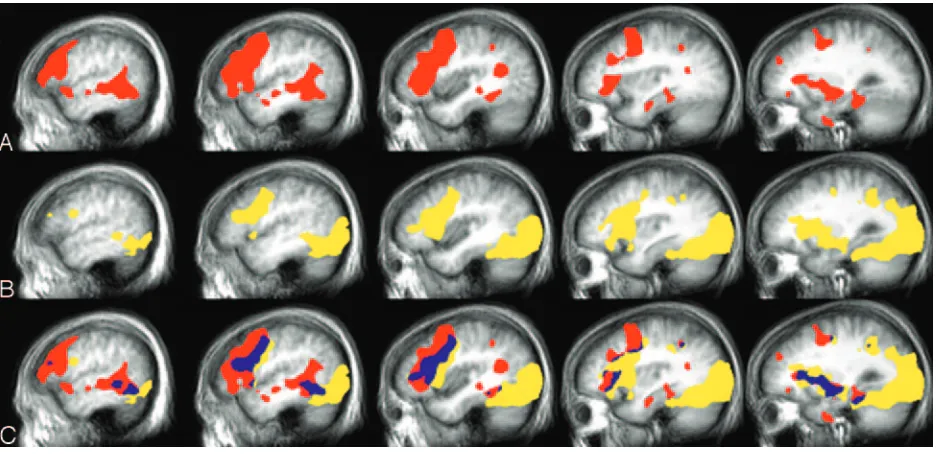
Figure 2 shows the group activation maps for both language tasks. Responsive naming produced more activation compared with the confrontation naming task within the temporal lobe, with the largest activa-tion cluster extending from the posterior superior temporal gyrus through the middle temporal gyrus to anterior and inferior portions of the temporal lobe. Table 1 also includes the Talairach coordinates that are associated with significant differences between the group means for responsive versus confrontation naming within the temporal lobe. The responsive naming versus confrontation naming contrast re-sulted in significant activation within the inferior, middle, and superior temporal gyri of the dominant hemisphere. Other areas outside of the temporal lobe were also identified in this contrast, most notably in the inferior frontal lobe (BA 9/46; data not shown). The confrontation naming versus responsive naming contrast indicated that confrontation naming was not associated with increased activation anywhere in the temporal lobe as compared with responsive naming. The confrontation versus responsive naming contrast did result in significant activation in several extratem-poral sites, including the cuneus and middle occipital gyri, as well as some other areas, including the insula, cingulate, parahippocampal, and inferior parietal gyri (data not shown). In general, the confrontation nam-ing task was also associated with greater activation of the right hemisphere compared with the responsive naming task.
There were several areas of activation that were associated with both naming tasks. Figure 2 (row C) also depicts the overlapping activation for the group analyses across the 2 naming tasks. In the dominant hemisphere, task activation overlap included areas in the middle occipital and middle temporal gyri, the inferior frontal and precentral gyri, the medial fron-tal, middle frontal and cingulate gyri, the hippocam-pus and parahippocampal gyrus, the inferior parietal gyrus, and the basal ganglia. Areas of overlap in the right hemisphere were largest in the inferior frontal gyrus, parahippocampal gyrus, basal ganglia, insula, and cerebellum.
Individual Analysis. To further investigate differ-ences in activation patterns between the 2 naming tasks at an individual subject level, the number of FIG 1. Temporal regions of interest.
[image:3.585.53.283.58.247.2]participants producing activation within each region of interest and the volume of activation in each region of interest were examined. Responsive naming was associated with activation in 90% of the participants within the anterior temporal region of interest and 100% of participants within the posterior temporal region of interest. Confrontation naming also pro-duced activation within 100% of the subjects in the posterior temporal region of interest, but only 60% of subjects in the anterior temporal region of interest.
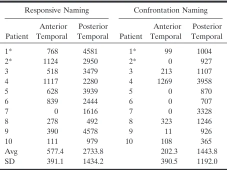
Table 2 includes the average volume of activation within the regions of interest for each task. Analysis of variance was used to examine differences in acti-vation for each task across the 2 regions of interest. There was a significant effect for both the region of interest (P⫽3.0⫻10⫺6) and the task (P⫽.01). The main effect for task indicates that, overall, responsive
naming produced more activation within the tempo-ral lobe than the confrontation naming task. The main effect for region of interest indicates that, re-gardless of task, there was more activation within the posterior temporal region of interest than in the an-terior temporal region of interest. There was only a nonsignificant trend for an interaction between task and region of interest (P⫽ .15), which provides just weak evidence for a possible difference in activation between anterior and posterior regions for confron-tation naming compared with responsive naming (with confrontation naming showing somewhat greater of a difference in activation between the 2 regions of interest).
Comparison of Activation in Subjects Who Com-pleted Both Tasks. Table 2 includes the activation volume for each individual subject under the naming FIG 2. Group activation, left hemisphere.
[image:4.585.53.542.69.233.2]A,Responsive naming;B,confrontation naming;C,overlap: yellow, confrontation naming; red, responsive naming; blue, overlap.
TABLE 1: Group results: left hemisphere temporal lobe
Responsive naming Confrontation naming
Cortex BA x y z zscore BA x y z zscore
Superior temporal 38/22 ⫺50 5 ⫺10 3.5 Middle temporal 21/22 ⫺48 ⫺12 ⫺6 3.9
Middle temporal 21/22/39 ⫺50 ⫺41 ⫺1 5.3 22 ⫺50 ⫺42 ⫺1 4.0 Inferior temporal 20 ⫺27 1 ⫺33 3.0 21 ⫺59 ⫺27 ⫺14 3.1
Responsive naming⬎Confrontation naming
Cortex BA x y z zscore
Middle superior temporal 22/39 ⫺41 ⫺55 19 4.0 Middle temp/fusiform 20/21 ⫺48 ⫺4 ⫺19 3.9 Superior temporal 22 ⫺60 ⫺42 6 2.8 Inferior temporal 20 ⫺32 ⫺1 ⫺38 2.9
Note.—BA indicates Brodmann area.
[image:4.585.62.530.293.520.2]conditions in the anterior and posterior temporal regions of interest, including the 2 subjects who com-pleted both tasks. The comparison of responsive nam-ing to confrontation namnam-ing for the 2 individual sub-jects who participated in both experiments were similar to the comparison of the tasks for the non-overlapping subjects; there was more activation de-tected within both regions of interest for responsive naming compared with confrontation naming. There was minimal activation within the anterior temporal region of interest for both subjects during confronta-tion naming. In fact, within the anterior temporal region of interest, activation associated with confron-tation naming was not detected at all for Subject 2 and the activation detected for Subject 1 was on the outside borders of the anterior temporal region of interest. Figure 3 shows the activation patterns for each task for these 2 subjects. Both subjects produced activation within the anterior temporal region of in-terest for responsive naming that was near regions detected in the group analyses.
Discussion
The results of this study suggest that there are some anatomically distinct sites of activation for responsive naming and confrontation naming. Group activation associated with the responsive naming task produced more widespread activation of the dominant temporal lobe, particularly within the superior and middle tem-poral gyri, which extended into anterior portions of the temporal lobe. This degree of mid- to anterior temporal activation was not observed in association with the confrontation naming task. Even at the indi-vidual subject level, activation associated with con-frontation naming was not as consistently produced in the anterior temporal region of interest as it was with responsive naming (60% vs 90% of the samples showed activation, respectively). As such, our findings by using fMRI are quite similar to the direct cortical stimulation studies, which showed that auditory re-sponsive naming, but not confrontation naming, was disrupted during stimulation of locations within the
anterior portions of the temporal lobe (extending about 4 –5 cm from the temporal pole).
The results of the fMRI activation for each task within the posterior temporal region of interest, in comparison to the previous direct cortical stimulation studies, are not as straightforward. The 2 previous stimulation studies reported somewhat different find-ings with respect to the pattern of disruption associ-ated with the 2 naming tasks within the posterior temporal lobe. Malow et al (2) reported that respon-sive naming was more disrupted than confrontation naming in the posterior part of the superior and middle temporal gyri, whereas there was a fairly equal degree of disruption of function across the 2 naming tasks in the posterior part of the inferior temporal gyrus. Hamberger et al (3) reported that stimulation within the posterior temporal lobe most often dis-rupted both confrontation and responsive naming. Our findings revealed some degree of activation within the posterior temporal region of interest dur-ing both tasks in all subjects. Responsive namdur-ing, however, produced more overall activation in both temporal regions of interest, including the posterior region of interest.
Unfortunately, we were limited to having only 2 subjects who completed both the responsive and con-frontation naming tasks. The individual results of the 2 subjects who completed both naming tasks parallel the group results in showing more activation detected in both regions of interest during responsive naming, with confrontation naming producing little if any ac-tivation of the anterior temporal region of interest. There have now been a number of studies examining fMRI-related activation associated with various ver-sions of confrontation naming paradigms. Results of the current study are consistent with previous studies showing activation associated with confrontation naming most often involves temporal-occipital corti-ces (BAs 37, 19, and 18) and the inferior frontal gyrus (7, 27). Most studies have used covert responses dur-ing confrontation namdur-ing, as done in the current study. Studies that have used overt responses within the scanner, however, have also produced highly sim-ilar results (28).
We are aware of only a single study that has exam-ined fMRI-related activation associated with a re-sponsive naming task. Similar to the current study, Balsamo et al (29) reported strong activation within the superior and middle temporal gyri associated with their responsive naming task, with the group analysis appearing to show activation extending into the supe-rior aspects of the temporal lobe. The Balsamo et al study, however, involved young children (mean age, 8.5 years) whose language and semantic network or-ganization may be quite different from those of adults. Furthermore, these authors used a “rest” baseline, which can be problematic (30, 31). Finally, the previous study did not include a confrontation naming task, so activation associated with responsive and confrontation naming could not be compared.
[image:5.585.54.281.69.239.2]Previous studies have shown that the hippocampus of the speech-dominant hemisphere is a significant
TABLE 2: Individual results
Responsive Naming Confrontation Naming
Patient
Anterior Temporal
Posterior
Temporal Patient
Anterior Temporal
Posterior Temporal
1* 768 4581 1* 99 1004 2* 1124 2950 2* 0 927 3 518 3479 3 213 1107 4 1117 2280 4 1269 3958 5 628 3939 5 0 870 6 839 2444 6 0 707 7 0 1616 7 0 3328 8 278 492 8 323 1246 9 390 4578 9 11 926 10 111 979 10 108 365 Avg 577.4 2733.8 202.3 1443.8 SD 391.1 1434.2 390.5 1192.0
component of the neuroanatomic network involved in naming (32–34). In line with such research, the cur-rent study also showed that both naming tasks pro-duced hippocampal and parahippocampal activation. The dissociation between responsive and confron-tation naming may reflect modality-specific sub-systems involved in word retrieval. Despite the use of an auditory baseline to control for low-level auditory processing, the responsive naming task produced ac-tivation throughout much of the temporal lobe (in-cluding, but not limited to, primary and secondary auditory cortex). Such activation was strongly lateral-ized to the left, which suggests that auditory process-ing was specific to the lexical nature of the stimuli. Activation of inferior frontal regions by both tasks is consistent with other fMRI studies that have com-pared areas of common activation across different language tasks and suggests that, though word selec-tion and retrieval may be somewhat modality specific (11, 35), other aspects of language such as articulatory planning activate similar areas of the inferior frontal lobe. The finding that both tasks activated a posterior region within the fusiform gyrus is also consistent with previous research that has suggested that BAs 37 and 20 are important in word selection and are believed to be multimodal (28, 36 –39), receiving input from au-ditory, visual, and somatosensory cortices (10, 40 – 43).
Unlike the confrontation naming task, responsive naming, in addition to word retrieval, requires sen-tence comprehension. A number of neuroimaging studies have now shown that sentence comprehension involves a distributed frontal and temporoparietal neural network—particularly within the dominant hemisphere—including the temporal pole (44, 45). The anterior temporal lobe may be particularly sen-sitive to both the semantic and the syntactic demands of the task and may help account for the increased anterior temporal activation during responsive nam-ing as compared with durnam-ing confrontation namnam-ing (44, 46).
Both Hamberger and Tamny (47) and Bell et al (1) have found that responsive naming is more sensitive than confrontation naming to the word retrieval def-icits associated with TLE of the dominant hemi-sphere. Positron-emission tomography (PET) studies have shown that, even in epilepsy patients with hip-pocampal sclerosis, hypometabolism is not restricted to mesial temporal structures, but generally extends into most of the temporal lobe (48 –50). It is this lateral temporal lobe involvement that, in addition to the hippocampal dysfunction, likely produces the mild naming deficits often associated with dominant TLE. The larger area of temporal lobe recruitment during responsive naming observed both in this fMRI study and in the direct cortical stimulation studies FIG 3. Individual activation, left hemisphere.
may account for the increased sensitivity of this task (compared with confrontation naming) in detecting the language deficits within the TLE population.
There are some limitations to this study. First, all behavioral responses in the scanner were made co-vertly, so it is not possible to confirm that the subjects were performing the task correctly. However, partic-ipants received extensive training before scanning and were administered an alternate form of the respon-sive naming task after scanning. These results showed that participants were highly accurate in their re-sponses (accurately named approximately 97% of the items on an alternate form of the task). We do not have behavioral data on the confrontation naming test that would enable a direct comparison of perfor-mance levels, but normative studies also indicate that healthy controls of similar age to our sample name approximately 94% of the items on the Boston Nam-ing Test (19). Use of namNam-ing tasks strictly matched on difficulty level should be used in future studies.
Group activation associated with responsive nam-ing was minimal in the inferior regions of the tempo-ral pole. This appears to be at odds with the results of Hamberger et al, who reported disruption of this task at multiple sites in this region (3). Limited activation of this area may be related to the MR imaging signal intensity loss due to magnetic susceptibility artifacts from the nearby auditory canal, mastoid air cells, and petrous bone (51). In fact, a study by Devlin et al (52) compared PET activation with fMRI activation for a semantic task and found temporal pole activation with PET but not fMRI. Those researchers also found that analysis of the echo-planar images showed 82% of the voxels in the temporal pole had a signal inten-sity loss ⬎25%. The use of shimming or acquiring extra images are approaches that may be used to compensate for, or reduce these artifacts (53, 54) and should be the focus of future research. Another lim-itation to our study findings, which could affect clin-ical applicability, was that responsive naming, though producing activation in the anterior temporal region of interest in 90% of our participants, fell short of producing activation in the entire sample. Ideally, fMRI tasks that are going to be used to map functions in clinical samples should produces activation within a given cortical region in all healthy controls. Anterior temporal regions are generally associated with low fMRI signal intensity to noise. Increased detection of activation would likely be achieved by increasing the degrees of freedom associated with the statistical tests. This can be done by repeating the same tasks in 2 different fMRI acquisitions and then combining the tasks or increasing the time points within the fMRI acquisition. Further investigation of the activation associated with responsive naming in a larger sample would help elucidate what percent of healthy controls produce activation in the anterior temporal lobe dur-ing this task.
In summary, results of this study support the 2 previous cortical stimulation studies suggesting that responsive naming is, in part, subserved by areas of the temporal cortex that are anterior to those areas
associated with confrontation naming (2, 3). Ade-quate assessment of anterior temporal lobe functions through the identification of tasks such as responsive naming is critical to surgical planning, because they are likely to be useful in predicting the risk of the word-finding changes that can occur after a temporal lobectomy of the dominant hemisphere. Many of the fMRI language paradigms currently in use do not produce activation within the anterior temporal lobe. The selection of language paradigms must be chosen carefully and will vary as a function of the location of the planned surgical resection. We hypothesize that fMRI activation patterns within the anterior temporal lobe associated with responsive naming or other tasks that activate this region may be better predictors of post–temporal lobectomy language outcome than other fMRI paradigms that do not produce activation in this region in healthy individuals. In support of this prediction, the results of Sabsevitz et al (55) suggest that language-related fMRI activation of a temporal region of interest better predicted postsurgical lan-guage changes than activation in an inferior frontal region of interest. Future studies examining patterns of temporal lobe fMRI activation associated with re-sponsive naming and other language tasks and de-tailed postoperative neuropsychological assessment will be useful in determining if they yield differential information in predicting language outcome after temporal lobectomy.
References
1. Bell BD, Seidenberg M, Hermann BP, Douville K. Visual and auditory naming in patients with left or bilateral temporal lobe epilepsy.Epilepsy Res2003;55:29 –37
2. Malow BA, Blaxton TA, Sato S, et al.Cortical stimulation elicits regional distinctions in auditory and visual naming. Epilepsia
1996;37:245–252
3. Hamberger MJ, Goodman RR, Perrine K, Tamny T.Anatomic dissociation of auditory and visual naming in the lateral temporal cortex.Neurology2001;56:56 – 61
4. Rausch R, Boone K, Ary CM.Right-hemisphere language domi-nance in temporal lobe epilepsy: clinical and neuropsychological correlates.J Clin Exp Neuropsychol1991;13:217–231
5. Devinsky O, Perrine K, Llinas R, et al.Preoperative predictors of anterior temporal language areas.J Neurosurg1998;34:727–732 6. Binder JR, Frost JA, Hammeke TA, et al.Human brain language
areas identified by functional magnetic resonance imaging.J Neu-rosci1997;17:353–362
7. Votaw JR, Faber TL, Popp CA, et al.A confrontational naming task produces congruent increases and decreases in PET and fMRI.Neuroimage1999;10:347–356
8. Binder JR.Functional MRI of the language system.In: Moonen CTW, Bandettine PA, eds.Functional MRI.Berlin and Heidelberg: Springer-Verlag;1999:407– 419
9. Price CJ.The anatomy of language: contributions from functional neuroimaging.J Anat2000;197:335–359
10. Cabeza R, Nyberg L.Imaging cognition. II. An empirical review of 275 PET and fMRI studies.J Cogn Neurosci2000;12:1– 47 11. Carpentier A, Pugh K, Westerveld M, et al. Functional MRI of
language processing: dependence on input modality and temporal lobe epilepsy.Epilepsia2001;42:1241–1254
12. Gaillard WD, Pugliese M, Grandin CB, et al.Cortical localization of reading in normal children: an fMRI language study.Neurology
2001;57:47–54
13. Ogawa S, Tank DW, Menon R, et al. Intrinsic signal changes accompanying sensory stimulation: functional brain mapping with magnetic resonance imaging. Proc Natl Acad Sci U S A
1992;89:5951–5955
15. Doyle WK, Spencer DD.Anterior temporal resections.In: Engle J Jr, Pedley TA, eds.Epilepsy: a comprehensive textbook.Philadelphia: Lippincott-Raven;1997:1807–1817
16. Kim R, Spencer D. Surgery for mesial temporal sclerosis. In: Luders HO, Comair YG, eds.Epilepsy surgery.Philadelphia: Lip-pincott, Williams & Wilkins;2001:643– 652
17. Kaplan E, Goodglass H, Weintraub S.Boston naming test.2nd ed. Philadelphia: Lea and Febiger;1983
18. Hamberger MJ, Seidel WT. Auditory and visual naming tests: normative and patient data for accuracy, response time, and tip-of-the-tongue.J Int Neuropsychol Soc2003;9:479 – 489
19. Spreen O, Strauss E. A compendium of neuropsychological tests.
New York: Oxford University Press;1998
20. Buonocore MH, Gao L.Ghost artifact reduction for echo-planar imaging using image phase correction. Magn Reson Med
1997;38:89 –100
21. Cox RW, Jesmanowicz A. Real-time 3D image registration for functional MRI.Magn Reson Med1999;42:1014 –1018
22. Cox R.AFNI: Software for analysis and visualization of functional magnetic resonance neuroimages. Comp Biomed Res 1996;29: 162–173
23. Forman SD, Cohen JD, Fitzgerald M, et al.Improved assessment of significant activation in functional magnetic resonance imaging (fMRI): use of a cluster-size threshold. Magn Reson Med
1995;33:636 – 647
24. Xiong J, Gao J-H, Lancaster JL, Fox PT.Clustered pixels analysis for functional MRI activation studies of the human brain.Hum Brain Mapping1995;3:287–301
25. Talairach J, Tournoux P.Co-planar stereotaxic atlas of the human brain.New York: Thieme Medical;1988
26. Lancaster JL, Woldorff MG, Parsons LM, et al. Automated Ta-lairach atlas labels for functional brain mapping.Hum Brain Mapp
2000;10:120 –131
27. Murtha S, Chertkow H, Beauregard M, Evans A.The neural sub-strate of picture naming.J Cogn Neurosci1999;11:399 – 423 28. Abrahams S, Goldstein LH, Simmons A, et al.Functional magnetic
resonance imaging of verbal fluency and confrontation naming using compressed image acquisition to permit overt responses. Hum Brain Mapp2003;20:29 – 40
29. Balsamo LM, Xu B, Grandin CB, et al. A functional magnetic resonance imaging study of left hemisphere language dominance in children.Arch Neurol2002;59:1168 –1174
30. Newman SD, Twieg DB, Carpenter PA.Baseline conditions and subtractive logic in neuroimaging. Hum Brain Mapp
2001;14:228 –235
31. Stark CE, Squire LR. When zero is not zero: the problem of ambiguous baseline conditions in fMRI.Proc Natl Acad Sci U S A
2001;98:12760 –12766.
32. Martin R, Sawrie S, Hugg J, et al. Cognitive correlates of 1H MRSI-detected hippocampal abnormalities in temporal lobe epi-lepsy.Neurology1999;53:2052–2058
33. Sawrie S, Martin RC, Gilliam, FG, et al. Visual confrontation naming and hippocampal function: A neural network study using quantitative (1)H magnetic resonance spectroscopy. Brain2000; 124:770 –780
34. Aldenkamp A, Boon P, Deblaere K, et al.Usefulness of language
and memory testing during intracarotid amobarbital testing: ob-servations from an fMRI study. Acta Neurol Scand 2003;108: 147–152
35. Lehericy S, Cohen L, Bazin B, et al.Functional MR evaluation of temporal and frontal language dominance compared with the Wada test.Neurology2000;54:1625–1633
36. Benson D, ed.Neurological correlates of anomia.New York: Oxford University Press;1979
37. Damasio A.Aphasia.N Engl J Med1992;59:531–539
38. Warburton E, Wise RJ, Price CJ, et al.Noun and verb retrieval by normal subjects: studies with PET.Brain1996;119:159 –179 39. Vandenberghe R, Price C, Wise R, et al.Functional anatomy of a
common semantic system for words and pictures. Nature
1996;383:254 –256
40. Luders H, Lesser RP, Hahn J, et al.Basal temporal language area. Brain1991;114:743–754
41. Suzuki W, Amaral D.Topographic organization of the reciprocal connections between the monkey entorhinal cortex and the perirhi-nal and parahippocampal cortices.J Neurosci1994;14:1856 –1877 42. Price CJ. The functional anatomy of word comprehension and
production.Trends Cogn Sci1998;2:1201–1207
43. Buchel C, Price C, Friston K.A multimodal language region in the ventral visual pathway.Nature1998;394:274 –277
44. Noppeney U, Price CJ.An FMRI study of syntactic adaptation.J Cogn Neurosci2004;16:702–713
45. Vandenberghe R, Nobre AC, Price CJ.The response of left tem-poral cortex to sentences.J Cogn Neurosci2002;14:550 –560 46. Cohen L, Jobert A, Le Bihan D, Dehaene S.Distinct unimodal and
multimodal regions for word processing in the left temporal cortex. Neuroimage2004;23:1256 –1270
47. Hamberger MJ, Tamny TR.Auditory naming and temporal lobe epilepsy.Epilepsy Res1999;35:229 –243
48. Arnold S, Schlaug G, Niemann H, et al.Tomography of interictal glucose hypometabolism in unilateral mesiotemporal epilepsy. Neurology1996;46:1422–1430
49. Theodore W, Sat S, Kufta CV, et al. Temporal lobectomy for uncontrolled seizures: the role of positron emission tomography. Ann Neurol1992;32:789 –794
50. Chassoux F, Semah F, Bouilleret L, et al.Metabolic changes and electro-clinical patterns in mesio-temporal lobe epilepsy: a correl-ative study.Brain2004;127:164 –174
51. Ojemann JG, Akbudak E, Snyder AZ, et al.Anatomic localization and quantitative analysis of gradient refocused echo-planar fMRI susceptibility artifacts.Neuroimage1997;6:156 –167
52. Devlin JT, Russell RP, Davis MH, et al.Susceptibility-induced loss of signal: comparing PET and fMRI on a semantic task. Neuroim-age2000;11:589 – 600
53. Jezzard P, Clare S.Sources of distortion in functional MRI data. Hum Brain Mapp1999;8:80 – 85
54. Deichmann R, Josephs O, Hutton C, et al.Compensation of sus-ceptibility-induced BOLD sensitivity losses in echo-planar fMRI imaging.Neuroimage2002;15:120 –135