NHE gene family
The mammalian NHE (Na/H exchanger) gene family (SLC9 family) consists of nine isoforms which fall into two major subdivisions (Brett et al., 2005a): isoforms that function primarily on the plasma membrane (NHEs 1, 2, 3, 4 and 5) and those that are present primarily on intracellular organelles (NHEs 6, 7 and 9). The plasma membrane NHEs, NHE3 and 5, continually cycle between an intracellular juxtanuclear location which includes the recycling system and the plasma membrane (D’Souza et al., 1998). NHEs 1, 2 and 4 are static on the plasma membrane, although data for NHE4 are not thorough. Of the intraorganellar NHEs, NHE 6, 7 and 9 have been localized primarily to the recycling system (NHE6), trans-Golgi network (NHE7) and late endosomes (NHE9), respectively; although it is possible that these NHEs all move among intracellular organelles (Brett et al., 2002; Numata and Orlowski, 2001; Nakamura et al., 2005; Orlowski and Grinstein, 2007). The two classes of NHEs can be separated by the amino acids which make up their transport domains (Mukherjee et al., 2006). NHE8 seems to resemble organellar NHEs by its amino acid composition, but it has been suggested that it resides on the plasma membrane as well (Goyal et al., 2003; Xu et al., 2005; Becker et al., 2007; Xu et al., 2008). Progress has been made in understanding structure/function relationships and regulation of the plasma membrane NHEs. A major limitation to progress in the study of the organellar NHEs and NHE8 has been the failure to be able to detect organellar NHE activity in simple cell systems. Evidence from yeast studies of the endosomal (vacuolar) exchanger Nhx1 (the first organellar NHE identified) suggests that this NHE class functions as K–Na/H exchangers. These appear to act to remove intraorganellar H pumped in via the V-ATPase and serve to regulate organellar H by acting as a ‘leak pathway’ (Xu et al., 2008; Brett et al., 2005b).
Physiological roles of NHE3
NHE3, the major focus of this review, is an epithelial brush border Na/H exchanger (Zachos et al., 2005; Donowitz and Li, 2007; Orlowski and Grinstein, 2004). NHE3 is present most abundantly in epithelial cells in the GI tract and kidney where it is responsible for the majority of Na absorption in these two organ systems that are responsible for most of the water and Na homeostasis of the body (Zachos et al., 2005). In addition, NHE3 is present in the epidydimus, ovary, thymus, prostate, and in some respiratory neurons in the ventrolateral medulla (Zachos et al., 2005). At a subcellular level in epithelial cells, NHE3 is found in the brush border/apical membrane of duodenum, jejunum, ileum, proximal colon, gall bladder, proximal tubule and thick ascending limb as well as the proximal portion of the long descending thin limb of Henle (Zachos et al., 2005). However, in parietal cells of some species, NHE3 localizes to the basolateral membrane (Zachos et al., 2005). In the intestine and proximal tubule, NHE3 functions on the apical membrane to allow absorption of large amounts of Na by a high capacity, low efficiency process called neutral NaCl absorption (Brett et al., 2005b). In the intestine, NHE3 is linked functionally with SLC26A family members DRA (SLC26A3) and PAT-1 (SLC26A6), predominantly through small changes in intracellular pH; although it remains possible that there is an additional physical linkage (Lamprecht et al., 2002; Seidler et al., 2008; Petrovic et al., 2008; Walker et al., 2008; Musch et al., 2008) (Fig. 1). Which SLC26A member(s) is coupled to NHE3 in the small intestine remains in question, although in the colon it appears to be DRA (Lamprecht et al., 2002). In the small intestine, some functional data suggest linkage primarily with DRA; however, other results suggest a link to PAT-1. In addition, the amount and localization of small intestinal DRA does not appear to correlate with NHE3 localization along the crypt/villus axis (Seidler et al.,
The Journal of Experimental Biology 212, 1638-1646 Published by The Company of Biologists 2009 doi:10.1242/jeb.028605
Review
NHE3 regulatory complexes
Mark Donowitz*, Sachin Mohan, Cindy Xinjun Zhu, Tian-E Chen, Rong Lin, Boyoung Cha,
Nicholas C. Zachos, Rakhilya Murtazina, Rafiquel Sarker and Xuhang Li
Johns Hopkins University School of Medicine, 720 Rutland Avenue
Baltimore, MD 21205, USA
*Author for correspondence (e-mail: [email protected])
Accepted 12 February 2009
Summary
The epithelial brush border Na/H exchanger NHE3 is active under basal conditions and functions as part of neutral NaCl absorption in the intestine and renal proximal tubule, where it accounts for the majority of total Na absorbed. NHE3 is highly regulated. Both stimulation and inhibition occur post-prandially. This digestion related regulation of NHE3 is mimicked by multiple extracellular agonists and intracellular second messengers. The regulation of NHE3 depends on its C-terminal cytoplasmic domain, which acts as a scaffold to bind multiple regulatory proteins and links NHE3 to the cytoskeleton. The cytoskeletal association occurs by both direct binding to ezrin and by indirect binding viaezrin binding to the C-terminus of the multi-PDZ domain containing proteins NHERF1 and NHERF2. This is a review of the domain structure of NHE3 and of the scaffolding function and role in the regulation of NHE3 of the NHE3 C-terminal domain.
2008; Petrovic et al., 2008; Walker et al., 2008; Musch et al., 2008). In the intestine, Na absorption linked to Cl absorption is the major way neutral NaCl absorption functions, although NHE3 activity can be uncoupled from Cl/HCO3exchange, at least in the mouse small
intestine (Walker et al., 2008). An important characteristic of NHE3 activity in all cell types studied is that it is in a partially activated state under basal conditions (Brett et al., 2005b). As part of normal digestive physiology, NHE3 is thought to be inhibited early in the digestive period. This presumably leads to increased luminal water and serves to spread digestive enzymes secreted by the GI organs over the intestinal surface to allow increased efficiency of digestion. Later in digestion, NHE3 activity is stimulated, to maintain volume homeostasis. In the kidney, NHE3, in addition to functioning in neutral NaCl absorption, is important for HCO3
absorption and NH4+secretion (Bobulescu and Moe, 2006).
Besides its function in absorption of Na and water from the intestinal lumen, NHE3 is also responsible for the absorption of other nutrients which use an acid outside H+ gradient (acidic
microclimate at the apical surface) (Thwaites and Anderson, 2007; Anderson and Thwaites, 2005; Anderson et al., 2004). The most clearly linked substrates include dipeptides and amino acids viathe transporters PepT1 (di/tripeptides) and several L-amino acid
transporters, but might include many other transporters. The mechanism of coupling has not been established but is likely to include the apical acidic microclimate generated by NHE3. In addition to these functions on the apical membrane, NHE3 appears to cause acidification of an early endosomal compartment, using a gradient generated by Na engulfed from the apical extracellular environment to exchange for cytosolic H (Gekle et al., 1999; Gekle et al., 2001). Why this process is able to acidify an organelle, apparently in the presence of a V-ATPase, is not clear. Also, the specific compartment and its constituent transport proteins have not been defined adequately. NHE3 knockout mice have increased stool water and, while initially viable, have a shortened life span and die rapidly when placed on a low Na diet (Schultheis et al., 1998; Noonan et al., 2005). They have modest diarrhea and increased fluid in the small intestine and especially the colon, abnormal acid/base balance (acidemia), low blood pressure and reduced body fat. Of interest is the observation that these mice have distal colitis (Laubitz et al., 2008).
Mimicking NHE3 inhibition and stimulation which occurs as part of digestive and renal physiology, various neurohumoral agonists, growth factors, chronically altered states of systemic acid/base balance, and drugs have been shown to stimulate or inhibit NHE3 activity. These effects occur within minutes to hours (Zachos et al., 2005; Bobulescu and Moe, 2006). Short-term regulation consists largely of changes in the amount of plasma membrane NHE3 as a result of changes in NHE3 trafficking, although changes in the complex affinity construct for intracellular H+[K⬘(H+)
i] occur as well. Abnormal inhibition of NHE3 occurs
in diarrheal diseases and with chronic inflammatory bowel diseases (IBD) being associated with changes in amount of NHE3 while acute inflammation is associated with reduced NHE3 activity in the presence of normal amounts of NHE3 (Sullivan et al., 2008; Hecht et al., 2004).
As shown in Fig. 1, there are two major types of Na linked transport processes in the proximal small intestine. In addition to neutral NaCl absorption, there are transporters which take up end products of digestion. End products of carbohydrate digestion are primarily taken up by the Na linked D-glucose or D-galactose
transporter SGLT1 and by GLUT5 (a fructose transporter). There are also multiple Na linked L-amino acid transporters. These
transporters have been used to stimulate intestinal Na absorption in patients with diarrhea in preparations called oral rehydration solutions (ORS). In enterotoxin related diarrheal diseases, such as cholera, which activates the adenylate cyclase/cAMP system, there is increased stimulation of electrogenic Cl (apical cystic fibrosis transmembrane conductance regulator, CFTR) and K (basolateral channels) secretion as well as inhibition of neutral NaCl absorption. In contrast, in inflammation related diarrheal diseases there appears to be inhibition of neutral NaCl absorption without much stimulation of Cl secretion (Hecht et al., 2004). In spite of inhibition of neutral NaCl absorption, predominantly due to inhibition of NHE3 activity, ORS have proven effective in rehydrating patients because in most diarrheal diseases SGLT1 and the basolateral membrane Na–K-ATPase function normally as long as there is not extensive destruction of Na absorptive cells. Still not understood, however, is why these solutions are so effective in rehydrating patients and saving lives, with the amount of Na absorption seeming to exceed the established SGLT1 stoichiometry of 2 Na:1 Glu (Mackenzie et al., 1998). A model to explain this increased water absorption argued that Na–D-glucose absorption caused a
change in myosin related control of tight junctions leading to increased permeability and net water absorption (Madara and Pappenheimer, 1987). This concept, however, was challenged based on the fact that the luminal concentration of D-glucose needed
to change permeability in the intact intestine seemed to be greater than occurred physiologically (Ferraris et al., 1990). Another possible explanation was suggested via the work of Turner and colleagues (Turner et al., 2000; Turner and Black, 2001; Zhao et al., 2004; Shiue et al., 2005). They showed in Caco-2 cells, that luminal D-glucose initiates apical signaling pathways which result
in the translocation of NHE3 from intracellular stores to the apical membrane. This signaling pathway in Caco-2 cells was shown to involve p38 MAP kinase, MAP kinase kinase, Akt2 and ezrin. This linking of increased D-glucose uptake to translocation of
intracellular NHE3 to the plasma membrane would identify NHE3 as the major small intestinal Na absorptive protein and could partially explain the great effectiveness of ORS, as well as provide another protein (NHE3) to target in improving ORS solutions. In this strategy it must be considered that NHE3 is inhibited in many diarrheal diseases, although increased exocytosis induced by ORS
Brush border
Small intestinal sodium absorption
NaCl
ATP
ADP Na+/glucose
Amino acids
Na+
Na+
pump
15 mmol l–1 Na+
–40 mV
H+
HCO3–
Cl–
Electrogenic
Neutral
[image:2.612.51.284.66.242.2]K+
might reverse that inhibition. These studies have only been reported to date in Caco-2 cells, a colon cancer cell line which is a Na absorptive cell line. However, preliminary studies indicate that the same linking of D-glucose uptake and increased small intestinal
NHE3 activity under basal conditions appears to exist in mouse proximal small intestine (R.L., R.M., O. Kovbasnjuk and M.D., unpublished). Further studies of this linking of SGLT1 and NHE3 must be carried out in animal diarrheal disease models before any suggestion of possible relevance to humans should be considered.
NHE3 molecular physiology: transport domain Studies of the molecular physiology of NHE3 have attempted to relate NHE3 function to specific parts of the N- and C-termini. NHE3 is thought to consist of two functionally distinct domains, the N-terminus, which carries out the Na and H exchange (human NHE3, amino acid L456) and the C-terminus, which regulates the transport rate (human NHE3 amino acids 457–834). While we discuss these two domains separately, there are suggestions that there is more than one physical interaction between the two domains. The structure of the NHE3 transport domain has not been solved. The most detailed characterization of the NHE transport domain has been carried out by the group of S. Wakabayashi, using Cys mutagenesis (Wakabayashi et al., 2000; Hisamitsu et al., 2007; Kemp et al., 2008). They have proposed a model for the transport domain which includes 12 membrane spanning domains (MSD) and two re-entry loops which dip into the membrane (Wakabayashi et al., 2000; Hisamitsu et al., 2007). However, the structure has been solved for a bacterial (E. coli) Na/H exchanger, EcNhaA, by Padan and co-workers (Hunte et al., 2005; Kozachkov et al., 2007; Padan, 2008). This protein is <10% identical to the NHEs at an amino acid level. However, it has been suggested that it is very similar at a tertiary structural level in the NHE3 N-terminus, particularly in the area of the transport fold, based on bioinformatics analyses with fold alignment plus some select mutagenesis (Landau et al., 2007). This was suggested in spite of major functional differences between the two families (Nha and NHE). For instance, NhaA transports Na out of the cell in exchange for H, has a stoichiometry of 2:1, and a modifier site such that it is inactive at acidic pH. Please note that these characteristics differ from those of the NHE family in which the Na:H exchange stoichiometry is 1:1 or 2:2, Na is generally transported into and H exported from the cells, transport activity is maximum at acidic pH and the exchangers are inactive at alkaline pH. The NhaA structure consists of 12 MSD and is characterized by a unique double funnel in the middle of the protein that is the site of Na and H exchange and occurs viatwo split membrane spanning domains with charged dipoles being critical for the physical transport (Hunte et al., 2005; Kozachkov et al., 2007; Padan, 2008). These structural insights into NhaA have been applied to NHE1 and a homology model for NHE1 has been proposed (Landau et al., 2007). In this model the cytoplasmic facing funnel is formed by TM2, TM4, TM5 and TM9 and the extracellular facing funnel is formed by TM8, TM11 and TM2 (Fig. 2A). In Fig. 2B (left) we show a figure taken from the Padan model for the structure of NhaA, emphasizing the areas that transport Na and H with the critical amino acid highlighted (Landau et al., 2007). Padan and her collaborators Landau, Herz and Ben-Tal (Landau et al., 2007) compared this structure with homologous amino acids in NHE1 (Fig. 2B, right) (Landau et al., 2007). Noteworthy are conserved, functionally important amino acids: NhaA/NHE1: D133/D238; D163/E262; D164/D267; K300/R425. Fliegel has analyzed the evidence supporting the NhaA and Wakabayashi models for NHE1 and suggested that most evidence
supports conclusions based on the solved structure (Kemp et al., 2008). He argues, however, that the first two NHE MSD may be more accurately represented by the scanning Cys mutagenesis model, and that the final understanding of the structure of the NHE transport domain is incomplete. This model of split membrane spanning domains with dipoles involved in transport has been shown to be a widely present transport fold, including that of the recently crystallized Na–glucose co-transporter (Faham et al., 2008).
Is there evidence supporting a similar ‘solved structure’ based model for the NHE3 transport domain? No formal analysis has been done comparing the secondary structure of NHE3 with that of NhaA. Of note, while NhaA and NHE1 are thought to have 12 membrane spanning domains, NHE3 and Nhx1 have been shown to have cleaved signal peptides consistent with them having only 11 membrane spanning domains (Zizak et al., 2000; Wells and Rao, 2001). In Fig. 2C, we show that four amino acids in NhaA and the homologous amino acids in NHE1 which are critical for Na and H transport activity are also conserved in NHE3.
A very recent elegant study of steady state function of NHE1 by an oscillating pH-sensitive microelectrode was used to quantify H fluxes and characterize transport kinetics (Fuster et al., 2008). This study challenged the concept that while the NHEs exist as dimers, they function as monomers with a 1:1 Na/H exchange stoichiometry. This study concluded that NHE1 (and likely NHE3) had 2 Na:2 H stoichiometry with function as a dimer dynamically based on a regulatory function of intracellular pH. At low pH, the two constituent monomers act independently and the NHE acts as a monomer, while at alkaline pH they function in a coupled manner and the NHE functions as a dimer. In addition, it was shown that Na dependence of exchange activity can be cooperative from both inside and outside the cells showing a further complexity in the kinetic function of the exchanger.
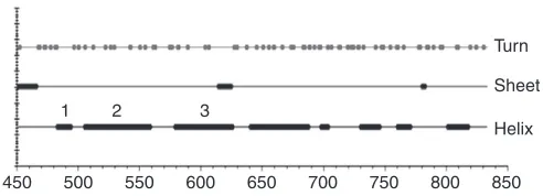
NHE3 molecular physiology: regulatory domain While the relationship between the transport domain of NhaA and the NHEs appears to be an example of convergent evolution, the same is not true for their C-termini. The NhaA C-terminus is very small, consisting of only a few amino acids (Hunte et al., 2005) while that of human NHE3 is large, being 378 amino acids in length. This appears to be where evolution has separated the two classes of transporters, allowing regulation at a more complex manner in eukaryotic cells. While the amino acid make up of the transport domains is highly homologous among the NHE isoforms, there is far greater divergence among the NHEs in the C-terminus (Table 1: based on secondary structure predictions, the cytoplasmic region of NHE3 has significant structure, in particular being predicted to be α-helical in the domains closest to the transport domain, and far less so in the downstream C-terminal area; Fig. 3) (Chou and Fasman, 1974). To date only one domain of the NHE C-terminus has been solved structurally. NHE1 between amino acids 516 and 540 has been shown to be α-helical and to include intimate binding with CHP (calcineurin homologous protein) as part of the structure (Chou and Fasman, 1974; Pang et al., 2001; Ben Ammar et al., 2005). NHE3 amino acids 473–497 are highly homologous to the CHP binding domain of NHE1. As immunoprecipitating NHE3 co-precipitates CHP (R.L. and M.D., unpublished), we assume the secondary structure prediction of an
The NHE3 C-terminus is required for all NHE3 regulation described (Zachos et al., 2005; Donowitz and Li, 2007). If rabbit NHE3 is truncated at amino acid 454, the protein transports Na and H, although with much reduced activity compared with the intact protein; however, it is not regulated (Levine et al., 1995). Truncation studies of the NHE3 C-terminal domain have identified distinct regions which are required for specific aspects of acute regulation of NHE3 activity (Fig. 4). In addition, the C-terminus of NHE3 acts as a scaffold by binding multiple proteins that are involved in its regulation, some of which are scaffolds in their own right (Donowitz and Li, 2007). Fig. 4 illustrates two aspects of this scaffolding function of the NHE3 C-terminus. (1) NHE3 is linked to the cytoskeleton by binding at two areas, viadirect ezrin binding
at amino acids 509–529 (Cha et al., 2006a; Cha and Donowitz, 2008) and viaindirect ezrin binding to the NHERF family of multi-PDZ domain proteins at amino acids 586–605 (Yun et al., 1998). (2) The C-terminus binds multiple proteins which take part in NHE3 regulation. Fig. 4 illustrates the proteins shown to directly bind to the NHE3 C-terminus. These include CHP, NHERF1, 2, 3, 4, CaM kinase II (CaM KII), CaM, CK2, phospholipase Cγ(PLCγ), ezrin, megalin, IRBIT (IP3 receptor binding protein), Shank2, and perhaps PP2A (Pang et al., 2001; Ben Ammar et al., 2005; Ammar et al., 2006; Di Sole et al., 2004; Yun et al., 1997; Thompson et al., 2005; Sarker et al., 2008; Biemesderfer et al., 1999; Girardi et al., 2004; Girardi et al., 2001; Girardi et al., 2008; He et al., 2008; Han et al., 2006). NHE3 additionally interacts with DPPIV, although it
Extracellular Intracellular
EcNhaA crystal structure 3D-model of NHE1
B
A
C
Fig. 2. (A) Model of the transport domain of NHE1 showing the area of exchange as two opposing funnel structures facing the intracellular and extracellular surfaces with membrane spanning domains indicated by roman numerals with critical amino acids shown. With permission from Landau et al. (Landau et al., 2007). (B) Comparison of NhaA and NHE1 transport domains, modeled from the crystal structure of NhaA. The homologus amino acids crucial to exchange of NhaA and NHE1 are shown. With permission from Landau et al. (Landau et al., 2007). (C) Amino acid alignment of human NHE1 (SLC9A1) and human NHE3 (SLC9A3) with arrows designating the amino acids shown in Fig. 2B,
demonstrating identity. *, identical amino acids;
[image:4.612.48.367.65.581.2]appears that this is not a direct association. In particular, in one area (amino acids 586–605), multiple proteins associate with NHE3, suggesting that the involved complex consists of proteins interacting not only with NHE3 but also with each other (Fig. 5). This is discussed later in this review.
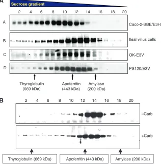
If NHE3 associates with at least the number of proteins shown in Fig. 4, it might be predicted to exist as a large complex or series of complexes. In fact this is true for all cases in which the size of NHE3 has been analyzed, from intact small intestinal Na absorptive cells that express NHE3 endogenously to cultured cells expressing NHE3 endogenously or exogenously to fibroblasts expressing NHE3 exogenously (Li et al., 2001; Akhter et al., 2002) (Fig. 5). However, as we reported, NHE3 is found in very small amounts in its predicted size as a monomer or as a dimer, but, rather, in OptiPrep or sucrose density gradients (solubilized in Triton X-100 1% initially) NHE3 occurs as trains of complexes up to 1200 kDa in size (Donowitz and Li, 2007; Li et al., 2001; Akhter et al., 2002; Li et al., 2004). Assuming that NHE3 exists as a homodimer and that an average sized protein is 50–70 kDa, we predict that under basal conditions NHE3 associates with ~20 proteins of 50 kDa or 15 proteins of 70 kDa. We do not know the meaning of the trains of NHE3 complexes identified in a single cell type at one point in time. However, this observation would suggest that NHE3 exists in many sized complexes at one time in a single cell, or that, as proteins bind to NHE3 with differing affinities, some proteins may come off differentially during the long centrifugation steps in separating the proteins on the density gradients. What is clear, however, is that the NHE3 complexes are dynamic upon signaling, as conditions that acutely stimulate or inhibit NHE3 activity have been associated with changes in the sizes of the NHE3 complexes. As an example, Fig. 5B shows that when rabbit ileal mucosa was
exposed to carbachol (10μmol l–1, 10 min), NHE3 complexes
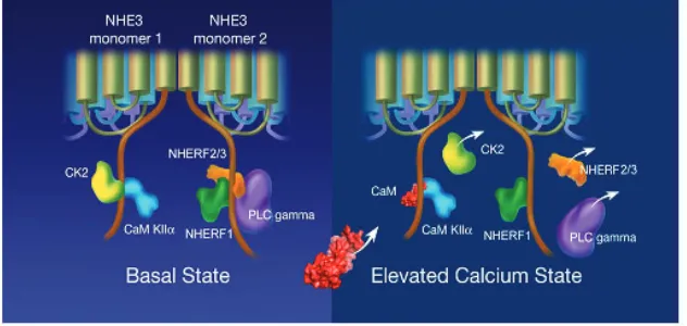
enlarged significantly compared with untreated controls (Li et al., 2004). That NHE3 complexes increase in size with changes in signaling is consistent with the proteins associating with NHE3 changing as part of signaling, an observation confirmed by co-precipitation studies. For instance, with elevation of Ca2+, NHE3/NHERF2 is joined by α-actinin-4 and PKCα(Kim et al., 2002; Lee-Kwon et al., 2003). It must be emphasized that not all changes in protein association with NHE3 are discernible as changes in NHE3 complex size. Additions and subtractions of similarly sized proteins or low affinity associations which dissociate during gradient preparation would be predicted to obscure changes in associating proteins. Of note, when the size of NHE3 complexes was examined in a single cell type, comparing the size of complexes at the plasma membrane with intracellular complexes (using cell surface biotinylation), the plasma membrane complexes were larger than the intracellular complexes (Donowitz and Li, 2007). This was interpreted to indicate that the NHE3 complexes changed with basal trafficking to different subcellular locations, with new binding proteins being added as NHE3 approached or left the plasma membrane. This result is a strong indication that not all NHE3 complexes are formed in the endoplasmic reticulum/Golgi. Demonstrated dynamic changes in NHE3 complexes with regulation of NHE3 activity include conditions in which the NHE3 complexes enlarge [carbachol inhibition, lysophosphatidic acid (LPA) stimulation of NHE3 activity] and get smaller (elevated Ca2+ in cells expressing NHERF4 which is associated with the stimulation of NHE3 activity).
[image:5.612.51.563.80.191.2]Recent studies have suggested that the dynamic NHE3 regulatory complexes formed on the NHE3 C-terminus do not just consist of isolated proteins scaffolded to the terminus or brought to the C-terminus viabinding to other scaffolds on the C-terminus, such as the NHERF proteins. Rather it appears that the regulatory proteins associating with the NHE3 C-terminus may be arranged in larger, more organized complexes which regulate NHE3 by changing their association with NHE3 and perhaps with each other. We illustrate this concept by describing the multiple proteins which bind to a small, putative α-helical domain of NHE3, between amino acids 586 and 605. The proteins which associate with NHE3 in this domain both stimulate and inhibit NHE3 activity. The interactions of many of these proteins are dynamic, often changing with signal transduction that affects NHE3 activity. Because the up and down regulation of NHE3 is so characteristic of NHE3 function, we hypothesize that this domain of NHE3 is involved in this characteristic of NHE3 function and suggest that this domain may
Table 1. Comparsion of NHE3 with other human NHEs (1–9)
N-terminal region (% amino acids) C-terminal region (% amino acids)
Identical with HsNHE3 Positive with HsNHE3 Identical with HsNHE3 Positive with HsNHE3
HsNHE 1 47 65 21 36
HsNHE 2 49 65 18 29
HsNHE 4 43 62 16 26
HsNHE 5 59 70 28 38
HsNHE 6 27 42 n.s. n.s.
HsNHE 7 24 38 n.s. n.s.
HsNHE 8 27 46 n.s. n.s.
HsNHE 9 25 40 n.s. n.s.
Comparison was done for N-terminal and C-terminal regions using Blast P (BLOSUM 62 Matrix) from http://www.ncbi.nlm.nih.gov/blast/bl2seq/wblast2.cgi. Percentage scores were calculated by dividing the number of identical and positive (conserved) amino acids by the total number of subject amino acids in HsNHE3. Sequence for all isoforms was obtained from Uniprot database http://www.uniprot.org/uniprot/.
n.s., not significant.
1 2
450 500 550 600 650 700 750 800 850 Turn
Sheet
Helix
3
[image:5.612.48.294.622.711.2]act as the ‘switch domain’ involved in setting NHE3 activity (Fig. 6).
NHERF proteins bind to NHE3 predominantly between amino acids 586 and 605. The C-terminus of NHE3 is a class I PDZ domain binding sequence. It has been shown that this domain of NHE3 binds NHERF1 and NHERF2 by yeast two-hybrid and ubiquitin hybrid studies, and also binds NHERF3 (Thomson et al., 2005). It is unusual that a single protein associates with NHERF
proteins at two sites. Of note, no regulatory significance has been identified yet for the C-terminal association of NHE3 with the NHERF proteins, while NHERF interactions with amino acids 586–605 have been shown to be involved in multiple aspects of NHE3 regulation (Donowitz and Li, 2007). This is an area requiring further study. The role of NHERF family binding to NHE3 in NHE3 regulation has been reviewed in detail (Donowitz and Li, 2007).
Ezrin
Hyper Osm
FBS
455 475 509 585 702
FGF S/T phosphorylation
756 OA
832
689
640
CaM CaM
KII
CHP
PKA, PKC
Regulating signals NHE3 binding partners
COOH PMA ATP depletion
Vmax increase Vmax decrease
Ez
rin
Actin cytoskeleton
Tyrosine kinase
Squalam
ine (Genis
tein )
Tyrosine kinase
Squalam
ine (Genis
tein ) PI?
CK2
NHERF1/2
605
PLC
γ
Megalin
IRB
[image:6.612.53.405.65.277.2]IT
Fig. 4. Cartoon of domain structure of rabbit NHE3. The N-terminus includes amino acids 1–454. The C-terminus is the regulatory domain and in blue are some of the extracellular and second messenger regulators with the parts of the C-terminus necessary for their effects shown. In green is the area of attachment of NHE3 to the cytoskeleton via ezrin/radixin/moesin (ERM) proteins which associate with NHE3. In yellow are shown the proteins binding to and regulating the NHE3 C-terminus.
Caco-2-BBE/E3HA
PS120/E3V OK-E3V Ileal villus cells
2 4 6 8 10 12 14 16 18 20
Sucrose gradient
A
B
C
D
Amylase (200 kDa) Apoferritin
(443 kDa) Thyroglobulin
(669 kDa)
2 4 6 8 10 12 14 16 18 20
–Carb
+Carb
Thyroglobulin (669 kDa) Apoferritin (443 kDa) Amylase (200 kDa)
A
B
[image:6.612.48.364.425.747.2]The NHERFs are involved with anchoring NHE3 to the cytoskeleton. Under basal conditions, brush border NHE3 has a limited mobile fraction (~30%) and this requires the presence of the NHERFs (Cha et al., 2004). Mobile fraction refers to the percentage of apical domain GFP tagged NHE3 which recovers after being bleached, as studied by ‘fluorescence recovery after photobleaching’. A two amino acid point mutation of NHE3 between amino acids 586 and 605 abolishes NHERF association with NHE3 and increases the mobile fraction to ~75%, a similar value to glycosylphosphatidylinositol (GPI) which is present only in the outer leaflet of the plasma membrane and is a control for free membrane mobility. As part of stimulation (LPA) and inhibition (elevation of Ca2+) of NHE3, the NHE3 mobility transiently increases, presumably freeing up the NHE3 initially in the microvilli to allow endocytosis and to accept further NHE3 trafficking to the brush border free from the NHERFs and cytoskeleton (Cha et al., 2006b). The dynamic aspects of NHERF interactions with their substrates were reported by Weinman, who showed that NHERF1 phosphorylation at S77 led to dissociation from some of its substrates, including NaPi2a, CFTR, platelet-derived growth factor (PDGF) and the β2-adrenergic receptor (Voltz et al., 2007; Weinman et al., 2007). We have shown that the NHE3 interaction with NHERF2 and NHERF3 is dynamic, decreasing with elevation in intracellular Ca2+(Cha et al., 2006b).
What determines the ligand specificity of the dynamic interaction of NHERFs with its substrates and how that is regulated for NHERF2, which has not been shown to be phosphorylated, remains unknown.
The other proteins shown to directly bind this domain of NHE3 under basal conditions are CK2, CaM KII and PLCγ(Figs 4 and 6), as discussed below.
CK2 binds NHE3 between amino acids 586 and 605 and phosphorylates it at another site, S719 (Sarker et al., 2008). Phosphorylation of S719 accounted for 67–75% of basal NHE3 activity [effect on Vmax and K⬘(H+)i]. Inhibition of CK2 reduces
plasma membrane expression of NHE3 to a similar extent to the decrease in NHE3 activity, indicating that the majority of the CK2 effect is via regulation of NHE3 trafficking. This has been evaluated by either mutating S719 to A or to D or exposing NHE3 to a CK2 inhibitor such as DMAT. CK2 phosphorylation of NHE3 increases NHE3 trafficking to the plasma membrane from the recycling pool of NHE3 and delivery of newly synthesized NHE3. The CK2 α-subunits, but not the β-subunits, are associated with NHE3. In addition, the association of CK2 and NHE3 was dynamic and decreased with elevated Ca2+(Sarker et al., 2008). Whether and how much of Ca2+inhibition of NHE3 is due to removal of the CK2
stimulation and the subsequent decrease in basal phosphorylation of NHE3 S719 is unknown.
As CK2 binds to and stimulates basal NHE3 activity, CaM KII binds to the same α-helical domain but inhibits basal NHE3 activity (Zizak et al., 2003). The role of CaM KII was examined by study of the CaM KII inhibitor KN-62. The CaM KII effect is also due to a decrease in NHE3 Vmax. However, unlike the effect of CK2,
CaM KII regulation was not associated with a change in NHE3 plasma membrane expression but, rather, it exerted its effects by altering NHE3 turnover number. CaM KII directly bound NHE3 between amino acids 586 and 605 and phosphorylated NHE3 downstream of this site. The association of CaM KII with NHE3 was not dependent on Ca2+. The mechanism by which CaM KII inhibits NHE3 basal activity at basal Ca2+is not known.
PLCγ also binds and stimulates NHE3 under basal conditions (Zachos et al., 2008). Previous studies demonstrated that elevating Ca2+ with carbachol exposure in ileal Na absorptive cells was associated with translocation of PLCγto the apical membrane by a process in which the translocated PLCγwas active but not tyrosine phosphorylated (Khurana et al., 1996; Khurana et al., 1997). This translocation was associated with a carbachol-induced increase in free intracellular Ca2+, which appeared first at the apical surface,
and seemed to occur viaa phosopholipase dependent process. In addition to this role of PLCγ, this phospholipase appears to play an additional but separate role in the regulation of NHE3 which occurs viaa phospholipase independent mechanism (Zachos et al., 2008). Under basal conditions PLCγ co-precipitates with NHE3, interacting with NHE3 C-terminal amino acids 586–605. This co-precipitation was demonstrated both in fibroblasts and in the polarized Na absorptive Caco-2 cells. This association with NHE3 was dynamic, decreasing after Ca2+ elevation via carbachol exposure. The part of PLCγwhich associates with NHE3 is the PHc domain, which is consistent with involvement with a part of PLCγ that takes part in phospholipase independent effects. Moreover, this was supported by ability of a peptide that is a competitive inhibitor of the PLCγSH2 domains to reverse the contribution of PLCγto NHE3 regulation (inhibited basal NHE3 activity and prevented Ca2+inhibition of NHE3 activity in fibroblasts).
While at least seven proteins associate with NHE3 in this single
[image:7.612.45.361.70.220.2]α-helical domain of the NHE3 C-terminus, we assume that the interactions involve the NHE3 dimeric cytoplasmic domains, which are likely to interact with each other to accommodate NHERFs 1, 2, 3 and 4, CK2, CaM KII and PLCγ. This interaction is dynamic with at least CK2, NHERFs 2 and 3 and PLCγbut not CaM KII dissociating with elevated Ca2+ (Fig. 6). The physical nature of these multiple proteins simultaneously interacting with
the NHE3 C-terminus and whether they interact physically with each other is not known. Also unknown is whether they exist in separate NHE3 complexes. Similarly unknown is how their regulation of NHE3 changes with digestive physiology to switch NHE3 activity from basally active to inactive as digestion is initiated and then to a higher state of activity than basal later during digestion. What appears likely, however, is that study of this small
α-helical area of NHE3, the ‘switch domain’, will provide insights into the defining aspects of NHE3 function in intestinal Na absorption.
We would like to thank Dr Sandra B. Gabelli from the Department of Biophysical Chemistry, Johns Hopkins University School of Medicine, Baltimore, MD, for helpful discussions and Virginia Ferrante, Ferrante Medical Media for artistic assistance. Supported in part by the National Institutes of Health, NIDDK Grants RO1-DK26523, RO1-DK61765, PO1-DK072084 and R24-DK64388 (The Hopkins Basic Research Digestive Diseases Development Core Center) and the Hopkins Center for Epithelial Disorders. Deposited in PMC for release after 12 months.
References
Akhter, S., Kovbasnjuk, O., Li, X., Cavet, M., Noel, J., Arpin, M., Hubbard, A. L.
and Donowitz, M.(2002). Na+/H+exchanger 3 is in large centrally located
complexes on the brush border of proximal tubule-derived OK cells. Am. J. Physiol. 283, C927-C940.
Ammar, Y. B., Takeda, S., Hisamitsu, T., Mori, H. and Wakabayashi, S.(2006).
Crystal structure of CHP2 complexed with NHE1-cytosolic region and an implication for pH regulation. EMBO J. 25, 2315-2325.
Anderson, C. M. and Thwaites, D. T.(2005). Indirect regulation of the intestinal H+
-coupled amino acid transporter hPAT1 (SLC36A1). J. Cell Physiol. 204, 604-613. Anderson, C. M., Grenade, D. S., Boll, M., Foltz, M., Wake, K. A., Kennedy, D. J.,
Munck, L. K., Miyauchi, S. and Taylor, P. M.(2004). H+/amino acid transporter 1
(PAT1) is the imino acid carrier: an intestinal nutrient/drug transporter in human and rat. Gastroenterology 127, 1410-1422.
Becker, A. M., Zhang, J., Goyal, S., Dwarakanath, V., Aronson, P. S., Moe, O. W.
and Baum, M.(2007). Ontogeny of NHE8 in the rat proximal tubule. Am. J. Physiol.
Renal Physiol. 293,F255-F261.
Ben Ammar, Y., Takeda, S., Sugawara, M., Miyano, M., Mori, H., Wakabayashi, S. (2005). Crystallization and preliminary crystallographic analysis of the human calcineurin homologous protein CHP2 bound to the cytoplasmic region of the Na+/H+ exchanger NHE1. Acta Crystallogr. Sect. F Struct. Biol. Cryst. Commun. 61, 956-958.
Biemesderfer, D., Nagy, T., DeGray, B. and Aronson, P. S.(1999). Specific
association of megalin and the Na+/H+exchanger isoform NHE3 in the proximal tubule. J. Biol. Chem. 274, 17518-17524.
Bobulescu, I. A. and Moe, O. W.(2006). Na+/H+exchangers in renal regulation of
acid-base balance. Semin. Nephrol. 26, 334-344.
Brett, C. L., Wei, Y., Donowitz, M. and Rao, R.(2002). Human Na+/H+exchanger
isoform 6 is found in the recycling endosomes of cells, not mitochondria. Am. J. Physiol. 282, 1031-1041.
Brett, C. L., Donowitz, M. and Rao, R.(2005a). The evolutionary origins of eukaryotic
sodium/proton exchangers. Am. J. Physiol. Cell288, C223-C229.
Brett, C. L., Tukaye, D. N., Mukherjee, S. and Rao, R.(2005b). The yeast
endosomal Na+K+H+exchanger Nhx1 regulates cellular pH to control vesicle trafficking. Mol. Biol. Cell16, 1396-1405.
Cha, B. and Donowitz, M.(2008). The epithelial brush border Na+/H+exchanger
NHE3 associates with the actin cytoskeleton by binding to ezrin directly and via PDZ domain-containing Na+/H+exchanger regulatory factor (NHERF) proteins. Clin. Exp. Pharmacol. Physiol. 35, 863-871.
Cha, B., Kenworthy, A., Murtazina, R. and Donowitz, M.(2004). The lateral mobility
of NHE3 on the apical membrane of renal epithelial OK cells is limited by the PDZ domain proteins NHERF1/2, but is dependent on an intact actin cytoskeleton as determined by FRAP. J. Cell Sci. 117, 3353-3365.
Cha, B., Tse, M., Yun, C., Kovbasnjuk, O., Mohan, S., Hubbard, A., Arpin, M. and
Donowitz, M.(2006a). The NHE3 juxtamembrane cytoplasmic domain directly binds
ezrin: dual role in NHE3 trafficking and mobility in the brush border. Mol. Biol. Cell 17, 2661-2673.
Cha, B., Kenworthy, A., Tse, M. and Donowitz, M.(2006b). NHE3 has restricted
brush border (BB) mobility due to cytoskeletal association which is dynamic as part of acute stimulation and inhibition. Gastroenterology 132,A100.
Chou, P. Y. and Fasman, G. D.(1974). Prediction of protein conformation.
Biochemistry13, 222-245.
Di Sole, F., Cerull, R., Babich, V., Quiñones, H., Gisler, S. M., Biber, J., Murer, H.,
Burckhardt, G., Helmle-Kolb, C. and Moe, O. W.(2004). Acute regulation of Na/H
exchanger NHE3 by adenosine A(1) receptors is mediated by calcineurin homologous protein. J. Biol. Chem. 279, 2962-2974.
Donowitz, M. and Li, X.(2007). Regulatory binding partners and complexes of NHE3.
Physiol. Rev. 87, 825-887.
D’Souza, S., Garcia-Cabado, A., Yu, F., Teter, K., Lukacs, G., Skorecki, K., Moore,
H. P., Orlowski, J. and Grinstein, S.(1998). The epithelial sodium-hydrogen
antiporter Na+/H+exchanger 3 accumulates and is functional in recycling endosomes. J. Biol. Chem. 273, 2035-2043.
Faham, S., Watanabe, A., Besserer, G. M., Cascio, D., Specht, A., Hirayama, B. A.,
Wright, E. M. and Abramson, J.(2008). The crystal structure of a sodium
galactose transporter reveals mechanistic insights into Na+/sugar symport. Science 321, 810-814.
Ferraris, R. P., Yasharpour, S., Lloyd, K. C., Mirzayan, R. and Diamond, J. M. (1990). Luminal glucose concentrations in the gut under normal conditions.Am. J. Physiol. 259,G822-G837.
Fuster, D., Moe, O. W. and Hilgemann, D. W.(2008). Steady-state function of the
ubiquitous mammalian Na/H exchanger (NHE1) in relation to dimmer coupling models with 2Na/2H stoichiometry. J. Gen. Physiol. 132, 465-480.
Gekle, M., Drumm, K., Mildenberger, S., Freudinger, R., Gassner, B. and
Silbernagll, S.(1999). Inhibition of Na+/H+exchange impairs receptor-mediated
albumin endocytosis in renal proximal tubule-derived epithelial cells from opossum. J. Physiol. 520, 709-721.
Gekle, M., Freudinger, R. and Mildenberger, S.(2001). Inhibition of Na+/H+
exchanger 3 interferes with apical receptor-mediated endocytosis via vesicle fusion. J. Physiol. 531, 619-629.
Girardi, A. C., Degray, B. C., Nagy, T., Biemesderfer, D. and Aronson, P. S. (2001). Association of Na(+)-H(+) exchanger isoform NHE3 and dipeptidyl peptidase IV in the renal proximal tubule. J. Biol. Chem. 276, 46671-46677.
Girardi, A. C., Knauf, F., Demuth, H. U. and Aronson, P. S.(2004). Role of
dipeptidyl peptidase IV in regulating activity of Na+/H+ exchanger isoform NHE3 in proximal tubule cells. Am. J. Physiol. Cell Physiol. 287,C1238-C1245.
Girardi, A. C., Fukuda, L. E., Rossoni, L. V., Malnic, G. and Rebouças, N. A. (2008). Dipeptidyl peptidase IV inhibition downregulates Na+-H+exchanger NHE3 in rat renal proximal tubule. Am. J. Physiol. Renal Physiol. 294, F414-F422.
Goyal, S., Vanden Heuvel, G. and Aronson, P. S.(2003). Renal expression of novel
Na+/H+exchanger isoforms NHE3. Am. J. Physiol. Renal Physiol. 284, F467-F473. Han, W., Kim, K. H., Jo, M. J., Lee, J. H., Yang, J., Doctor, R. B., Moe, O. W., Lee,
J., Kim, E. and Lee, M. G.(2006). Shank2 associates with and regulates Na+/H+
exchanger 3. J. Biol. Chem. 281, 1461-1469.
He, P., Zhang, H. and Yun, C. C.(2008). IRBIT, Inositol 1,4,5-Triphosphate (IP3)
Receptor-binding protein released with IP3, binds Na+/H+exchanger NHE3 and activates NHE3 activity in response to calcium. J. Biol. Chem. 283, 33544-33553. Hecht, G., Hodges, K., Gill, R. K., Kear, F., Tyagi, S., Malakooti, J., Ramaswamy,
K. and Dudeja, P. K.(2004). Differential regulation of Na+/H+exchange isoforms
activities by enteropathogenic E. coli in human intestinal epithelial cells. Am. J. Physiol. Gastrointest. Liver Physiol. 287,G370-G377.
Hisamitsu, T., Yamada, K., Nakamura, Y. and Wakabayashi, S.(2007). Functional
importance of charged residues within the putative intracellular loops in pH regulation by Na+/H+exchanger NHE1. FEBS J. 274, 4326-4335.
Hunte, C., Screpanti, E., Venturi, M., Rimon, A., Padan, E. and Michel, H.(2005).
Structure of a Na+/H+antiporter and insights into mechanism of action and regulation by pH. Nature435, 1197-1202.
Kemp, G., Young, H. and Fliegel, L.(2008). Structure and function of the human
Na+/H+exchanger isoform 1. Channels (in press).
Khurana, S., Kreydiyyeh, S., Aronzon, A., Hoogerwerf, W. A., Rhee, S. G.,
Donowitz, M. and Cohen, M. E.(1996). Asymmetric signal transduction in polarized
ileal Na+absorbing cells: carbachol activates brush border but not basolateral membrane PIP2-PLC and translocates PLC gamma 1 only to the brush border. Biochem. J. 313, 509-518.
Khurana, S., Arpin, M., Patterson, R. and Donowitz, M.(1997). Ileal microvillar
protein villin is tyrosine-phosphorylated and associates with PLC-gamma1: role of cytoskeletal rearrangement in the carbachol-induced inhibition of ileal NaCl absorption. J. Biol. Chem. 272, 30115-30121.
Kim, J. H., Lee-Kwon, W., Park, J. B., Ryu, S. H., Yun, C. H. and Donowitz, M. (2002). Ca2+-dependent inhibition of Na+/H+exchanger 3 (NHE3) requires an NHE3-E3KARP-alpha-actinin-4 complex for oligomerization and endocytosis. J. Biol. Chem. 277, 23714-23724.
Kozachkov, L., Herz, K. and Padan, E.(2007). Functional and structural interactions
of the transmembrane domain X of NhaA, Na+/H+antiporter of Escherichia coli, at physiological pH. Biochemistry46, 2419-2430.
Lamprecht, G., Heil, A., Baisch, S., Lin-Wu, E., Yun, C. C., Kalbacher, H., Gregor,
M. and Seidler, U.(2002). The down regulated in adenoma (dra) gene product
binds to the second PDZ domain of the NHE3 kinase A regulatory protein (E3KARP), potentially linking intestinal Cl-HCO3-exchange to Na+/H+exchange. Biochemistry 41, 12336-12342.
Landau, M., Herz, K., Padan, E. and Ben-Tal, N.(2007). Model structure of the
Na+/H+exchanger 1 (NHE1): functional and clinical implications. J. Biol. Chem. 282, 37854-37863.
Laubitz, D., Larmonier, C. B., Bai, A., Midura-Kiela, M. T., Lipko, M. A., Thurston,
R. D., Kiela, P. R. and Ghishan, F. K.(2008). Colonic gene expression profile in
NHE3-deficient mice: evidence for spontaneous distal colitis.Am. J. Physiol. Gastrointest. Liver Physiol. 295,G63-G77.
Lee-Kwon, W., Kim, J. H., Choi, J. W., Kawano, K., Cha, B., Dartt, D. A., Zoukhri,
D. and Donowitz, M.(2003). Ca2+-dependent inhibition of NHE3 requires PKC alpha
which binds to E3KARP to decrease surface NHE3 containing plasma membrane complexes. Am. J. Physiol. Cell Physiol. 285,C1527-C1536.
Levine, S. A., Nath, S. K., Yun, C. H., Yip, J. W., Montrose, M., Donowitz, M. and
Tse, C. M.(1995). Separate C-terminal domains of the epithelial specific brush
border Na+/H+exchanger isoform NHE3 are involved in stimulation and inhibition by protein kinases/growth factors. J. Biol. Chem. 270, 13716-13725.
Li, X., Galli, T., Leu, S., Wade, J. B., Weinman, E. J., Leung, G., Cheong, A.,
Louvard, D. and Donowitz, M.(2001). Na+/H+exchanger 3 (NHE3) is present in
lipid rafts in the rabbit ileal brush border: a role for rafts in trafficking and rapid stimulation of NHE3. J. Physiol. 535, 537-552.
Mackenzie, B., Loo, D. D. and Wright, E. M.(1998). Relationships between Na+/glucose cotransporter (SGLT1) currents and fluxes. J. Membr. Biol. 162, 101-106.
Madara, J. L. and Pappenheimer, J. R.(1987). Structural basis for physiological
regulation of paracellular pathways in intestinal epithelia. J. Membr. Biol. 100, 149-164.
Mukherjee, S., Kallay, L., Brett, C. L. and Rao, R.(2006). Mutational analysis of the
intramembranous H10 loop of yeast Nhx1 reveals a critical role in ion homeostasis and vesicle trafficking. Biochem. J. 398, 97-105.
Musch, M. W., Arvans, D. L., Wu, G. D. and Chang, E. B.(2008). Functional
coupling of the down regulated in adenoma Cl-/base exchanger DRA and the apical sodium/hydrogen exchangers NHE2 and NHE3. Am. J. Physiol. Gastrointest. Liver Physiol. 296, G202-G210.
Nakamura, N., Tanaka, S., Teko, Y., Mitsui, K. and Kanazawa, H.(2005). Four
Na+/H+exchange isoforms are distributed to Golgi and post-Golgi compartments and are involved in organelle pH regulation. J. Biol. Chem. 280, 1561-1572.
Noonan, W. T., Woo, A. L., Nieman, M. L., Prasad, V., Schultheis, P. J., Shull, G.
E. and Lorenz, J. N.(2005). Blood pressure maintenance in NHE3-deficient mice
with transgenic expression of NHE3 in small intestine.Am. J. Regul. Integr. Comp. Physiol. 288,R685-R689.
Numata, M. and Orlowski, J.(2001). Molecular cloning and characterization of a
novel (Na+,K+)/H+exchanger localized to the trans-Golgi network. J. Biol. Chem. 280, 1561-1572.
Orlowski, J. and Grinstein, S.(2004). Diversity of the mammalian sodium/proton
exchanger SLC9 gene family. Pflugers Arch. 447, 549-565.
Orlowski, J. and Grinstein, S.(2007). Emerging roles of alkali cation/proton
exchangers in organellar homeostasis. Curr. Opin. Cell Biol. 1, 483-492.
Padan, E.(2008). The enlightening encounter between structure and function in the
NhaA Na+/H+antiporter. Trends Biochem. Sci. 33, 435-443.
Pang, T., Su, X., Wakabayashi, S. and Shigekawa, M.(2001). Calcineurin
homologous protein as an essential cofactor for Na+/H+exchangers. J. Biol. Chem. 276, 17367, 17372.
Petrovic, S., Barone, S., Wang, Z., McDonough, A. A., Amlal, H. and Soleimani, M. (2008). Slc26a6 (PAT1) deletion downregulates the apical Na+/H+exchanger in the straight segment of the proximal tubule. Am. J. Nephrol. 28, 330-338.
Sarker, R., Grønborg, M., Cha, B., Mohan, S., Chen, Y., Pandey, A., Litchfield, D.,
Donowitz, M. and Li, X.(2008). Casein kinase 2 binds to the C terminus of Na+/H+
exchanger 3 (NHE3) and stimulates NHE3 basal activity by phosphorylating a separate site in NHE3. Mol. Biol. Cell 19, 3859-3870.
Schultheis, P. J., Clarke, L. L., Meneton, P., Miller, M. L., Soleimani, M., Gawenis,
L. R., Riddle, T. M., Duffy, J. J. and Doetschman, T.(1998). Renal and intestinal
absorptive defects in mice lacking the NHE3 Na+/H+exchanger.Nat. Genet. 19, 282-285.
Seidler, U., Rottinghaus, I., Hillesheim, J., Chen, M., Riederer, B., Krabbenhöft, A.,
Engelhardt, R., Wiemann, M., Wang, Z., Barone, S. et al.(2008). Sodium and
chloride absorptive defects in the small intestine in Slc26a6 null mice. Pflugers Arch. 455, 757-766.
Shiue, H., Musch, M. W., Wang, Y., Chang, E. B. and Turner, J. R.(2005). Akt2
phosphorylates ezrin to trigger NHE3 translocation and activation. J. Biol. Chem. 280, 1688-1695.
Sullivan, S., Alex, P., Dassopoulos, T., Zachos, N. C., Iacobuzio-Donahue, C.,
Donowitz, M., Brant, S. R., Cuffari, C., Harris, M. L., Datta, L. W. et al. (2008).
Downregulation of sodium transporters and NHERF proteins in IBD patients and mouse colitis models: potential contributors to IBD-associated diarrhea. Inflamm. Bowel Dis. 15, 261-274.
Thomson, R. B., Wang, T., Thomson, B. R., Tarrats, L., Girardi, A., Mentone, S.,
Soleimani, M., Kocher, O. and Aronson, P. S.(2005). Role of PDZK1 in
membrane expression of renal brush border ion exchangers. Proc. Natl. Acad. Sci. USA 102, 13331-13336.
Thwaites, D. T. and Anderson, C. M.(2007). H+-coupled nutrient, micronutrient and
drug transporters in the mammalian small intestine. Exp. Physiol. 92, 603-619.
Turner, J. R. and Black, E. D.(2001). NHE3-dependent cytoplasmic alkalinization is
triggered by Na+glucose cotransport in intestinal epithelia.Am. J. Cell Physiol. 281, C1533-C1541.
Turner, J. R., Black, E. D., Ward, J., Tse, C. M., Uchwat, F. A., Alli, H. A.,
Donowitz, M., Madara, J. L. and Angle, J. M.(2000). Transepithelial resistance
can be regulated by the intestinal brush border Na+/H+exchanger NHE3. Am. J. Cell Physiol. 279,C1918-C1924.
Voltz, J. W., Brush, M., Sikes, S., Steplock, D., Weinman, E. J. and Shenolikar, S.. (2007). Phosphorylation of PDZ1 domain attenuates NHERF-1 binding to cellular targets. J. Biol. Chem. 282, 33879-33887.
Wakabayashi, S., Pang, T., Su, X. and Shigekawa, M.(2000). A novel topology
model of the human Na+/H+exchanger isoforms 1. J. Biol. Chem. 275, 7942-7949. Walker, N. M., Simpson, J. E., Yen, P. F., Gill, R. K., Rigsby, E. V., Brazill, J. M.,
Dudeja, P. K., Schweinfest, C. W. and Clarke, L. L.(2008). Down-regulated in
adenoma Cl/HCO3exchanger couples with Na/H exchanger 3 for NaCl absorption in murine small intestine.Gastroenterology135, 1645-1653.
Weinman, E. J., Biswas, R. S., Peng, G., Shen, L., Turner, C. L., E, X., Steplock,
D., Shenolikar, S. and Cunningham, R.(2007). Parathyroid hormone inhibits renal
phosphate transport by phosphorylation of serine 77 of sodium-hydrogen exchanger regulatory factor-1. J. Clin. Invest. 117, 3412-3420.
Wells, K. M. and Rao, R.(2001). The yeast Na+/H+exchanger Nhx1 is an N-linked
glycoprotein. Topological implications. J. Biol. Chem. 276, 3401-3407.
Xu, H., Chen, R. and Ghishan, F. K.(2005). Subcloning, localization, and expression
of the rat intestinal sodium-hydrogen exchanger isoforms 8. Am. J. Physiol. Gastrointest. Liver Physiol. 289,G36-G41.
Xu, H., Chen, H., Dong, J., Lynch, R. and Ghishan, F. K.(2008). Gastrointestinal
distribution and kinetic characterization of the sodium-hydrogen exchanger isoforms 9 (NHE8). Cell Physiol. Biochem. 21, 109-116.
Yun, C. H., Oh, S., Zizak, M., Steplock, D., Tsao, S., Tse, C. M., Weinman, E. J.
and Donowitz, M.(1997). cAMP-mediated inhibition of the epithelial brush border
Na+/H+exchanger, NHE3, requires an associated regulatory protein. Proc. Natl. Acad. Sci. USA 94, 3010-3015.
Yun, C. H., Lamprecht, G., Forster, D. V. and Sidor, A.(1998). NHE3 kinase A
regulatory protein E3KARP binds the epithelial brush border Na+/H+exchanger NHE3 and the cytoskeletal protein ezrin. J. Biol. Chem. 273, 25856-25863.
Zachos, N. C., Tse, M. and Donowitz(2005). M. Molecular physiology of intestinal
Na+/H+exchange. Annu. Rev. Physiol. 67, 411-443.
Zachos, N. C., van Rossum, D. B., Patterson, R., Snyder, S. and Donowitz, M. (2008). Phospholipase Cγ(PLCγ) directly binds to the Na+/H+exchanger 3 (NHE3) C-terminus which is necessary for basal and calcium-mediated inhibition of NHE3 activity. Gastroenterology 134,A107.
Zhao, H., Shiue, H., Palkon, S., Wang, Y., Cullinan, P., Burkhardt, J. K., Musch,
M. W., Chang, E. B. and Turner, J. R.(2004). Ezrin regulates NHE3 translocation
and activation after Na+-glucose cotransport. Proc. Natl. Acad. Sci. USA 101, 9485-9490.
Zizak, M., Cavet, M. E., Bayle, D., Tse, C. M., Hallen, S., Sachs, G. and Donowitz, M.(2000). Na+/H+exchanger NHE3 has 11 membrane spanning domains and a cleaved signal peptide: topology analysis using in vitro transcription/translation. Biochemistry39, 8102-8112.
Zizak, M., Bartonicek, D., Cha, B., Murtazina, R., Kim, J. H., Lee-Kwon, W.,
Gorelick, F., Tse, M. and Donowitz, M.(2003). Calcium/calmodulin dependent