University of Pennsylvania
ScholarlyCommons
Publicly Accessible Penn Dissertations
1-1-2016
Proteostasis Responses to Endogenous
Alpha-Synuclein Aggregation in the Brain
Scott Edward Ugras
University of Pennsylvania, [email protected]
Follow this and additional works at:
http://repository.upenn.edu/edissertations
Part of the
Biochemistry Commons
This paper is posted at ScholarlyCommons.http://repository.upenn.edu/edissertations/2068 For more information, please [email protected].
Recommended Citation
Ugras, Scott Edward, "Proteostasis Responses to Endogenous Alpha-Synuclein Aggregation in the Brain" (2016).Publicly Accessible Penn Dissertations. 2068.
Proteostasis Responses to Endogenous Alpha-Synuclein Aggregation in
the Brain
Abstract
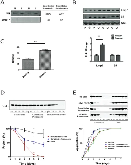
α-Synuclein aggregation is implicated in several neurodegenerative diseases, including Parkinson’s disease (PD) and dementia with Lewy Bodies (DLB). Changes in cellular signaling pathways induced by this aggregation may contribute to cell death and disease pathogenesis. To investigate this, we used quantitative proteomics to measure the relative abundance changes of the proteome and phosphoproteome in response to aggregation of endogenous α-synuclein in the brain of a mouse model. Aggregation in this model is induced by the intrastriatal injection of α-synuclein pre-formed fibrils and recapitulates several cardinal features of human PD, including progressive aggregation concomitant with dopaminergic degeneration and motor symptoms. We quantified the relative abundance changes of 5,290 proteins and 2,763 phosphosites in wildtype mice and found significant changes in vesicle-mediated transport, RNA processing and the immune response. The immunoproteasome, an altered form of the constitutive proteasome that is induced in response to stress, was elevated in response to α-synuclein aggregation. Increased levels and activity of the
immunoproteasome were found in human DLB compared with age-matched healthy controls. Additionally, the immunoproteasome degrades α-synuclein fibrils more efficiently than the constitutive proteasome. This is the first documented role of the immunoproteasome in synucleinopathies.
Degree Type
Dissertation
Degree Name
Doctor of Philosophy (PhD)
Graduate Group
Biochemistry & Molecular Biophysics
First Advisor
Harry Ischiropoulos
Keywords
Parkinson's, Proteomics, Proteostasis, Synuclein
Subject Categories
Biochemistry
1
PROTEOSTASIS RESPONSES TO ENDOGENOUS ALPHA-SYNUCLEIN AGGREGATION IN
THE BRAIN
Scott Edward Ugras
A DISSERTATION
in
Biochemistry and Molecular Biophysics
Presented to the Faculties of the University of Pennsylvania
in
Partial Fulfillment of the Requirements for the
Degree of Doctor of Philosophy
2016
Supervisor of Dissertation
__________________________________________________________
Harry Ischiropoulos, Ph.D.
Research Professor, Department of Pediatrics, Children's Hospital of Philadelphia; Department of Systems Pharmacology and Translational Therapeutics, Perelman School of Medicine, University of Pennsylvania
Graduate Group Chairperson
__________________________________________________________
Kim A. Sharp, Ph.D.
Professor, Department of Biochemistry and Biophysics
Dissertation Committee
Yair Argon, Ph.D. Professor, Department of Pathology and Laboratory Medicine (Chair)
Walter Englander, Ph.D. Professor, Department of Biochemistry and Biophysics Rahul Kohli, M.D., Ph.D. Assistant Professor, Department of Medicine
ii
PROTEOSTASIS RESPONSES TO ENDOGENOUSE ALPHA-SYNUCLEIN AGGREGATION IN THE BRAIN COPYRIGHT
2016
iii
Eppur si muove
iv
ACKNOWLEDGMENTS
First and foremost, I would like to thank Harry Ischiropoulos for his guidance over the
past several years. Harry has consistently pushed me to be the best thinker and problem
solver I can be, and has truly become a mentor to me. I look forward to continuing to
receive mentorship and guidance from him in the years to come.
I would like to thank the members of my thesis committee, including Yair Argon, Rahul
Kohli, Walter Englander, and James Petersson, who have helped me both inside and
outside of my committee meetings.
I would like to thank members of the Ischiropoulos lab who have been scientific peers
and have helped make doing research in lab fun, especially members of the ‘Synuclein
Data Club’ including Danielle Mor, Malcolm Daniels and Dick Lightfoot.
I would like to thank my friends in the BMB program who have helped make my graduate
school experience enjoyable, including Chris Yarosh, Michael Soo, Bengi Turegun, Dan
Ricketts and Eric Babiash.
I am especially grateful for the support my family has provided me throughout my life,
and particularly throughout graduate school. The older I get the more I appreciate all the
love, support and guidance I have received from all members of my family, especially my
parents, and my siblings Sandra, Steven and Stacy.
Finally I would like to thank my girlfriend Hilary. Having you in my life has taught me so
much about finding balance, being confident and pursuing my passions, things I could
not have learned in the classroom or at the bench. Thank for you challenging me to
v
ABSTRACT
PROTEOSTASIS RESPONSES TO ENDOGENOUS ALPHA-SYNUCLEIN AGGREGATION IN THE BRAIN
Scott Edward Ugras
Harry Ischiropoulos
α-Synuclein aggregation is implicated in several neurodegenerative diseases, including
Parkinson’s disease (PD) and dementia with Lewy Bodies (DLB). Changes in cellular signaling
pathways induced by this aggregation may contribute to cell death and disease pathogenesis. To
investigate this, we used quantitative proteomics to measure the relative abundance changes of
the proteome and phosphoproteome in response to aggregation of endogenous α-synuclein in
the brain of a mouse model. Aggregation in this model is induced by the intrastriatal injection of
α-synuclein pre-formed fibrils and recapitulates several cardinal features of human PD, including
progressive aggregation concomitant with dopaminergic degeneration and motor symptoms.
We quantified the relative abundance changes of 5,290 proteins and 2,763 phosphosites in
wildtype mice and found significant changes in vesicle-mediated transport, RNA processing and
the immune response. The immunoproteasome, an altered form of the constitutive proteasome
that is induced in response to stress, was elevated in response to α-synuclein aggregation.
Increased levels and activity of the immunoproteasome were found in human DLB compared
with age-matched healthy controls. Additionally, the immunoproteasome degrades α-synuclein
fibrils more efficiently than the constitutive proteasome. This is the first documented role of the
vi
TABLE OF CONTENTS
ACKNOWLEDGMENTS ... II
ABSTRACT ... VI
TABLE OF CONTENTS ... VII
LIST OF TABLES ... VIII
LIST OF FIGURES ... VIII
CHAPTER 1: INTRODUCTION ... 1
1.1 Proteostasis ... 2
1.2 Protein Misfolding in Neurodegeneration ... 7
1.3 Parkinson’s disease ... 11
References ... 12
CHAPTER 2: DYNAMIC STRUCTURAL FLEXIBILITY OF A-SYNUCLEIN ... 19
2.1 Abstract... 21
2.2 Introduction ... 21
2.3 The physiological function(s) of α-synuclein ... 23
2.4 α-Synuclein structural flexibility ... 26
2.5 Concluding remarks and perspectives. ... 37
References ... 40
CHAPTER 3 ... 53
3.1 Abstract... 55
3.2 Introduction ... 55
vii
3.4 Results ... 66
3.5 Discussion ... 75
References ... 80
CHAPTER 4: QUANTITATIVE PHOSPHOPROTEOMICS REVEALS CHANGES
IN CELLULAR SIGNALING IN RESPONSE TO ENDOGENOUS
ALPHA-SYNUCLEIN AGGREGATION ... 97
4.1 Abstract... 99
4.2 Introduction ... 99
4.3 Experimental Procedures ... 101
4.4 Results ... 103
4.5 Discussion ... 107
References ... 107
CHAPTER 5: CONCLUSIONS ... 115
5.1 Summary and Conclusions ... 116
References ... 123
LIST OF TABLES Table 3.1 List of Antibodies………...………66
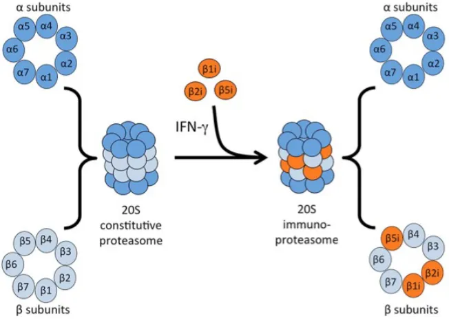
LIST OF FIGURES Figure 1.1Subunit composition of the constitutive proteasome and the immunoproteasome...…..6
Figure 2.1Primary sequence of human α-synuclein.………50
Figure 2.2 Alpha synuclein publications……...…...………51
viii
Figure 3.1 Quantitative Proteomic Workflow and Characterization of WT and Snca -/- Injected
Mice……….………..………85
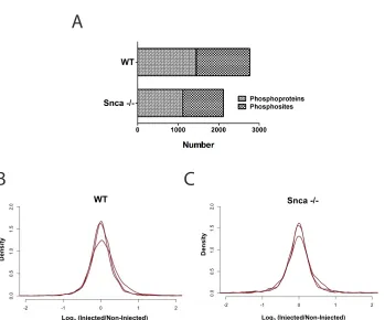
Figure 3.2 Analysis of Quantified Proteins and Identification of α-Synuclein Responsive
Proteins……….………87
Figure 3.3 Validation of Changes in Dopamine Neuron Specific Proteins……….89
Figure 3.4 Enrichment Analysis of α-Synuclein Responsive Proteins………90
Figure 3.5 The Immunoproteasome is Implicated in Human Disease Driven by α-Synuclein
Aggregation.……….………91
Figure S3.1Greater Insoluble α-Synuclein in the Injected Side than the Non-Injected Side…...93
Figure S3.2 Breakdown of 311 α-Synuclein Responsive Proteins…………..…………...…………94
Figure S3.3 Relative Abundance of α-Synuclein Does Not Change in WT………...…………94
Figure S3.4 Myelin Basic Protein is More Rapidly Degraded by the Immunoproteasome than the
Constitutive Proteasome………95
Figure S3.5Amyloid Characteristics of α-Synuclein Fibrils Used for Pure Proteasome Assay.…96
Figure 4.1 Overview of Phosphoproteomic Workflow……….………110
Figure 4.2 Phosphosites Quantified in WT and Snca -/-……….………111
Figure 4.3 Significantly Changed Phosphosites………..………112
Figure 4.4 Properties, Biological Processes and Pathways of Significantly Altered
Phosphoproteins……….……..…113
1
CHAPTER 1
2
1.1 Proteostasis
Proteins have an intrinsic ability to adopt three-dimensional conformations that are necessary
to execute proper functions1,2. Proteins may fail to adopt or maintain their proper conformation,
however, due to errors in protein folding, mutations, or changes in binding partners, pH or
concentrations3. To combat these potentially toxic events, cells have evolved diverse and
integrated cellular machinery that maintains proper protein homeostasis through various
cellular pathways. These proteostasis networks regulate protein expression, degradation,
binding partners, locations and conformations4. A key element of proteostasis is its ability to
dynamically regulate the proteome, through transcriptional and post-translational
modifications4,5.
Perturbations in the proteostasis networks, whether through age-related decline, environmental
factors, or mutations, can contribute to diseases including neurodegenerative diseases6, aging7–
9, cystic fibrosis10, cancer11,12 and Diabetes13. Intriguingly, attempts to modulate central nodes
within proteostasis networks, such as the molecular chaperone Hsp90, have shown some
therapeutic promise14. As our knowledge of proteostasis continues to advance, several other
targets will emerge that have therapeutic potential12.
The three major elements of the proteostasis network include chaperones that assist in proper
protein folding15,16, chaperone-mediated autophagy that clears misfolded proteins by
translocating them into the lysosomal lumen, and the ubiquitin-proteasome system that targets
specific proteins for degradation.
3
Despite the intrinsic ability of proteins to spontaneously adopt their native three-dimensional
conformations, molecular chaperones often assist protein folding17. The complexity of protein
folding in vivo, especially due to the crowded molecular environment within cells, increases the
likelihood of aberrant protein aggregation18. Furthermore, large, multidomain proteins have a
greater propensity to fold into off-pathway, misfolded intermediates19.
Molecular chaperones assist in protein folding without being part of the final protein structure16.
The Heat Shock Proteins (HSPs) are upregulated in times of stress or increased protein
aggregation20. Several classes of HSPs exist and have different functions. The Hsp70 family is a
group of constitutively expressed, highly conserved, ATP-dependent chaperones that assist
folding of nascent proteins emerging from the ribosome and refold misfolded protein
aggregates21,22. The Hsp90 chaperones are ATP-dependent and assist in proper protein folding
events downstream of Hsp70s and are critical in many signaling events23–26. Another class of
chaperones, chaperonins, are ATP-dependent molecular machines that include the well-studied
GroEL/ES system27,28.
1.1.2 Chaperone-Mediated Autophagy
Chaperone-mediated autophagy (CMA) is a degradation pathway that targets cytosolic proteins
for lysosomal destruction29. CMA upregulation occurs in response to oxidative stress30 and
exposure to toxic compounds31. CMA substrates typically contain the pentapeptide KFERQ that
is recognized by the Hsp70 member hsc70 when the motif is surface exposed. Hsc70 is part of
the larger complex known as the CMA cargo recognition complex32. The newly formed
4
translocation. Lysosome-associated membrane protein type 2A (LAMP-2A) is a single-span
membrane protein that participates in substrate binding and translocation into the lumen of the
lysosome29,33. The highly acidic intraluminal environment and lysosomal proteases then rapidly
degrade substrate proteins.
CMA has been implicated in human pathology, including aging34 and neurodegenerative
diseases35. Age-related disruptions in lysosomal membrane dynamics cause a decline in total
LAMP-2A levels that contribute to the decreased rate of CMA with age36,37. In Parkinson’s
disease (PD), where mutations and aggregation of α-synuclein are implicated in disease
pathology, wildtype α-synuclein but not PD-linked mutant forms can be degraded by CMA38,39.
Wildtype forms of leucine-rich repeat kinase 2 (LRKK2), another protein that is mutated in some
cases of familial PD, are efficiently degraded by CMA but PD-linked mutant forms are not40. In
Alzheimer’s disease, the protein tau aggregates in the brain and forms intracellular tangles41.
Normally, wildtype tau undergoes degradation by CMA but mutant tau undergoes differential
degradation that may contribute to AD42. These findings suggest that the CMA arm of the
proteostasis network plays a key role in maintaining functional protein networks.
1.1.3 Ubiquitin-Proteasome System
1.1.3.1 Substrate Targeting
The ubiquitin proteasome system (UPS) is the major cellular system that degrades misfolded
proteins43–45. Proteins are targeted for degradation through a serious of sequential steps
5
protein. First, the E1 Ub-activating enzyme covalently binds to Ub in an ATP-dependent process.
Then, this Ub is transferred to a cysteine residue on the E2 Ub-conjugating enzyme. Then this
activated E2 collaborates with the E3 enzyme to transfer the Ub to the target substrate. This
mono-Ub substrate can then undergo further ubiquitination resulting in a polyubiquitinated
protein. Substrates with at least four Ub are then recognized by the mature proteasome for
degradation.
1.1.3.2 Constitutive Proteasome
The proteasome is composed of a 20S core catalytic subunit and two 19S regulatory caps that
are responsible for substrate recognition46–48. The 20S core is composed of four seven-subunit
rings, including two outer rings composed of seven α subunits and two inner rings composed of
seven β subunits. These subunits are arranged in a α1-7β1-7β1-7α1-7 configuration. The β1, β2 and β5
subunits confer the catalytic activity and specificity of the proteasome. The 19S caps regulate
several steps in proteasomal degradation of substrate targets in an ATP-dependent manner,
including entry into the 20S core, disassembly of the polyubiquitin chains, and binding to
substrate46.
1.1.3.3 Immunoproteasome
The immunoproteasome is an alternative form of the constitutive proteasome that is expressed
under conditions of proinflammatory stimuli or oxidative stress49–51. In the 20S core of the
6
and Lmp7 (also known as β1i, β2i and β5i), respectively52 (Figure 1.1). β2 and MECL-1 each exhibit
trypsin-like activity and β5 and Lmp7 each exhibit chymotrypsin-like activity. Lmp2 and β1 differ,
however, in that Lmp2 exhibits chymotrypsin-like activity whereas β1 exhibits caspase-like
activity53. Assembly of the immunoproteasome by incorporation of the catalytic subunits occurs
in response to interferon gamma (IFNγ) and is 4-fold faster than assembly of the constitutive
proteasome54. Additionally, the immunoproteasome has a greater degradation efficiency of
basic proteins compared with the constitutive proteasome55. These differences result in distinct
kinetics and cleavage site preference of the immunoproteasome, resulting in the generation of
antigenic peptides that are presented to major histocompatibility (MHC) class I molecules56,57.
Figure 1.1 Subunit composition of the constitutive proteasome and the immunoproteasome.
7
The immunoproteasome has been implicated in a number of human diseases59, including
autoimmune diseases60 and neurodegenerative diseases. Mutations in the PSMB8 gene that
encodes Lmp7 have been found to cause chronic atypical neutrophilic dermatosis with
lipodystrophy and elevated temperature (CANDLE)61 and joint contractures, muscle atrophy,
microcytic anemia, and panniculitis-induced childhood-onset lipodystrophy (JMP)62.
Immunoproteasome levels and activity were found to be elevated in the brains of patients who
had Huntington’s disease63. Lmp2-positive staining was also found predominantly within
neurons and overlapped with 5% of cortical aggregates. Levels of the immunoproteasome have
been found to be increased in the Alzheimer’s disease (AD) brain compared with age-matched
healthy brain64. Subsequent studies found that this increase is predominantly found in reactive
glia that surrounds amyloid-β plaques in the AD brain65.
1.2 Protein Misfolding in Neurodegeneration
1.2.1 Spreading of Protein Aggregates
Neurodegenerative diseases include a broad-array of progressive diseases afflicting the central
nervous system (CNS)66. They are a major burden on older people and are predicted to increase
in prevalence as populations continue to age67. Though these diseases affect different neuronal
populations and manifest with different symptoms, they share a common feature of the
accumulation of insoluble protein aggregates. These culpable proteins include amyloid-β and
tau in AD, TDP43 and FUS in Amyotrophic Lateral Sclerosis (ALS), and α-synuclein in PD. These
8
While amorphous aggregates do not contain any regular structural characteristics, amyloid
fibrils are highly ordered polymers that are rich in β-sheets68. Proteins have an intrinsic ability to
adopt the amyloid conformation, though the proteostasis network prevents most from doing
so69. These fibrils contain filaments of parallel β-strands that run perpendicular to the fibrillar
axis, forming a cross-β structure70. The hydrogen bonds that form between the backbones of
these strands provide the stability that is a characteristic of amyloid fibrils. This stability makes
amyloid fibrils particularly recalcitrant to proteostasis pathways that degrade misfolded
proteins. Amyloid fibrils are generated through a self-templating process composed of a
nucleation phase followed by a growth phase71. In this process, oligomers of amyloid fibrils form
first, and then recruit soluble forms of the same protein to grow at their ends. Once a critical
nucleus is formed, growth rapidly proceeds until all available protein is consumed, resulting in a
thermodynamically stable amyloid fibril.
This self-templating process, also known as seeding, is thought to underlie the predictable
pathological spread of protein aggregates seen in neurodegenerative diseases, including AD, ALS
and PD67. Strong evidence for seeding in neurodegenerative diseases comes from studies in PD
patients who received fetal nigral transplants. In one study, human fetal neurons that were
transplanted into a 61-year old patient contained Lewy Body-like aggregates characteristic of
the PD brain only fourteen years after transplantation72. In another study, two patients who
received fetal mesencephalic dopaminergic neurons developed α-synuclein positive, Lewy
Body-like aggregates in the grafted neurons73. These studies provide evidence that in humans, protein
aggregates can spread from host to graft, supporting the theory of cell-to-cell transmission in
protein aggregation disorders. Additionally, studies in model organisms also support this theory,
9
model, preformed fibrils of α-synuclein are intrastriatally injected into the right striatum of a
non-transgenic mouse, resulting in progressive aggregation of endogenous α-synuclein
concomitant with dopaminergic degeneration and motor phenotype. Critically, injection into
α-synuclein-null mice does not result in degeneration or motor phenotype, providing evidence for
the progressive aggregation of endogenous α-synuclein as being critical for pathology. This
model of progressive aggregation of endogenous α-synuclein driven by a seeding mechanism
has been reproduced in both mice75,76 and rats77.
1.2.2 Neurodegenerative Disease
1.2.2.1 Alzheimer’s Disease
AD is the most common neurodegenerative disease worldwide, affecting over 45 million people
in 2015 and is expected to grow to 115 million people by 205078. An estimated 9.9 million new
cases of AD occur each year, and the risk of developing AD increases exponentially with age78.
Accumulation of extracellular aggregates of amyloid-β and cytoplasmic aggregates of tau are the
pathological hallmarks of AD79. There is strong evidence for the progressive spreading of
aggregates in the AD brain.
The major form of aggregated amyloid-β in the AD brain is a 42 amino acid peptide formed from
the cleavage of amyloid precursor protein (APP)80,81. Recombinant forms of this peptide form
amyloid fibrils, and these fibrils are capable of accelerating the aggregation of soluble forms of
the same protein82. Injection of brain extracts from AD patients into APP transgenic mice
10
amyloid-β aggregates induced aggregation in the APP transgenic mouse brain84. The aggregation
of tau, a protein that binds to and stabilizes microtubules, is linked to AD and other
tauopathies85,86. Injection of lysate from human brain that suffered from tauopathy into
transgenic mouse that expressed human tau resulted in the development of tau-positive
aggregates87. Collectively, these data support the notion that spreading of aggregates in the
brain contributes to the pathogenesis of AD.
1.2.2.2 Amyotrophic Lateral Sclerosis
ALS (also known as Lou Gehrig’s Disease) is a progressive motor degenerative disorder with a
prevalence of 4-6 per 100,000 that is uniformly fatal within 1-5 years of disease onset88.
Mutations in the RNA-binding proteins89 TDP4390–94 and FUS95,96 cause familial forms of ALS.
Cytoplasmic inclusions containing these proteins have been found in ALS patients, indicating
that aggregation may contribute to disease97. Recombinant TDP43 forms non-amyloid
aggregates in vitro under standard aggregating conditions98. The ALS-linked mutations Q331K
and M337V accelerate this aggregation in cell-free systems and are toxic when expressed in
cells98. Similarly, FUS also rapidly misfolds into non-amyloid, amorphous aggregates, and is toxic
when expressed in cells99. Postmortem analysis of ALS patients reveals that tissues directly
connected to the cortex develop TDP43 inclusions once the cortex does, but those tissues not
connected do not100. These findings support a role for protein aggregation and spreading in the
11
1.3 Parkinson’s disease
1.3.1 Disease Overview
PD is a neurodegenerative disorder of the CNS that afflicts over 50 million people worldwide101.
First described in an 1817 essay by the English surgeon James Parkinson102, it is currently the
second most common neurodegenerative disease worldwide103. Degeneration of dopamine
producing neurons in the substantia nigra pars compacta is a defining feature of PD104. This
degeneration contributes to both motor and non-motor symptoms that PD patients exhibit. The
five major motor symptoms of PD include bradykinesia, resting tremor, muscle rigidity, postural
change and gait. Non-motor symptoms include cognitive changes, sleep and sensory
deprivations, and constipation. PD patients may also experience dementia, particularly at later
stages of the disease105. Estimates of the prevalence of dementia with patients who are
diagnosed with PD vary, but most studies estimate 30%-40%106,107. Similar to other
neurodegenerative diseases, age is the greatest risk factor for developing PD. Approximately
10% of PD cases are familial versus 90% that are sporadic108.
1.3.2 Pathology of Parkinson’s disease
The histopathological hallmark of PD is dense, cytoplasmic inclusions known as Lewy Bodies
(LBs)104. The presence of LBs in different brain regions defines the progression of PD, starting
from the olfactory bulb in stage 1 and progressing to the neocortex in stage 6109. Though some
exceptions to this progression have been found110,111, most cases follow this staging
12
Chapter 2). The aggregation of α-synuclein and formation of LBs is thought to contribute to the
degeneration of dopamine-producing neurons in PD115. Additionally, α-synuclein mutations that
cause PD have been identified116–120, along with gene multiplications121–123. These data strongly
implicate a role for α-synuclein aggregation in the pathogenesis of PD.
1.3.3 Treatments
PD is currently incurable and no disease modifying therapies exist. Most current therapies aim
at elevating dopamine levels or potentiating dopamine pathways. The first therapy shown to be
effective at mitigating PD symptoms was levodopa in 1967124. Levodopa, a precursor to
dopamine that is able to cross the blood-brain barrier, is part of the current therapeutic regime.
It is typically administered along with carbidopa, a dopa decarboxylase inhibitor, and is often the
first line treatment for patients over 55 years of age. This regime is usually effective for 5 years
at delaying motor symptoms of PD125. For patients under 55 years of age, a dopamine agonist,
such as ropinirole or rotigotine, is typically the first line treatment. Selective type B monoamine
oxidase inhibitors can also be administered and function by decreasing the conversion of
dopamine to 3,4-Dihydroxyphenylacetic acid (DOPAC). At more advanced stages of PD, deep
brain stimulation of the subthalamic nucleus is more effective at managing symptoms in the
majority of patients compared with medical treatment alone, though the risk of adverse effects
is greater126.
References
13
2. Karplus, M., Dobson, C. M. & Andrej, S. Protein folding — a perspective from theory and experiment. Angew. Chem. Int. Ed.37, 868–893 (1998).
3. Díaz-villanueva, J. F., Díaz-molina, R. & García-gonzález, V. Protein Folding and Mechanisms of Proteostasis. Int. J. Mol. Sci.16, 17193–17230 (2015).
4. Balch, W. E., Morimoto, R. I., Dillin, A. & Kelly, J. W. Adapting Proteostasis for Disease Intervention. Science319, 916 – 919 (2008).
5. Powers, E. T. & Balch, W. E. Diversity in the origins of proteostasis networks — a driver for protein function in evolution. Nat. Rev. Mol. Cell Biol.14, 237–248 (2013).
6. Tanaka, K. & Matsuda, N. Proteostasis and neurodegeneration: The roles of proteasomal degradation and autophagy. BBA - Mol. Cell Res.1843, 197–204 (2014).
7. Douglas, P. M. & Dillin, A. Protein homeostasis and aging in neurodegeneration. J. Cell Biol.190, 719–729 (2010).
8. Kikis, E. A., Gidalevitz, T. & Morimoto, R. I. Protein Homeostasis in Models of Aging and Age-Related Conformational Disease. Adv Exp Med Biol694, 138 – 159 (2010).
9. Vilchez, D., Saez, I. & Dillin, A. Organismal ageing and age-related diseases. Nat. Commun. 5, 1–13 (2014).
10. Balch, W. E., Roth, D. M. & Hutt, D. M. Emergent Properties of Proteostasis in Managing Cystic Fibrosis. Cold Spring Harb. Perspect. Biol. 1–17 (2011).
11. Mendillo, M. L. et al. HSF1 Drives a Transcriptional Program Distinct from Heat Shock to Support Highly Malignant Human Cancers. Cell150, 549–562 (2012).
12. Powers, E. T., Morimoto, R. I., Dillin, A., Kelly, J. W. & Balch, W. E. Biological and Chemical Approaches to Diseases of Proteostasis Deficiency. Annu. Rev. Biochem.78, 959–991 (2009).
13. Back, S. H. & Kaufman, R. J. Endoplasmic Reticulum Stress and Type 2 Diabetes. Annu. Rev. Biochem.81, 767–793 (2012).
14. Trepel, J., Mollapour, M., Giaccone, G. & Neckers, L. Targeting the dynamic HSP90 complex in cancer. Nat. Rev. Cancer10, 537–549 (2010).
15. Hartl, F. U., Bracher, A. & Hayer-Hartl, M. Molecular chaperones in protein folding and proteostasis. Nature475, 324–32 (2011).
16. Kim, Y. E., Hipp, M. S., Bracher, A., Hayer-hartl, M. & Hartl, F. U. Molecular Chaperone Functions in Protein Folding and Proteostasis. Annu. Rev. Biochem.82, 323–355 (2013). 17. Doyle, S. M., Genest, O. & Wickner, S. Protein rescue from aggregates by powerful
molecular chaperone machines. Nat. Rev. Mol. Cell Biol.14, 617–629 (2013). 18. White, D. A., Buell, A. K., Knowles, T. P. J., Welland, M. E. & Dobson, C. M. Protein
Aggregation in Crowded Environments. J. Am. Chem. Soc.132, 5170–5175 (2010). 19. Herbst, R., Scha, U. & Seckler, R. Equilibrium Intermediates in the Reversible Unfolding of
Firefly (Photinus pyralis) Luciferase. J. Biol. Chem.272, 7099–7105 (1997).
20. Verghese, J., Abrams, J., Wang, Y. & Morano, K. A. Biology of the Heat Shock Response and Protein Chaperones: Budding Yeast (Saccharomyces cerevisiae) as a Model System. Microbiol. Mol. Biol. Rev.76, 115–158 (2012).
21. Mayer, M. P. & Bukau, B. Cellular and Molecular Life Sciences Hsp70 chaperones: Cellular functions and molecular mechanism. Cell. Mol. Life Sci.62, 670–684 (2005).
22. Kampinga, H. H. & Craig, E. A. The HSP70 chaperone machinery: J proteins as drivers of functional specificity. Nat. Rev. Mol. Cell Biol.11, 579 – 592 (2010).
14
24. Li, J., Soroka, J. & Buchner, J. The Hsp90 chaperone machinery: Conformational dynamics and regulation by. BBA - Mol. Cell Res.1823, 624–635 (2012).
25. Li, J. & Buchner, J. Structure , Function and Regulation of the Hsp90 Machinery. Biomed J. 36, 106–117 (2012).
26. Taipale, M., Jarosz, D. F. & Lindquist, S. HSP90 at the hub of protein homeostasis: emerging mechanistic insights. Nat. Rev. Mol. Cell Biol.11, 515–528 (2010).
27. Chen, Dong-Hua, Madan, Damian, Weaver, Jeremy, Lin, Zong, Schroder, Gunnar, Chiu, Wah, Rye, H. Visualizing GroEL / ES in the Act of Encapsulating a Folding Protein. Cell153,
1354–1365 (2008).
28. Georgescauld, F. et al. GroEL / ES Chaperonin Modulates the Mechanism and Accelerates the Rate of TIM-Barrel Domain Folding. Cell157, 922–934 (2014).
29. Kon, M. & Cuervo, A. M. Chaperone-mediated autophagy in health and disease. FEBS Lett.584, 1399–1404 (2010).
30. Kiffin, R., Christian, C., Knecht, E. & Cuervo, A. M. Activation of Chaperone-mediated Autophagy during Oxidative Stress. Mol. Biol. Cell15, 4829–4840 (2004).
31. Fred, J., Cuervo, A. N. A. M., Knecht, E. & Maria, A. Activation proteolysis of a selective pathway of lysosomal in rat liver by prolonged starvation. Am. J. Physiol. Cell Physiol. 200–208 (1995).
32. Chiang, H., SR, T., Plant, C. & Dice, J. A role for a 70-kilodalton heat shock protein in lysosomal degradation of intracellular proteins. Science246, 382–385 (1989). 33. Cuervo, A. M. & Dice, J. F. A Receptor for the Selective Uptake and Degradation of
Proteins by Lysosomes. Sci. Reports273, 501 – 503 (1996).
34. Cuervo, A. M. & Wong, E. Chaperone-mediated autophagy: roles in disease and aging. Cell Res.24, 92–104 (2013).
35. Wang, G. & Mao, Z. Chaperone-mediated autophagy: roles in neurodegeneration. Transl. Neurodegener.3, 1–7 (2014).
36. Kiffin, R. et al. Altered dynamics of the lysosomal receptor for chaperone-mediated autophagy with age. J. Cell Sci. 782–791 (2007). doi:10.1242/jcs.001073
37. Cuervo, A. M. & Dice, J. F. Age-related Decline in Chaperone-mediated Autophagy. J. Biol. Chem.275, 31505–31513 (2000).
38. Cuervo, A. M., Stefanis, L., Fredenburg, R., Lansbury, P. T. & Sulzer, D. Impaired
Degradation of Mutant α-Synuclein by Chaperone-Mediated Autophagy. Sci. Reports305,
1292 – 1295 (2004).
39. Vogiatzi, T., Xilouri, M., Vekrellis, K. & Stefanis, L. Wild Type a-Synuclein Is Degraded by Chaperone-mediated Autophagy and Macroautophagy in Neuronal Cells. J. Biol. Chem. 283, 23542–23556 (2008).
40. Orenstein, S. J. et al. Interplay of LRRK2 with chaperone-mediated autophagy. Nat. Neurosci.16, 394–406 (2013).
41. Lee, V. M., Goedert, M. & Trojanowski, J. Q. Neurodegenerative tauopathies. Annu. Rev. Neurosci.24, 1121 – 1159 (2001).
42. Kaushik, S. et al. Tau fragmentation , aggregation and clearance : the dual role of lysosomal processing. Hum. Mol. Genet.18, 4153–4170 (2009).
43. Schwartz, A. L. & Ciechanover, A. The Ubiquitin-Proteasome Pathway and Pathogenesis. Ann. Rev. Med.50, 57–74 (1999).
15
45. Wang, J. & Maldonado, M. A. The Ubiquitin-Proteasome System and Its Role in Inflammatory and Autoimmune Diseases. Cell. Mol. Immunol.3, 255–261 (2006). 46. Besche, H. C., Peth, A. & Goldberg, A. L. Getting to First Base in Proteasome Assembly.
Cell3, 26–29 (2009).
47. Murata, S., Yashiroda, H. & Tanaka, K. Molecular Mechanisms of Proteasome Assembly. Nat. Rev. Mol. Cell Biol.10, 104 – 115 (2009).
48. Xie, Y. Structure, Assembly and Homeostatic Regulation of the 26 S Proteasome. J. Mol. Cell Biol. 308–317 (2010).
49. Ciehanover, A., Hod, Y. & Rershkol, A. A heat-stable polypeptide component of an ATP-dependent proteolytic system from reiculocytes. Biochem. Biophys. Res. Commun.81,
1100–1105 (1978).
50. Etlinger, J. D. & Goldberg, A. L. A soluble ATP-dependent proteolytic system responsible for the degradation of abnormal proteins in reticulocytes. Proc. Natl. Acad. Sci. U. S. A. 74, 54–58 (1977).
51. Brown, Michael, Driscoll, James, Monaco, J. Structural and serological similarity of MHC-linked LMP and proteasome (multicatalytic proteinase) complexes. Nature353, 355 – 357 (1991).
52. Ferrington, D. A. & Gregerson, D. S. Immunoproteasomes: Structure, Function, and Antigen Presentation. Prog. Mol. Biol. Transl. Sci.109, 75–112 (2012).
53. Marques, J., Palanimurugan, R., Matias, A. C., Ramos, P. C. & Ju, R. Catalytic Mechanism and Assembly of the Proteasome. Chem. Rev.109, 1509–1536 (2009).
54. Heink, S., Ludwig, D., Kloetzel, P. & Kru, E. IFN-y-induced immune adaptation of the proteasome system is an accelerated and transient response. Proc. Natl. Acad. Sci. U. S. A.102, 9241–9246 (2005).
55. Raule, M., Cerruti, F. & Cascio, P. Enhanced rate of degradation of basic proteins by 26S immunoproteasomes. BBA - Mol. Cell Res.1843, 1942–1947 (2014).
56. Gaczynska, Maria, Rock, Kenneth, Goldberg, A. y-Interferon and expression of MHC genes regulate peptide hydrolysis by proteasomes. Nature 264 – 267 (1993).
57. Rock, K. L. et al. Inhibitors of the Proteasome Block the Degradation of Most Cell Proteins and the Generation of Peptides Presented on MHC Class I Molecules. Cell78, 761–771 (1994).
58. Mccarthy, M. K. & Weinberg, J. B. The immunoproteasome and viral infection: a complex regulator of inflammation. Front. Mol. Biol.6, 1–16 (2015).
59. Huber, E. M. & Groll, M. Inhibitors for the Immuno- and Constitutive Proteasome: Current and Future Trends in Drug Development Angewandte. Angew. Chemie51, 8708– 8720 (2012).
60. Basler, M., Mundt, S., Bitzer, A., Schmidt, C. & Groettrup, M. The immunoproteasome: a novel drug target for autoimmune diseases. Clin. Exp. Rheumatol. 74–79 (2015).
61. Liu, Y. et al. Mutations in Proteasome Subunit B Type 8 Cause Chronic Atypical
Neutrophilic Dermatosis With Lipodystrophy and Elevated Temperature With Evidence of Genetic and Phenotypic Heterogeneity. Arthritis Rheum.64, 895–907 (2012).
62. Atrophy, M. et al. An Autosomal Recessive Syndrome of Joint Contractures, Muscular Atrophy, Microcytic Anemia, and Panniculitis-Associated Lipodystrophy. JCEM95, 58–63 (2010).
16
64. Mishto, M. et al. Immunoproteasome and LMP2 polymorphism in aged and Alzheimer’s disease brains. Neurobiol. Aging27, 54–66 (2006).
65. Orre, M. et al. Reactive glia show increased immunoproteasome activity in Alzheimer’s disease. Brain136, 1415–1431 (2013).
66. Skovronsky, D. M., Lee, V. M. & Trojanowski, J. Q. Neurodegenerative Diseases: New Concepts of Pathogenesis and Their Therapeutic Implications. Annu. Rev. Pathol.1, 151 – 170 (2006).
67. Brettschneider, J., Tredici, K. Del & Lee, V. M. Spreading of pathology in
neurodegenerative diseases: a focus on human studies. Nat. Rev. Neurosci.16, 109–120 (2015).
68. Jucker, M. & Walker, L. C. Self-propagation of pathogenic protein aggregates in neurodegenerative diseases. Nature501, 45 – 51 (2013).
69. Dobson, C. M. Protein misfolding, evolution and disease. Trends Biochem. Sci.24, 329– 332 (1999).
70. Nelson, R. et al. Structure of the cross-B spine of amyloid-like fibrils. Nature435, 773– 778 (2005).
71. Wiltzius, J. J. W. et al. Molecular mechanisms for protein-encoded inheritance. Nat. Struct. Mol. Biol.16, 973–978 (2009).
72. Kordower, J. H., Chu, Y., Hauser, R. A., Freeman, T. B. & Olanow, C. W. Lewy body – like pathology in long-term embryonic nigral transplants in Parkinson’s disease. Nat. Med.14,
504–506 (2008).
73. Li, J. et al. Lewy bodies in grafted neurons in subjects with Parkinson’s disease suggest host-to-graft disease propagation. Nat. Med.14, 501–503 (2008).
74. Luk, K. C. et al. Pathological α-synuclein transmission initiates Parkinson-like neurodegeneration in nontransgenic mice. Science338, 949–53 (2012).
75. Osterberg, V. R. et al. Progressive Aggregation of Alpha-Synuclein and Selective
Degeneration of Lewy Inclusion-Bearing Neurons in a Mouse Model of Parkinsonism. Cell Rep.10, 1252–1260 (2015).
76. Sacino, A. N. et al. Brain Injection of a-Synuclein Induces Multiple Proteinopathies, Gliosis, and a Neuronal Injury Marker. J. Neurosci.34, 12368–12378 (2014).
77. Paumier, K. L. et al. Intrastriatal injection of pre-formed mouse α-synuclein fibrils into rats triggers α-synuclein pathology and bilateral nigrostriatal degeneration. Neurobiol. Dis.82, 185–199 (2015).
78. Prince, M. et al. World Alzheimer Report 2015. Alzheimer’s Dis. Int. 1 – 87 (2015). 79. Duyckaerts, C., Delatour, B. & Potier, M. Classification and basic pathology of Alzheimer
disease. Acta Neuropathol118, 5–36 (2009).
80. Iwatsubo, T., Odaka, A., Suzuki, N. & Mizusawa, H. Visualization of AB42(43) and AP40 in Senile Plaques with End-Specific AB Monoclonals: Evidence That an Initially Deposited Species Is AB42(43). Neuron13, 45–53 (1994).
81. Glenner, G. G. & Wong, C. W. Alzheimer’s Disease: Initial Report of the Purification and Charactrization of a Novel Cerebrovascular Amyloid Protein. Biochem. Biophys. Res. Commun.120, 885–890 (1984).
82. Kenyon, C. et al. Self-Propagating, Molecular-Level Polymorphism in Alzheimer’s Beta-Amyloid Fibrils. Science307, 262 – 265 (2005).
17
84. Stöhr, J. et al. Purified and synthetic Alzheimer’s amyloid beta (Aβ) prions. PNAS109,
11025 – 11030 (2012).
85. Cleveland, D. W., Hwo, S.-Y. & Kirschner, M. W. Purification of Tau, a Microtubule-associated Protein that Induces Assembly of Microtubules from Purified Tubulin. J. Mol. Biol.116, 207 – 225 (1977).
86. Ballatore, C., Lee, V. M.-Y. & Trojanowski, J. Q. Tau-mediated neurodegeneration in Alzheimer’s disease and related disorders. Nat. Rev. Neurosci.8, 663 – 672 (2007). 87. Clavaguera, F. et al. Brain homogenates from human tauopathies induce tau inclusions in
mouse brain. Proc. Natl. Acad. Sci. U. S. A.110, 9535–9540 (2013).
88. Pasinelli, P. & Brown, R. H. Molecular biology of amyotrophic lateral sclerosis: insights from genetics. Nat. Rev. Neurosci.7, 18–23 (2006).
89. Ugras, S. E. & Shorter, J. RNA-Binding Proteins in Amyotrophic Lateral Sclerosis and Neurodegeneration. Neurol. Res. Int.2012, 1 – 5 (2012).
90. Bo, R. Del et al. TARDBP (TDP-43) sequence analysis in patients with familial and sporadic ALS: identification of two novel mutations. Eur. J. Neurol.16, 727–732 (2009).
91. Deerlin, V. M. Van et al. TARDBP mutations in amyotrophic lateral sclerosis with TDP-43 neuropathology: a genetic and histopathological analysis. Lancet7, 409–416 (2008). 92. Pesiridis, G. S., Lee, V. M. & Trojanowski, J. Q. Mutations in TDP-43 link glycine-rich
domain functions to amyotrophic lateral sclerosis. Hum. Mol. Genet.18, 156–162 (2009). 93. Rutherford, N. J. et al. Novel Mutations in TARDBP (TDP-43) in Patients with Familial
Amyotrophic Lateral Sclerosis. PLoS Genet.4, 1–8 (2008).
94. Sreedharan, J. et al. TDP-43 Mutations in Familial and Sporadic Amyotrophic Lateral Sclerosis. Science319, 1668 –1672 (2008).
95. Hosler, B. A., Cortelli, P., Jong, P. J. De, Yoshinaga, Y. & Haines, J. L. Mutations in the FUS/TLS Gene on Chromosome 16 Cause Familial Amyotrophic Lateral Sclerosis. Science 323, 1205–1209 (2009).
96. Nishimura, A. L. et al. Mutations in FUS, an RNA Processing Protein, Cause Familial Amyotrophic Lateral Sclerosis Type 6. Science323, 1208 –1211 (2009).
97. Parakh, S. & Atkin, J. D. Protein folding alterations in amyotrophic lateral sclerosis. Brain Res. 1–17 (2016).
98. Johnson, B. S. et al. TDP-43 Is Intrinsically Aggregation-prone, and Amyotrophic Lateral Sclerosis-linked Mutations Accelerate Aggregation and Increase Toxicity. J. Biol. Chem. 284, 20329–20339 (2009).
99. Sun, Z. et al. Molecular Determinants and Genetic Modifiers of Aggregation and Toxicity for the ALS Disease Protein FUS / TLS. PLoS Biol.9, 1 – 25 (2011).
100. Brettschneider, J. et al. Stages of pTDP-43 Pathology in Amyotrophic Lateral Sclerosis. Ann. Neurol.74, 20–38 (2013).
101. Mortality, G. B. D. & Collaborators, D. Global, regional, and national age – sex specific all-cause and all-cause-specific mortality for 240 all-causes of death, 1990 – 2013: a systematic analysis for the Global Burden of Disease Study 2013. Lancet385, 117–171 (2014). 102. Parkinson, J. An essay on shaking palsy. Whiitngham Rowl. London (1817).
103. de Lau, L. & Breteler, N. Epidemiology of Parkinson’s disease. Lancet Neurol.6, 525–535 (2006).
104. Goedert, M., Spillantini, M. G., Tredici, K. Del & Braak, H. 100 years of Lewy pathology. Nat. Rev. Neurol.9, 13–24 (2012).
18
α-synuclein, tau and amyloid‑β pathologies. Nat. Rev. Neurosci.14, 626–636 (2013). 106. Aarsland, D., Zaccai, J. & Brayne, C. A Systematic Review of Prevalence Studies of
Dementia in Parkinson’s Disease. Mov. Disord20, 1255–1263 (2005).
107. Aarsland, D. & Kurz, M. W. The Epidemiology of Dementia Associated with Parkinson’s Disease. Brain Pathol.20, 633–639 (2010).
108. Elbaz, A. et al. Familial aggregation of Parkinson’s disease: a population-based case-control study in Europe. Neurology52, 1876 – 1882 (1999).
109. Braak, H. et al. Staging of brain pathology related to sporadic Parkinson’s disease. Neurobiol. Aging24, 197–211 (2003).
110. Braak, H. & Al, E. Pathology associated with sporadic Parkinson’s disease—where does it end? J. Neural Transm.70, 89–97 (2006).
111. Uchikado, H., Lin, W., Delucia, M. W. & Dickson, D. W. Alzheimer Disease With Amygdala Lewy Bodies: A Distinct Form of a-Synucleinopathy. J. Neuropathol. Exp. Neurol.65, 685– 697 (2006).
112. Dickson, D. W., Uchikado, H., Fujishiro, H. & Tsuboi, Y. Evidence in favour of Braak staging of Parkinson’s disease. Mov. Disord25, S78–S82 (2010).
113. Halliday, G., McCann, H. & Shepherd, C. Evaluation of the Braak hypothesis: how far can it explain the pathogenesis of Parkinson’s disease? Expert Rev. Neurother.12, 673–686 (2012).
114. Spillantini, M. et al. Alpha-Synuclein in Lewy bodies. Nature 839–840 (1997).
115. Bendor, J. T., Logan, T. P. & Edwards, R. H. The Function of a-Synuclein. Neuron79, 1044– 1066 (2013).
116. Zarranz, J. J. et al. The new mutation, E46K, of alpha-synuclein causes Parkinson and Lewy body dementia. Ann. Neurol.55, 164–73 (2004).
117. Polymeropoulos, M. H. et al. Mutation in the α-Synuclein Gene Identified in Families with Parkinson’s Disease. Sci. Reports276, 2045 – 2047 (1997).
118. Lesage, S. et al. G51D α-synuclein mutation causes a novel parkinsonian-pyramidal syndrome. Ann. Neurol.73, 459–471 (2013).
119. Appel-Cresswell, S. et al. Alpha-synuclein p.H50Q, a novel pathogenic mutation for Parkinson’s disease. Mov. Disord.28, 811–3 (2013).
120. Krüger, R. et al. Ala30Pro mutation in the gene encoding α-synuclein in Parkinson’s disease. Nat. Genet.18, 106–108 (1998).
121. Chartier-Harlin, M.-C. et al. A-synuclein locus duplication as a cause of familial Parkinson’s disease. Lancet07, 1167–1169 (2004).
122. Singleton, A. B. et al. Alpha-Synuclein locus triplication causes Parkinson’s disease. Science302, 841 (2003).
123. Ferese, R. et al. Four Copies of SNCA Responsible for Autosomal Dominant Parkinson’s Disease in Two Italian Siblings. Parkinsons. Dis.2015, 1–6 (2015).
124. Cotzias, G. C., Van Woert, M. H. & Schiffer, L. M. Aromatic amino acids and modification of parkinsonism. NEJM276, 374 – 379 (1967).
125. Fahn, S. et al. Levodopa and the progression of Parkinson’s disease. NEJM351, 2498 – 2508 (2004).
19
20
Dynamic Structural Flexibility of α-Synuclein
Danielle E. Mor1*, Scott E. Ugras2*, Malcolm J. Daniels3* and Harry Ischiropoulos1,2,3,4#
Biomedical graduate studies in 1Neuroscience, 2Biochemistry and Molecular Biophysics and 3Pharmacology, Raymond and Ruth Perelman School of Medicine at the University of
Pennsylvania, PA 19104. 4Children’s Hospital of Philadelphia Research Institute and
Departments of Pediatrics and Systems Pharmacology and Translational Therapeutics, the Raymond and Ruth Perelman School of Medicine at the University of Pennsylvania, PA 19104
*Authors contributed equally.
#to whom correspondence should be addressed. E-mail: [email protected]
21
2.1 Abstract
α-Synuclein is a conserved, abundantly expressed protein that is partially localized in
pre-synaptic terminals in the central nervous system. The precise biological function(s) and
structure of α-synuclein are under investigation. Recently, the native conformation and the
presence of naturally occurring multimeric assemblies have come under debate. These are
important deliberations because α-synuclein assembles into highly organized amyloid-like fibrils
and non-amyloid amorphous aggregates that constitute the neuronal inclusions in Parkinson’s
disease and related disorders. Therefore understanding the nature of the native and
pathological conformations is pivotal from the standpoint of therapeutic interventions that
could maintain α-synuclein in its physiological state. In this review, we will discuss the existing
evidence that define the physiological states of α-synuclein and highlight how the inherent
structural flexibility of this protein may be important in health and disease.
2.2 Introduction
α-Synuclein is a soluble protein that is highly conserved in vertebrates and abundantly
expressed in nervous tissue (Jakes et al., 1994). It was first discovered in 1988 in association
with purified synaptic vesicles from the Torpedo electric ray (Maroteaux et al., 1988). Soon
afterward α-synuclein was found to be widely distributed across the mammalian brain and
localized to presynaptic nerve terminals, suggesting functions related to neurotransmission (Iwai
et al., 1995). Independent of these reports, α-synuclein was identified as the precursor to a
hydrophobic peptide found in Alzheimer’s disease senile plaques, termed the non-Aβ
22
also dynamically regulated during song learning in zebra finch, supporting a role in synaptic
plasticity (George et al., 1995).
The discovery of a mutation in the α-synuclein gene that was associated with autosomal
dominant inheritance of Parkinson’s disease (PD) provided the impetus for a major shift in
α-synuclein research (Polymeropoulos et al., 1997). PD is a neurodegenerative disorder primarily
characterized by the loss of dopamine-producing neurons in the substantia nigra pars compacta
resulting in motor impairment. Since the original publication of the A53T mutation, several
mutations, as well as multiplications of the α-synuclein gene have been linked to PD
(Chartier-Harlin et al., 2004; Krüger et al., 1998; Lesage et al., 2013; Pasanen et al., 2014; Proukakis et al.,
2013; Singleton et al., 2003; Zarranz et al., 2004; Ferese et al. 2015) Furthermore, several
antibodies against α-synuclein robustly detect the well-known pathoanatomical features of PD,
Lewy bodies and Lewy neurites, in postmortem brain tissue from patients with sporadic PD as
well as other related neurodegenerative disorders (Baba et al., 1998; Spillantini et al., 1997;
Takeda et al., 1998). The finding that wildtype α-synuclein was detected in Lewy bodies and
Lewy neurites prompted the publication of numerous studies that investigated the biochemistry
and biology of α-synuclein. Despite the rather impressive body of work several fundamental
questions remain: What is the physiological function of α-synuclein? What is the structure of
native α-synuclein? What factors contribute to the induction of aggregation-competent
conformational states of α-synuclein? In this review, we will briefly review the evidence for the
different biological functions and discuss ongoing efforts to precisely define physiological
23
2.3 The physiological function(s) of α-synuclein
The initial studies indicated that α-synuclein is not required for neuronal development or
synapse formation, but instead may modulate synaptic activity. In rodents, α-synuclein is
detected close to the time of birth and continues to increase until one month of age, when it
reaches a steady-state level that is maintained throughout adulthood (Shibayama-Imazu et al.,
1993). Similarly, in cultured rat neurons the development of synapses precedes α-synuclein
expression and translocation to axonal terminals (Murphy et al., 2000; Withers et al., 1997). The
hypothesis that synuclein regulates synaptic activity was directly tested in mice lacking
α-synuclein. α-Synuclein null mice develop normal brain architecture and synaptic contacts, and
do not exhibit gross behavioral phenotypes (Abeliovich et al., 2000). However, subtle
abnormalities in activity-dependent neurotransmitter release have been observed. Upon
repeated stimulation, dopaminergic synapses from α-synuclein null mice sustain highly elevated
dopamine release (Abeliovich et al., 2000; Yavich et al., 2004). Functional redundancy among
α-synuclein and the other α-synuclein family members, β- and γ-α-synuclein, may account for the mild
phenotypes observed in the single knockout. In α/β-synuclein double knockout mice, synaptic
plasticity appears unaltered relative to α-synuclein single knockouts, although dopamine levels
in the striatum are reduced (Chandra et al., 2004). The importance of synucleins is particularly
highlighted by α/β/γ-synuclein triple knockouts, which have decreased life span and late-onset
synaptic dysfunction compared with wildtype mice (Burré et al., 2010; Greten-Harrison et al.,
2010). Triple knockouts in another study had motor deficits and decreased striatal dopamine,
along with abnormal dopamine neurotransmission (Anwar et al., 2011). Collectively, these
reports emphasize the important role of the synucleins in long-term synaptic maintenance and
24
Synaptic vesicle trafficking. Examination of the role of α-synuclein in the synaptic vesicle cycle
has yielded conflicting results. Depletion of α-synuclein from rodent hippocampal neurons both
in vivo and in vitro induces a significant loss of undocked synaptic vesicles, suggesting that
α-synuclein acts to replenish or maintain the resting and/or reserve vesicle pools (Cabin et al.,
2002; Murphy et al., 2000). In contrast, another study found that increasing α-synuclein in
rodent hippocampal neurons reduces the recycling pool of vesicles (Nemani et al., 2010). The
effect of α-synuclein on vesicles docked at the plasma membrane prior to exocytosis is similarly
unclear. Knockout or knockdown of α-synuclein in rodent hippocampal neurons results in either
a decrease or no change in the number of docked vesicles (Cabin et al., 2002; Murphy et al.,
2000). Conversely α-synuclein expression in PC12 cells causes an accumulation of vesicles at the
plasma membrane and impairment of exocytosis (Larsen et al., 2006). However, in mice
modestly overexpressing α-synuclein (levels are not associated with neurotoxicity), hippocampal
synapses display a redistribution of vesicles away from the active zone. The density of vesicles
in synaptic boutons is also reduced, consistent with α-synuclein-mediated inhibition of vesicle
clustering. This is supported by α-synuclein-induced defects in vesicle re-clustering following
endocytosis in rat hippocampal neurons (Nemani et al., 2010). Still, opposing results have been
obtained from yeast, in which α-synuclein expression results in massive accumulations of
vesicles that co-localize with Rab GTPases (Gitler et al., 2008; Soper et al., 2008). Likewise,
α-synuclein has been shown to restrict vesicle diffusion away from synapses in mouse
hippocampal neurons (Wang et al., 2014). Several lines of evidence, therefore, support the
participation of α-synuclein in synaptic vesicle trafficking, though the specific steps for which it
25
Chaperone-like activity and neurotransmitter release. α-Synuclein and the other synuclein
family members may act as molecular chaperones, facilitating neurotransmitter release.
Cysteine-string protein α (CSPα) is a chaperone that is essential for synaptic health; its deletion
in mice leads to a decrease in SNARE protein complexes, nerve terminal degeneration, motor
impairment and death. When expressed in CSPα-deficient mice, α-synuclein is able to rescue
this degenerative phenotype and restore levels of SNARE complexes in synaptic terminals.
Moreover, mice lacking both α-synuclein and CSPα exhibit an exacerbated phenotypic decline
(Chandra et al., 2005). These findings suggest that α-synuclein is able to complement the
activity of CSPα in promoting synapse integrity. Direct evidence for the interaction of
α-synuclein with SNARE complexes was documented by co-immunoprecipitation of α-α-synuclein
with SNARE proteins and specific binding to the vesicle-associated SNARE protein
synaptobrevin-2. In mammalian cells and purified in vitro systems, α-synuclein
dose-dependently facilitates SNARE complex assembly (Burré et al., 2010). Additional support for
chaperone-like activity includes sequence homology between α-synuclein and 14-3-3 protein
chaperones as well as the association of α-synuclein with 14-3-3 and its binding partners in rat
brain (Ostrerova et al., 1999). α-, β-, and γ-synucleins are also able to prevent the aggregation
of denatured proteins in vitro (Souza et al., 2000a), further supporting a conserved
chaperone-like function of synucleins and the existence of several protein-protein interactions that
facilitate synaptic function.
Putative role in neurotransmitter synthesis and reuptake. Published evidence indicates that
α-synuclein-mediated protein-protein interactions may modulate dopamine synthesis and
recycling. α-Synuclein may inhibit the activity of tyrosine hydroxylase (TH), the rate-limiting
26
tissue and MN9D dopaminergic cells and α-synuclein was shown to inhibit TH activity in MN9D
and PC12 cells, potentially through PP2A phosphatase-mediated reduction of serine 40
phosphorylation of TH (Peng et al., 2005; Perez et al., 2002). α-Synuclein may also interact with
and inhibit the activity of aromatic amino acid decarboxylase, which catalyzes the conversion of
L-DOPA to dopamine (Tehranian et al., 2006). Thus, α-synuclein may serve as a negative
regulator of dopamine synthesis, though further validation of these findings is necessary.
Several reports have also implicated α-synuclein in the regulation of the dopamine transporter
(DAT), though the evidence is conflicting with regards to the functional consequences. Direct
binding of α-synuclein to DAT has been demonstrated in multiple studies. However, α-synuclein
does not appear to alter DAT function, but rather in various cellular contexts can promote or
inhibit DAT trafficking to the plasma membrane (Oaks and Sidhu, 2011). Elucidating the
relationship between α-synuclein and DAT requires further investigation.
2.4 α-Synuclein structural flexibility
Primary sequence. The primary sequence of α-synuclein consists of 140 amino acids with a
predicted molecular mass of 14,460.16 Da and an isoelectric point of 4.67 (figure 1). The
sequence of α-synuclein is composed of three functionally defined domains. The N-terminal
region (amino acids 1-60) is characterized by the presence of unique and highly conserved
sequence of imperfect tandem repeats with a central consensus motif of K(A)-T(A,V)-K
(V)-E(Q,T)-G(Q)-V(A). These motifs spanning residues 10-86 are projected to form two amphipathic
α-helices and are characteristic of several proteins such as apolipoproteins that bind reversibly
to membranes (George et al., 1995; Maroteaux et al., 1988). Indeed the structure of membrane
27
arrangement with a short linking region (Ulmer et al., 2005). These helices are stabilized by
interaction with a variety of phospholipid bilayers, though α-synuclein interacts preferentially
with membranes of high curvature and an abundance of acidic phospholipids, properties
consistent with those of synaptic vesicles (Davidson et al., 1998; Zhu et al., 2003). Upon
interaction with membranes of low curvature α-synuclein adopts a distinct secondary structure
characterized by a single extended helix that includes both previously described helical domains
and the linker region (amino acids 38-44) (Ferreon et al., 2009; Georgieva et al., 2010; Trexler
and Rhoades, 2009). All known mutations associated with familial PD (A30P, E46K, H50Q, G51D,
A53E, and A53T) are found in the N-terminal domain (Krüger et al., 2008; Lesage et al., 2013;
Pasanen et al., 2014; Polymeropoulos et al., 1997; Proukakis et al., 2013; Zarranz et al., 2004).
These mutations, with the exception of G51D, A53E, and A30P, increase the propensity of
α-synuclein to form insoluble aggregates and produce morphologically distinct aggregate species
(Ghosh et al., 2014; Giasson et al., 1999; Greenbaum et al., 2005; Lesage et al., 2013;
Mahul-Mellier et al., 2015; Narhi et al., 1999). Though the precise mechanism by which these
mutations promote aggregation has not been conclusively shown, evidence implicate an
accelerated formation of oligomers (Conway et al., 2000) likely due to the destabilization of the
native N-terminal conformation (Bertoncini et al., 2005a; Burré et al., 2015; Coskuner and
Wise-Scira, 2013; Dettmer et al., 2015).
Amino acids 61-95 compose the hydrophobic NAC domain (Uéda et al., 1993). This region
contains a sequence of amino acids (71-82) necessary and sufficient for α-synuclein
self-assembly into amyloid fibrils (Giasson et al., 2001). Recently the crystal structures of residues
micro-28
electron diffraction, revealing that strands in this region stack in-register into β-sheets that are
typical of amyloid assemblies (Rodriguez et al., 2015).
The C-terminal domain (96-140) is rich in negatively charged amino acids (contains 10 glutamate
and 5 aspartate residues) and was originally proposed to be essential for maintaining the
solubility of the protein. The presence of 5 proline residues, which are known to induce turns
and disrupt secondary protein structure, suggested that this region is devoid of secondary
structure (George et al., 1995; Ulmer et al., 2005). However, the C-terminus was shown to form
transient, long-range interactions with the N-terminus resulting in the formation of multiple
compact monomeric structures (Bertoncini et al., 2005a; Dedmon et al., 2005). These
compacted structures of α-synuclein are temperature sensitive and are resistant to aggregation.
The data also indicated that at elevated temperatures the C-terminus assumes an extended
conformation that liberates N-terminal associations and enables aggregation (Bertoncini et al.,
2005b; Dedmon et al., 2005). Moreover, C-terminally truncated forms of α-synuclein aggregate
faster than full length protein (Hoyer et al., 2004; Li et al., 2005). Truncated α-synuclein has
been detected in the brains of both control (non-disease) and PD patients. Cleavage of
full-length protein at residues D115, D119, N122, D125 and Y133 was documented in α-synuclein
extracted from LBs (Anderson et al., 2006).
The C-terminus appears to be important for the interaction of α-synuclein with other proteins
and for the interaction with small molecules (Burre et al., 2012; Burré et al., 2010; Conway et al.,
2001; Mazzulli et al., 2006; Souza et al., 2000b; Woods et al., 2007). Additionally, it contains the
major sites of metal binding and post-translational modifications. Binding of iron, copper, and
other metals has been shown to influence α-synuclein function and aggregation (Uversky et al.,
29
aggregation, raising the question of metal binding at different points during the aggregation
process (Kostka et al., 2008). Cu(II) is unique among metals at accelerating aggregation of
synuclein at physiologically relevant concentrations. The sole histidine residue H50 in
α-synuclein was found to be critical for Cu(II) binding (Rasia et al., 2005) whereas other divalent
metal ions, including Mn(II), Co(II), Ni(II) and Fe(II), preferentially bind to the C-terminus of
α-synuclein at residues D121, N122, and E123 (Binolfi et al., 2006).
Post-translational modifications. α-Synuclein undergoes a number of post-translational
modifications, including N-terminal acetylation, serine and tyrosine phosphorylation, lysine
ubiquitination and tyrosine nitration (Oueslati et al., 2010; Barrett & Greenamayer 2015).
α-Synuclein purified under mild conditions is acetylated in the N-terminus. The N-terminal
acetylation may account for the formation of an oligomeric form of the protein with partial
α-helical structure (Trexler and Rhoades, 2012). However, semisynthetic production of
N-terminally acetylated α-synuclein demonstrated that modified and unmodified versions of the
protein share similar secondary structure, aggregation propensities, and membrane binding
(Fauvet et al., 2012a). NMR studies indicated that the first 12 residues undergo a chemical shift
due to terminal acetylation. This modification also appears to stabilize the helicity of the
N-terminus within the context of the full-length protein, and increases the affinity of α-synuclein
for lipids (Dikiy and Eliezer, 2014).
Mass spectrometry-based methodologies revealed that α-synuclein extracted from human Lewy
bodies was phosphorylated at S129 (Fujiwara et al., 2002). An antibody raised against
phosphorylated S129 was then used to show that α-synuclein was phosphorylated at this site
only in subjects with disease and that S129 phosphorylated α-synuclein was present only in the