University of Pennsylvania
ScholarlyCommons
Publicly Accessible Penn Dissertations
1-1-2016
Multiple Roles of Ret Signaling in Mechanosensory
Neuron Development
Michael Scott Fleming
University of Pennsylvania, [email protected]
Follow this and additional works at:http://repository.upenn.edu/edissertations
Part of theMolecular Biology Commons, and theNeuroscience and Neurobiology Commons
This paper is posted at ScholarlyCommons.http://repository.upenn.edu/edissertations/1718 For more information, please [email protected].
Recommended Citation
Fleming, Michael Scott, "Multiple Roles of Ret Signaling in Mechanosensory Neuron Development" (2016).Publicly Accessible Penn Dissertations. 1718.
Multiple Roles of Ret Signaling in Mechanosensory Neuron Development
Abstract
Somatosensation is critical for interaction with the surrounding environment. Somatosensory stimuli are detected by primary somatosensory neurons of the dorsal root ganglia and trigeminal ganglia, which detect distinct classes of stimuli, such as temperature, pain, and pressure. In Chapters 2 and 3 of this thesis, we focus on rapidly adapting low-threshold mechanoreceptors (RALTMRs), which mediate the detection of light touch. RALTMRs are molecularly defined by the early embryonic expression of the receptor tyrosine kinase Ret. Ret is required for the development of central axonal projections of RALTMRs into the dorsal spinal cord. RET responds to the glial cell line-derived family of neurotrophic factors, which activate RET in combination with GPI-linked GFRα co-receptors. In vitro, RET can be activated by co-receptor expressed in the same cell (cis signaling) or by co-receptor expressed by neighboring cells (trans signaling), but previous studies suggest that trans RET signaling may not play a physiologically relevant role in vivo. Here, we show that RET in mouse RALTMRs can be activated by both GFRα2 expressed in the same cell (cis signaling) and GFRα1 expressed by neighboring cells (trans signaling), and that trans RET signaling is sufficient for the development of RALTMR central projections in vivo. Peripherally, Ret is required for the development of vibration sensitive Pacinian corpuscle RALTMR end organs. We show that Ret mediates the neuronal expression of the ETS transcription factor Er81, which is also required for Pacinian corpuscle development, and that deficient axon/Schwann cell communication is the primary deficit in Pacinian corpuscle
development in Er81 mutant mice. Furthermore, we show that Neuregulin-1, an important mediator of axon/ Schwann cell interactions, is required for Pacinian corpuscle development, and that Er81 regulates the expression of specific Neuregulin-1 isoforms. In total, we demonstrate that RET signaling drives the development of distinct developmental processes in both the central and peripheral axonal branches of RALTMRs. In Chapter 4, we describe the expression of itch-related neuropeptides GRP and NMB and their receptors in somatosensory neurons and the dorsal spinal cord.
Degree Type
Dissertation
Degree Name
Doctor of Philosophy (PhD)
Graduate Group
Neuroscience
First Advisor
Wenqin Luo
Keywords
Development, Molecular Biology, Neuroscience, Neurotrophic Factors
Subject Categories
Molecular Biology | Neuroscience and Neurobiology
MULTIPLE ROLES OF RET SIGNALING IN MECHANOSENSORY NEURON DEVELOPMENT
Michael S. Fleming
A DISSERTATION
in
Neuroscience
Presented to the Faculties of the University of Pennsylvania
in
Partial Fulfillment of the Requirements for the Degree of Doctor of Philosophy
2016
Supervisor of Dissertation
______________
Wenqin Luo, MD, PhD
Assistant Professor of Neuroscience
Graduate Group Chairperson
_________________
Joshua I. Gold, PhD Professor of Neuroscience
Dissertation Committee:
Gregory J. Bashaw, PhD, Professor of Neuroscience Douglas J. Epstein, PhD, Professor of Genetics
MULTIPLE ROLES OF RET SIGNALING IN MECHANOSENSORY NEURON
DEVELOPMENT
COPYRIGHT
2016
iii
ACKNOWLEDGEMENT
I would like to thank my mentor, Dr. Wenqin Luo. Wenqin has been an amazingly
supportive and inspiring mentor over the past five years. Her insights and encouragement
have been crucial in performing the work in this thesis. Wenqin is a brilliant scientist and
our countless conversations about my project and science in general have been invaluable
in teaching me how to think scientifically.
I would also like to thank members of the Luo lab for insightful comments, suggestions,
and friendship. Thank you to Dr. Jingwen Niu, Dr. Lian Cui, Dr. Ishmail Abdus-Saboor,
Dr. Nathan Fried, Will Olson, Peter Dong, Kim Kridsada, and Anna Vysochan for
making the Luo lab such a great environment. Thank you to Dr. Minghong Ma and the
members of her lab for helpful comments during joint lab meetings.
Many thanks to Drs. Greg Bashaw, Doug Epstein, Steve Scherer, and Robert Heuckeroth
for serving on my thesis committee. Their expertise has been a valuable resource in
designing and interpreting experiments, and their insights in and out of committee
meetings have strengthened my thesis work in many ways.
Thank you to my family and friends for all of your support. My parents have always
supported my interest in science and I want to thank them for encouraging me to pursue
my goals. Most importantly, thank you to my wife, Melissa, for constant love, support,
iv
ABSTRACT
MULTIPLE ROLES OF RET SIGNALING IN THE DEVELOPMENT OF
MECHANOSENSORY NEURONS
Michael S. Fleming
Wenqin Luo
Somatosensation is critical for interaction with the surrounding environment.
Somatosensory stimuli are detected by primary somatosensory neurons of the dorsal root
ganglia and trigeminal ganglia, which detect distinct classes of stimuli, such as
temperature, pain, and pressure. In Chapters 2 and 3 of this thesis, we focus on rapidly
adapting low-threshold mechanoreceptors (RALTMRs), which mediate the detection of
light touch. RALTMRs are molecularly defined by the early embryonic expression of the
receptor tyrosine kinase Ret. Ret is required for the development of central axonal
projections of RALTMRs into the dorsal spinal cord. RET responds to the glial cell
line-derived family of neurotrophic factors, which activate RET in combination with
GPI-linked GFRα co-receptors. In vitro, RET can be activated by co-receptor expressed in the
same cell (cis signaling) or by co-receptor expressed by neighboring cells (trans
signaling), but previous studies suggest that trans RET signaling may not play a
physiologically relevant role in vivo. Here, we show that RET in mouse RALTMRs can
be activated by both GFRα2 expressed in the same cell (cis signaling) and GFRα1
expressed by neighboring cells (trans signaling), and that trans RET signaling is
v
is required for the development of vibration sensitive Pacinian corpuscle RALTMR end
organs. We show that Ret mediates the neuronal expression of the ETS transcription
factor Er81, which is also required for Pacinian corpuscle development, and that deficient
axon/Schwann cell communication is the primary deficit in Pacinian corpuscle
development in Er81 mutant mice. Furthermore, we show that Neuregulin-1, an
important mediator of axon/Schwann cell interactions, is required for Pacinian corpuscle
development, and that Er81 regulates the expression of specific Neuregulin-1 isoforms.
In total, we demonstrate that RET signaling drives the development of distinct
developmental processes in both the central and peripheral axonal branches of
RALTMRs. In Chapter 4, we describe the expression of itch-related neuropeptides GRP
vi
TABLE OF CONTENTS
ACKNOWLEDGEMENT………..iii
ABSTRACT………...iv
LIST OF TABLES………...viii
LIST OF ILLUSTRATIONS………...ix
CHAPTER 1: INTRODUCTION………....1
MAMALIAN SOMATOSENSATION………2
MEISSNER’S CORPUSCLES……….4
PACINIAN CORPUSCLES……….9
LANCEOLATE ENDINGS………...14
MERKEL CELLS……….…………...20
RUFFINI CORPUSCLES………...29
RET STRUCTURE AND FUNCTION………..32
RET SIGNALING………..35
ER81……….…..38
SCHWANN CELLS AND NEUREGULIN-1………...41
BOMBESIN-RELATED PEPTIDES AND ITCH……….43
CHAPTER 2: CIS AND TRANS RET SIGNALING CONTROL THE SURVIVAL AND CENTRAL PROJECTION GROWTH OF RAPIDLY ADAPTING MECHANORECEPTORS……….47
ABSTRACT……….……..48
INTRODUCTION………..……49
RESULTS……….…..52
vii
MATERIALS AND METHODS………...78
CHAPTER 3: A RET-ER81-NEUREGULIN1 SIGNLAING PATHWAY DRIVES THE DEVELOPMENT OF PACINIAN CORPUSCLES………...117
ABSTRACT……….…118
INTRODUCTION………....119
RESULTS……….121
DISCUSSION………...135
MATERIALS AND METHODS……….138
CHAPTER 4:THE MAJORITY OF DORSAL SPINAL CORD GASTRIN RELEASING PEPTIDE IS SYNTHESIZED LOCALLY WHEREAS NEUROMEDIN B IS HIGHLY EXPRESSED IN PAIN- AND ITCH-SENSING SOMATOSENSORY NEURONS………...163
ABSTRACT……….……164
INTRODUCTION………..…..165
RESULTS……….…167
DISCUSSION………...181
MATERIALS AND METHODS………...186
CHAPTER 5: CONCLUSIONS AND FUTURE DIRECTIONS……….………..207
viii
LIST OF TABLES
Chapter 2:
Table 1: VGLUT1 dorsal spinal cord staining and RA mechanoreceptor number in P7
Gfrα2 mutants………..…109
Table 2: RA mechanoreceptor central projections and cell number in E13.5 Ret, Gfrα1,
Gfrα2, and Nrtn mutants………..110
Table 3: RA mechanoreceptor central projections and cell number in E15.5 Gfrα2
mutants……….111
Table 4: RA mechanoreceptor central projections and cell number in E18.5 Ret, Gfrα2,
Gfrα1, and Gfrα1;Gfrα2 mutants……….112
Table 5: QPCR of Gfrα1 in embryonic Gfrα2 null DRGs………...113
Table 6: Quantification of axonal growth in Ret mutant DRG explants………..114
Table 7: GFP+ neuron number in Gfrα2 null and Gfrα2;TrkA double null explants…...115
ix
LIST OF ILLUSTRATIONS
Chapter 1:
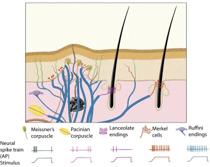
Figure 1: Illustration to demonstrate the morphologies and physiological properties of mammalian Aβ low-threshold mechanoreceptors……….46
Chapter 2:
Figure 1: P7 Gfra2 mutant mice show normal dSC VGLUT1 staining and Gfra1 null mice display normal RA mechanoreceptor central projections at E13.5………..88
Figure 2: Gfra2 and Nrtn null mice show reduced RA mechanoreceptor central
projections at E13.5………...89
Figure 3: Central projection growth deficit of Gfra2 null RA mechanoreceptors at E15.5………..90
Figure 4: Ret and Gfra2 null mice display different central projection and cell survival deficits at E18.5……….92
Figure 5: Gfra1;Gfra2 double null mice phenocopy Ret mutants at E18.5………...94
Figure 6: Gfra1 is not upregulated in Gfra2 null RA mechanoreceptors………..95
Figure 7: RA mechanoreceptors utilize GFRa1 produced by neighboring neurons to respond to GDNF………...96
Figure 8: Dynamic expression of GDNF during development………..98
Figure S1: Expression of Ret, Gfras, and Gfls in developing spinal cord and DRG…...100
Figure S2: Ret is required for the growth of RA mechanosensory 3rd order central
projections at E13.5……….102
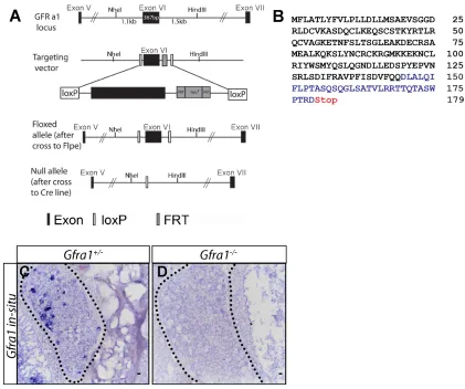
Figure S3: Generation of Gfra1 conditional and null alleles………...103
Figure S4: Generation of tandem RetCreERT;RosaTdt allele………...104
Figure S5: Gfra2 null RA mechanoreceptors retain phospho-S6 expression…………..105
Figure S6: Quantitative RT-PCR of Gfra1 in Gfra2 null DRGs……….106
Figure S7: RetCFPnull DRG explants lose responsiveness to GFLs………107
x
Chapter 3:
Figure 1: Er81 is not required for the development of Meissner’s corpuscles…………144
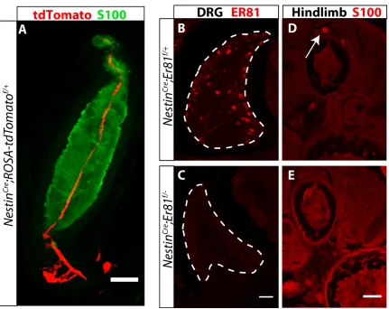
Figure 2: ER81 is expressed in Pacinian corpuscle innervating neurons………145
Figure 3: Er81 is required in neurons for Pacinian corpuscle development…………....146
Figure 4: Er81 in Schwann cells contributes to, but is not required for, Pacinian
corpuscle development……….147
Figure 5: Er81 in Schwann cells is not required for maintenance of Pacinian corpuscle following denervation………..149
Figure 6: The Ret null Pacinian corpuscle phenotype is independent of cell death…...150
Figure 7: The number of Pacinian corpuscles per limb is significantly reduced in
Ret;Er81 double heterozygotes………....151
Figure 8: Ret is required for the maintenance, but not initiation, of Er81 expression in RA mechanoreceptors………..…152
Figure 9: The Er81 Pacinian corpuscle phenotype is independent of cell death……….154
Figure 10: The primary deficit in Pacinian corpuscle formation in Er81 mutants is deficient axon/Schwann cell communication………..155
Figure 11: Interactions with myelinating Schwann cells are intact in Er81 mutant sensory neurons……….156
Figure 12: Nrg1 is required in neurons for Pacinian corpuscle formation………..157
Figure 13: Neuronal Nrg1 is not required for Meissner’s corpuscle development…….158
Figure 14: Nrg1-Ig, but not Nrg1-CRD, expression is reduced in Er81 null DRG
neurons……….159
Figure 15: Bace1 contributes to the development of Pacinian corpuscles………..160
Figure 16:Schwann cell derived Nrg1 is not required for maintenance of Pacinian
corpuscle following denervation………..…161
xi
Chapter 4:
Figure 1: Expression of Grp, Grpr, Nmb, and Nmbr mRNA in dSC and DRG
neurons……….…198
Figure 2: In vitro characterization of anti-bombesin antiserum specificity………….…199
Figure 3: Characterizing the localization of Grp in DRG, spinal cord, and dorsal root using anti-bombesin antiserum………200
Figure 4: Dorsal root rhizotomy causes a dramatic loss of Pap and Cgrp but significantly smaller changes of bombesin and Pkcγ in dSC………...202
Figure 5: Nmb is expressed in small and large diameter DRG neurons………..203
Figure 6: Nmb is expressed in peptidergic and nonpeptidergic nociceptors………204
Figure 7: Nmb is expressed in itch-sensitive DRG neurons………205
1 CHAPTER 1
Introduction
This chapter was written by Michael S. Fleming. Portions of this chapter have been
adapted from:
Fleming, MS and Luo W (2013). The anatomy, function, and development of
2 MAMALIAN SOMATOSENSATION
Light touch sensation is critical for our social interaction and daily lives. It allows for the
detection of diverse stimuli, like a breeze, a kiss, a hug, the texture of fabric, or a shape,
and is required for complex tasks, such as using a tool or reading Braille. These stimuli
cause vibration, indentation or stretching of the skin, the movement of hair follicles, or
some other physical change in the skin, which activate mechanosensory nerve fibers or
the specialized mechanosensory end organs in the skin. There are several different types
of mammalian low-threshold mechanosensory neurons (mechanoreceptors), including
Aβ, Aδ, and C (distinguished according to their transduction velocity), which mediate
various form of light touch sensation. In this introduction, I will focus on Aβ low
-threshold mechanoreceptors, which are the main type of primary sensory neurons that
mediate discriminative touch and tactile perception in mammals. Cell bodies of Aβ low
-threshold mechanoreceptors are located in the trigeminal (TGs) and dorsal root ganglia
(DRGs). Each neuron grows a single axon that bifurcates shortly after projecting from
the cell body, with the peripheral axon innervating mechanosensory end organs and the
central projection innervating the spinal cord and brain stem.
The morphologies and structures of Aβ low-threshold mechanosensory end organs have
been extensively examined since their first discovery in the 1800s. Based on
morphologies of these end organs, Aβ low-threshold mechanoreceptors are classified
3
lanceolate endings, Merkel cells, and Ruffini corpuscles (Iggo and Andres, 1982; Rice
and Albrecht, 2008). Physiological properties of Aβ low-threshold mechanoreceptors
started to be characterized around the middle of 20th century. According to their rates of
adaptation to sustained mechanical stimuli, Aβ low-threshold mechanoreceptors are
classified as either rapidly adapting (RA) or slowly adapting (SA) (Iggo, 1985;
Mountcastle, 1957). Interestingly, their end organ morphologies and physiological
properties are very well correlated. Meissner’s corpuscles, lanceolate endings, and
Pacinian corpuscles are the RA mechanoreceptors (Iggo, 1985; Iggo and Ogawa, 1977),
whereas Merkel cells and Ruffini corpuscles are the SA mechanoreceptors (Burgess,
1973; Iggo and Muir, 1969; Pare et al., 2002) (Figure 1). In the past two decades,
advances in mouse genetic techniques have enabled the dissection of key molecules,
mainly neurotrophic factors and transcription factors, involved in controlling the
specification and development of different subtypes of mammalian Aβ low-threshold
mechanoreceptors. In my thesis work, I have focused on the development of the RA
mechanoreceptors, and in particular, the Pacinian corpuscles.
Recently, a specific molecular marker which exclusively marks RA mechanoreceptors
has been identified. The receptor tyrosine kinase RET is expressed in 60% of adult
mouse DRG neurons. The majority of RET+ neurons are small diameter neurons which
begin to express Ret around E13.5 or later. However, a subset of large diameter neurons
begins to express RET around E10.5, but their function remained unknown (Molliver et
4
neurons are RA mechanoreceptors (Luo et al., 2009). Ret also has an important role in
the development of RA mechanoreceptors. In Ret mutants, central projections of RA
mechanoreceptors reach the dorsal spinal cord, but fail to extend interstitial branches to
innervate layers III-V of the dorsal horn (Bourane et al., 2009; Honma et al., 2010; Luo et
al., 2009). In Chapter 2 of this thesis, I examine the RET signaling mechanisms which
contribute to the growth of these central projections. Ret also has a role in the peripheral
development of RA mechanoreceptors. In neural crest specific Ret mutants, Pacinian
corpuscles do not form in the interosseous membrane surrounding the fibula, while
Meissner’s corpuscles and lanceolate endings appear mostly normal (Luo et al., 2009). In
Chapter 3 of this thesis, I further examine the role of Ret in the development of Pacinian
corpuscles.
MEISSNER’S CORPUSCLES
Anatomical location and morphology
Meissner’s corpuscles are present in the glabrous (hairless) skin, including palms of the
hand, soles of feet, and lips. They are located within the dermal papillae, which are
conical protrusions of the dermis into the epidermis, with their long axis perpendicular to
the surface of the skin (Munger and Ide, 1988). Meissner’s corpuscles were first
described in the fingers and palms of human hands by Georg Meissner and Rudolph
Wagner in 1852 (Cauna, 1956), but a high-resolution description of their morphology
5
and Ross, 1960; Pease and Pallie, 1959). The oval shaped corpuscle structure is
composed of disc-like lamellar stacks derived from Schwann cells, which is partially
surrounded by a thin fibroblast capsule. In most cases, the corpuscle is innervated by a
myelinated Aβ sensory fiber, which loses its myelination after traveling through the
bottom third of the corpuscle (Zelena, 1994). The sensory axon takes a circuitous route
within the corpuscle, weaving between stacks of lamellar cells. In addition to Aβ
innervation, some Meissner’s corpuscles are also innervated by C-fibers which have
molecular profiles similar to nociceptors (Pare et al., 2001), but the functional relevance
of this innervation is unclear.
A combination of physiological recording and horseradish peroxidase tracing has been
employed to reveal morphologies of the central projections of mammalian Aβ low
-threshold mechanoreceptors (Brown, 1981). Shortly after entering the spinal cord, their
central axon bifurcates and sends projections in the rostral and caudal directions in the
superficial layer of the spinal cord. Interstitial branches arise from these projections, and
innervate deeper layers of the dorsal horn (Brown, 1981). In cats, Meissner’s corpuscle
afferents synapse mostly in the medial aspect of layers III-IV of the dorsal horn, while in
rats they mostly innervate the medial aspect of layers III-V of the dorsal horn, with a
small number of synapses made in the inner part of layer II (Brown, 1981; Semba et al.,
6 Physiological properties and function
Meissner’s corpuscles are classified as type I RA (RAI) mechanoreceptors. In single unit
recordings from humans and monkeys, Meissner’s corpuscle innervating fibers display a
burst of activity upon the onset and offset of a stimulus, but remain silent during the static
phase of stimulation (Talbot et al., 1968). They have an extremely low threshold for
activation, responding to an indentation of the skin of less than 10μm (Iggo and Ogawa,
1977). In addition, Meissner’s corpuscles have relatively small receptive fields and are
most sensitive to low intensity stimuli of ~5-100Hz (Gardner and Palmer, 1990). When
stimulated at frequencies within their optimal range, Meissner’s corpuscle afferents
produce action potentials in a nearly perfect one-to-one relationship with the stimulus and
generate a “fluttering” feeling in human subjects (Talbot et al., 1968). Meissner’s
corpuscles may also function as velocity detectors to determine the rate of skin
indentation (Willis and Coggeshall, 2004).
At present, the precise mechanical transduction mechanism for Meissner’s corpuscles is
unknown. A recent study has identified the KCNQ4 potassium channel, which is
expressed in Meissner’s corpuscle innervating Aβ low-threshold mechanoreceptors, as an
important molecule to tune their sensitivity. KCNQ4 itself is not mechanically gated, but
is important for setting the resting potential of neurons. Both mice and humans with
KCNQ4 mutations display higher sensitivity to low frequency stimuli, which are
7 Development
Meissner’s corpuscle innervating axons reach the dermal papilla around birth. Schwann
cells associated with the axon begin to differentiate into the lamellar cells, which form
the corpuscle structure around one week in rodents. Innervation is required for the
development of the corpuscle structure, as corpuscles do not form if the footpad is
denervated at birth (Zelena et al., 1990).
Meissner’s corpuscles depend on the neurotrophic receptor tyrosine kinase TrkB for their
development. TrkB is expressed in a subset of RA mechanoreceptor neurons starting at
early developmental stages (Luo et al., 2009). In TrkB null mice, Meissner’s corpuscles
were not found in the dermal papilla of 2-3 week old mice (Gonzalez-Martinez et al.,
2004). Examination of micelacking the high affinity ligands of TrkB, brain derived
neurotrophic factor (BDNF) or neurotrophin-4 (NT4), revealed that BDNF, but not NT4,
is required for Meissner’s corpuscle development (Gonzalez-Martinez et al., 2005).
Since the number of DRG neurons is reduced by ~30% in TrkB and BDNF mutant mice
in the second postnatal week, it is possible that BDNF-TrkB signaling supports the
survival of Meissner’s corpuscle neurons and the loss of Meissner’s corpuscles is due to
the death of innervating neurons (Perez-Pinera et al., 2008). However, the mechanism of
TrkB signaling in controlling Meissner’s corpuscle formation could be more complicated.
8
the lamellar cells of the corpuscle, but not on the innervating axon (Calavia et al., 2010)
and the overexpression of NT4 or BDNF in the skin led to an increase in the size of
Meissner’s corpuscles, but no change in the number of sensory neurons in the DRG
(Krimm et al., 2006; LeMaster et al., 1999). In addition, myelinated axons are present in
the dermal papillae of neural crest specific TrkB mutants, even though no corpuscle
structure is formed, and the central projections of Aβ low-threshold mechanoreceptors
do not seem to be affected (Luo et al., 2009). These results suggests that at least some
Meissner’s corpuscle innervating neurons do not die in TrkB mutants, and the lack of
corpuscles may be due to a pro-survival independent function of TrkB signaling. Future
studies using tissue specific knockouts of TrkB and BDNF will be necessary to determine
the spatial and temporal requirement of TrkB and its ligands in Meissner’s corpuscle
formation.
Meissner’s corpuscles also express another neurotrophic receptor tyrosine kinase, Ret,
during early development (early RET+ RA mechanoreceptors) (Luo et al., 2009).
Surprisingly, RET signaling is not essential for Meissner’s corpuscle formation, as
corpuscles are present, although somewhat underdeveloped, in neural crest specific Ret
mutants. On the other hand, RET signaling is absolutely required for the central
projections of all RA mechanoreceptors. In Ret mutants, central projections of RA
mechanoreceptors reach the dorsal spinal cord, but fail to extend interstitial branches to
innervate layers III-V of the dorsal horn (Bourane et al., 2009; Honma et al., 2010; Luo et
9
In addition to extrinsic neurotrophic signaling, intrinsic transcriptional programs play
important roles to specify the neural identity of somatosensory neurons. One
transcription factor, Shox2, is critical for the innervation and development of Meissner’s
corpuscles. Ablation of Shox2 in mice leads to a lack of TrkB expression in two thirds of
the DRG neurons which normally express the receptor during embryonic development
(Abdo et al., 2011; Scott et al., 2011). As a result, Shox2 mutant mice lack Meissner’s
corpuscles and show reduced innervation of the dermal papillae, while the heterozygous
Shox2 adult mice display an increased threshold for light touch detection (Abdo et al.,
2011). Deletion of Shox2 also caused reduced touch sensory axonal innervation to layers
III/IV of the spinal cord (Scott et al., 2011). Additionally, two Maf family transcription
factors, MafA and c-Maf, are expressed in the RET+ RA mechanoreceptors. RET and
MafA reciprocally regulate the expression of each other, whereas c-Maf is upstream of
Ret expression (Bourane et al., 2009; Hu et al., 2012; Wende et al., 2012). In c-Maf
mutant mice, the number of Meissner’s corpuscles is drastically reduced and the
remaining corpuscles have a rudimentary structure, although the innervation of the
dermal papillae isn’t affected (Wende et al., 2012). A deficit in the formation of
Meissner’s corpuscles was not described in MafA mutant mice since these mutants were
not examined any later than postnatal day zero (P0) (Bourane et al., 2009).
10 Anatomical location and morphology
The anatomical location of Pacinian corpuscles varies greatly between species. In
primates, Pacinian corpuscles are most prominently located in the subcutaneous fat pads
of the fingers, palms, and soles (Zelena, 1994). They are also found in joints, tendons,
interosseous membrane, and around some muscles and internal organs, such as the
pancreas (Bell et al., 1994). Many early physiological and morphological studies of
Pacinian corpuscles were performed on corpuscles isolated from the cat mesentery (Sato,
1961). In rodents, Pacinian corpuscles are not present in the skin but are enriched in the
interosseous membrane around the fibula and ulna (Zelena, 1978). The Herbst corpuscle,
a structure similar to the Pacinian corpuscle, is found in beak and interosseous membrane
of birds (Saxod, 1996; Zelena et al., 1997).
Pacinian corpuscles were first described in the 18th century by Johannes Gottlieb
Lehmann, and later rediscovered by Fillipo Pacini in 1841 (Bentivoglio and Pacini,
1995). They are oval shaped end organs and can reach sizes of up to 4 mm in length in
adult human (Cauna and Mannan, 1958). Each Pacinian corpuscle is innervated by a
single myelinated Aβ somatosensory neuron, which loses its myelination and assumes a
relatively straight trajectory through the center of the corpuscle upon entering the
corpuscle’s inner core (Quilliam and Sato, 1955). Ultrastructural studies of the corpuscle
have revealed that the inner core is composed of two “hemilamellae” on either side of the
11
hemilamella of the mature corpuscle contains 40-80 layers of lamellar Schwann cells
while the outer core is composed of ~30 layers of perinurial epithelial cells (Cauna and
Mannan, 1958; Pease and Quilliam, 1957; Zelena, 1994). This layered construction
produces an onion-like appearance in cross sections of Pacinian corpuscles.
Central projections of Pacinian afferents form synapses in two distinct regions: a larger
dorsal region focused in layer III and outer layer IV with less dense innervation extending
dorsally to inner layer II and ventrally to outer layer V, and a smaller ventral region
concentrated in layer V but also sparsely innervating layers IV and VI (Brown, 1981;
Brown et al., 1980; Semba et al., 1984).
Physiological properties and function
Pacinian corpuscles are classified as type II RA (RAII) mechanoreceptors, which, like
Meissner’s corpuscles, respond to mechanical stimuli at the onset and offset of stimuli.
However, the RAI and RAII mechanoreceptors can be distinguished in two ways: 1)
RAII mechanoreceptors have larger, less defined receptive fields, suggesting a poor
ability of Pacinian corpuscles to localize stimuli (Palmer and Gardner, 1990); and 2)
RAII mechanoreceptors respond to a higher frequency of vibration, and are most
12
The precise mechanical transduction mechanism for Pacinian corpuscles is also
unknown. The RA properties of Pacinian corpuscles are partly due to the corpuscle
structure, which acts as a mechanical filter. Very low velocity or static stimuli cause
compression of the outer layers of the corpuscle, but this compression does not reach the
inner core. Instead, only the dynamic phase of compression results in deformation of the
corpuscle to the inner core, evoking a response from the innervating axon (Hubbard,
1958). This model was supported by experiments in which removing the capsule of
Pacinian corpuscles resulted in a prolonged generator potential upon a sustained
mechanical stimulus. However, despite the prolonged generator potential, the
innervating Aβ axon still only fired a few action potentials at the onset of stimulation.
This suggests that inherent properties of these Aβ axons prevents a steady outward
current from producing repetitive impulses, which is another potential mechanism
underlying the RA response (Loewenstein and Mendelson, 1965; Mendelson and
Lowenstein, 1964).
Pacinian corpuscles are exquisitely sensitive; in physiological preparations ambient
vibrations in the building resulted in a response from the innervating axon (Hunt, 1961).
These observations suggest that one potential function of Pacinian corpuscles, especially
those in the interosseous membrane, may be to sense vibration transmitted through the
skeletal system, either due to movement of the animal or due to external environmental
vibrations, possibly generated by predators or prey. In humans, Pacinian corpuscles in
13
the use of tools (Brisben et al., 1999). In addition, Pacinian corpuscles are important for
detecting the fine texture of objects. Experiments with biomimetic sensors have shown
that the normal spacing of fingerprints causes the amplification of vibrations in the ideal
detection range of Pacinian corpuscles when scanning across a finely textured surface
(Scheibert et al., 2009).
Development
Most studies regarding the development of Pacinian corpuscles have been performed on
the corpuscles in the interosseous membrane of rodents. In rats and mice, an immature
inner core and an outer capsule containing only a couple layers is present at birth (Zelena,
1994). The inner core becomes morphologically mature and more outer core layers are
added in the first postnatal week. Outer core layers continue to be added during the first
few weeks of life, and the corpuscle grows in size over the first few months. Innervation
is required for the development of the corpuscle, as Pacinian corpuscles won’t form if the
leg is neonatally denervated (Zelena et al., 1990).
Pacinian corpuscle neurons arise from the early RET+ RA mechanoreceptors and are
highly dependent on RET signaling for their development. In mice mutant for Ret, its
co-receptor Gfrα2, or its ligand Neurturin, no Pacinian corpuscles are formed (Luo et al.,
2009). The cause of the selective loss of Pacinian corpuscles, but not other subtypes of
14
In addition, several transcription factors play critical roles in the development of Pacinian
corpuscles. The ETS transcription factor Er81 is expressed in the inner core Schwann
cells of Pacinian corpuscles, and no Pacinian corpusclesare formed in Er81 null mice
(Sedy et al., 2006). In c-Maf mutant mice, both the number of Pacinian corpuscles and
axons in the interosseous nerve, which innervate Pacinian corpuscles, were greatly
reduced. In addition, the remaining corpuscles display abnormal morphology (Hu et al.,
2012; Wende et al., 2012). These results suggest that c-Maf is required in Pacinian
corpuscle neurons for axonal growth/targeting or corpuscle formation. Interestingly,
human patients with c-MAF missense mutations showed a somatosensory deficit
specifically related to Pacinian corpuscles. These patients have a decreased sensitivity to
high frequency vibration, which is detected by Pacinian corpuscles, while their detection
of lower frequencies, which is mediated by Meissner’s corpuscles, is not affected (Wende
et al., 2012).
LANCEOLATE ENDINGS
Anatomical location and morphology
The innervation of hair follicles by low-threshold mechanosensory neurons is very
complex. Aβ, Aδ, and C low-threshold mechanoreceptors all innervate hair follicles and
form a palisade structure surrounding the follicle (Li et al., 2011; Lou et al., 2013). In
15
fibers, and hair types vary by anatomical location and between species. In keeping with
the scope of this introduction, we will only discuss the Aβ low-threshold
mechanoreceptors in the hairy skin and whisker pad, which are Aβ lanceolate endings
and Merkel cells (see below). Aβ lanceolate endings are associated with the awl and
guard hairs of the hairy back skin and the whiskers, but not the zigzag hairs, which are
the most numerous hair type in mouse hairy back skin (Li et al., 2011; Mosconi et al.,
1993).
Hoggan and Hoggan originally described forked nerve endings surrounding hair follicles
in 1893 (Hoggan and Hoggan, 1893). The lanceolate endings form a palisade-like
structure which encircles the hair follicle. Individual endings have a flattened, oval
shape, with the thin aspect directly abutting the hair follicle. Each ending is composed of
a single oblong axonal fiber enclosed by flattened Schwann cells on either side of the
axon. Axonal spikes protruding through the sheath where the two Schwann cell faces
meet contact both the hair follicle and the surrounding connective tissue (Munger and
Ide, 1988). Sparse genetic labeling of hair follicle innervating axons shows that the
lanceolate endings which make up each palisade structure are innervated by more than
one sensory afferent, and that individual mechanoreceptive neurons can innervate
16
Centrally, Aβ hair follicle afferents form distinct “flame-shaped” collateral arbors, which
were first described by Ramon y Cajal and later characterized by Scheibel and Scheibel
in 1968 (Scheibel and Scheibel, 1968). Upon entering the dorsal horn, the afferent fiber
descends to layer IV or V, and then reverses direction and projects dorsally to the outer
layer IV and layer III, where the collateral undergoes extensive branching and forms
synapses with spinal neurons (Brown, 1981; Woodbury et al., 2001).
Physiological properties and function
Like Meissner’s and Pacinian corpuscles, Aβ lanceolate endings also display RA
properties. In recordings from cat whisker hair, two populations of RA afferents were
found. The more numerous population responded only to high velocity stimuli, such as a
flick of the hair or vibration from a tuning fork. This population could follow frequencies
up to 1000 Hz and was not affected by the direction of the stimulus. A much smaller low
velocity population of RA mechanoreceptors responded to stimuli from 5-200 Hz and
exhibited some direction selectivity. They responded to hair deformation in any
direction, but had a much lower activation threshold in the preferred direction
(Gottschaldt et al., 1973). The RA hair follicle afferents are also extremely sensitive; in
tests for the activation threshold using the skin nerve preparation, the activation threshold
of more than half of the units recorded was below the level of resolution (0.07 mN) in
17
Aβ lanceolate endings most likely function as velocity detectors for the deformation of
hair. The mechanical transduction mechanism is unclear, but ultrastructural studies
suggest that deformation of the lanceolate ending may occur upon movement of the hair,
due to the connections of the Schwann cell structure to the hair follicle and surrounding
tissue. This physical deformation may cause a response in the innervating axon
(Takahashi-Iwanaga, 2000).
Development
Due to the complex innervation of hair follicles by different types of sensory neurons, it
has been difficult to exclusively study the development of Aβ lanceolate endings. Most
studies described below depend on pan-neural or Schwann cell markers to identify
lanceolate endings. Therefore, the observed phenotypes in mutant mice may reflect
deficits in neurons other than Aβ low-threshold mechanoreceptors. Recently, new mouse
genetic tools have been developed to specifically label different classes of hair follicle
innervating neurons (Li et al., 2011), which will help to identify molecular mechanisms
that control development of different types of low-threshold mechanoreceptors in the
future.
The trigeminal nerve approaches the site of whisker hair follicles by embryonic day 12
(E12) in mice and the lanceolate endings start to appear at E17 (Maklad et al., 2010). In
18
reach the area of hair follicles around E14-E16 and immature lanceolate endings appear
around birth (Peters et al., 2002). These anatomical studies correlate well with
physiological recordings. Using the ex vivo skin nerve preparation, RA responses from
hair follicle innervating neurons could be recorded at P0, and backfilled neurons
displayed “flame-like” central innervation morphology typical of hair follicle receptors
by P2 (Woodbury et al., 2001).
The role of neurotrophic signaling in controlling the development of Aβ hair follicle
innervating neurons is less clear. Like the other low threshold RA mechanoreceptors, Aβ
hair follicle innervating neurons express Ret during early development. In P14 neural
crest specific Ret null mice, Luo et al. found that the morphology of lanceolate endings is
disorganized, but the percentage of hair follicles that receive lanceolate ending
innervation was not significantly different using a mixed pan-neuronal marker PGP9.5
and large diameter axon marker NFH (Luo et al., 2009). In P0 Ret null mice, Bourane et
al. found that the total innervated area of NFH+ lanceolate endings around the hair follicle
is significantly reduced (Bourane et al., 2009). Although the conclusions from these two
studies seem to be slightly different on the surface, which could well be explained by the
different staining and quantification method, both studies in fact suggest that the normal
19
Hair follicle innervation may also be dependent on TrkB signaling. In TrkB mutants, the
morphology of lanceolate endings is affected to a similar extent as seen in Ret mutants.
In addition, there was a reduction in the number of hair follicles innervated by lanceolate
endings in TrkB null mice (Perez-Pinera et al., 2008), but the number of endings was not
significantly changed in neural crest specific TrkB mutant mice (Luo et al., 2009). This
discrepancy could be due to either the differences between TrkB null and neural crest
conditional knockout mice or the technical difficulties of specifically identifying Aβ
lanceolate endings and quantifying them. On the other hand, overexpression of TrkB
ligands BDNF and NT4 in the skin led to an increased density of hair follicle innervation
(Krimm et al., 2006; LeMaster et al., 1999). Lastly, as discussed above, the transcription
factor Shox2promotes the expression of TrkB in DRG neurons. In Shox2 null mice,
lanceolate endings are disorganized, a phenotype very similar to the TrkB mutant
phenotype (Abdo et al., 2011).
The proper formation of lanceolate endings may also depend on the activity of the
innervating neurons. Woo et al. recently found that lanceolate ending innervating
neurons express the vesicular glutamate transporter VGLUT2, while the Schwann cells
which surround the innervating axons express NMDA receptors. Interestingly, ablating
VGLUT2 from somatosensory neurons leads to a reduction in the frequency and
organization of lanceolate forming Schwann cells at P0 (the latest stage the mice
survived). Moreover, pharmacologically blocking glutamatergic transmission by
20
organization of lanceolate Schwann cell processes in adult wild-type mice. Lastly,
physiological tests of antagonist treated mice revealed a decrease in the sensitivity and
conduction velocity of RA afferents innervating hair follicles (Woo et al., 2012).
Collectively, these results suggest that communication between the innervating axon and
the surrounding Schwann cell structure are crucial for the development and maintenance
of lanceolate endings.
MERKEL CELLS
Anatomical location and morphology
Merkel cells are located in the basal epidermis of both glabrous and hairy skin of
mammals. In glabrous skin, clusters of 4-40 Merkel cells are present in the epidermal
pegs, which are protrusions of the epidermis into the dermis that surrounds the dermal
papillae. In primates and marsupials, smaller clusters of Merkel cells are located at the
base of the epidermal ridges which are responsible for the fingerprint pattern of the hands
and feet (Halata et al., 2003). These clusters of Merkel cells in glabrous skin are often
referred to as “touch spots” (Boulais and Misery, 2007). In hairy skin, Merkel cells are
present in “touch domes”, which can be discerned by a slight elevation in the skin in
some species, and can contain up to 150 Merkel cells. Touch domes may or may not be
associated with a hair follicle (Zelena, 1994). In rodents, Merkel cells are closely
21
hair follicle, and whiskers, located in “cuffs” present underneath the glassy membrane of
the follicle (Halata and Munger, 1980b; Zelena, 1994).
Merkel cells were first described as “touch cells” by Freidrich Sigmund Merkel in 1875
(Merkel, 1875). They are oval in shape and 10-15 μm in length along the long axis,
which is the smallest among all mechanosensory endings discussed in this introduction.
They can be differentiated from the surrounding epidermal cells by their large,
multilobated nucleus which is oriented parallel to the dermis-epidermis junction. On the
basal side of the cell, numerous dense core vesicles measuring 70-180 nm in diameter are
located close to the cell membrane (Halata et al., 2003; Iggo and Muir, 1969; Tachibana
and Nawa, 2002; Winkelmann and Breathnach, 1973). Most Merkel cells are associated
with an innervating axon, forming a structure referred to as the Merkel cell-neurite
complex. A nerve plate, which is formed by a myelinated axon which loses its myelin
sheath upon entering the epidermis, directly opposes the vesicle dense basal membrane of
the Merkel cell. This plate is separated from the Merkel cell by 15 nm, but in small
regions they are separated by only 13 nm, and electron dense material is observed in both
the Merkel cell and the nerve plate at these points (Halata et al., 2003; Iggo and Muir,
1969). A small number of Merkel cells do not make contact with an innervating axon.
The function of these uninnervated Merkel cells is unknown, but it has been suggested
22
Central projections of Merkel cell innervating neurons bifurcate upon entering the spinal
cord, and send collaterals into the dorsal horn as these branches travel anteriorly and
posteriorly. The morphology of the collaterals innervating the dorsal horn are distinct
form the RA mechanoreceptors. Individual collaterals dive into the dorsal horn
perpendicular to the dorsal surface of the spinal cord. After reaching layer IV or V, the
collaterals make a C- or L-shaped turn and then travel medially. During and after this
turn, the collaterals gives off terminal arborizations in layers III-V (Brown, 1981).
Physiological properties and function
Merkel cell-neurite complexes are type I SA (SAI) mechanoreceptors. Unlike the RA
mechanoreceptors discussed above, SA mechanoreceptors remain active during the static
phase of stimuli. The innervating neuron is usually silent at rest, and responds to the
onset of stimulation with a burst of activity, which is proportional to the velocity and
displacement of the stimulus. After the initial phasic burst of activity, a tonic firing phase
occurs for the duration of the application of the stimulus. The firing pattern during the
tonic phase is irregular and can last for over 30 minutes (Iggo and Muir, 1969; Tapper,
1965; Willis and Coggeshall, 2004).
There has been a long-standing debate with regard to the exact role of Merkel cells in
light touch sensation. Many studies suggested that the Merkel cell is critical for
23
neurite, while other studies proposed that Merkel cells may play a modulatory role and
the neurite is primarily responsible for transducing the mechanical stimulus.
There is abundant biochemical evidence that Merkel cells produce neurotransmitters and
the machinery required for synaptic release (Haeberle et al., 2004; Maksimovic et al.,
2013; Tachibana and Nawa, 2002). Merkel cells also express voltage gated calcium
channels, and calcium induced calcium release from internal stores occurs upon the entry
of calcium into the cell, providing a potential mechanism for neurotransmitter release
(Senok and Baumann, 1997; Yamashita et al., 1992). In addition, blocking glutamatergic
transmission reduced the SA response evoked by activation of Merkel cell-neurite
complexes, suggesting that excitatory neurotransmission is required for transducing the
mechanical stimulus (Fagan and Cahusac, 2001). The most compelling evidence
suggesting a mechanosensory function of Merkel cells comes from Atoh1/Math1
conditional knockout mice, in which Merkel cells are not differentiated but the
innervating fibers are still present in touch domes. Strikingly, although the total number
of Aβ fibers is not significant changed, SAI responses could not be detected in these
animals using the ex vivo skin nerve preparation (Maricich et al., 2009). These results
suggest that Merkel cells are essential for mediating the SAI response. However, it is
unclear whether these remaining fibers completely lost their mechanosensitivity or
24
On the other hand, using both ultrastructural and electrophysiological evidence,
Gottschaldt and Vahle-Hinz argued that the ability of Merkel cell innervating neurites to
follow high frequency stimuli up to 1200 Hz with a one-to-one response for up to 500 ms
is incompatible with chemical communication, as neurotransmitter could not be released
and cleared from the synapse quickly enough to produce such a precise response. In
addition, the latency from application of stimulus to response in the innervating fiber was
too fast for chemical transmission (Gottschaldt and Vahle-Hinz, 1981), further supporting
their model that the innervating neurite acts as the mechanosensitive element.
Diamond’s group also argued that Merkel cells are dispensable for mechanosensation.
They found that touch domes were still mechanoresponsive after selective destruction of
Merkel cells using quinacrine loading and ultraviolet (UV) light irradiation (Diamond et
al., 1988). However, Ikeda et al. found that SAI responses evoked by touch dome
stimulation were lost using a different irradiation procedure to eliminate Merkel cells
(Ikeda et al., 1994). Further investigation revealed that quinacrine loading/UV irradiation
is not selective and incomplete, leaving some Merkel cells relatively intact and damaging
other nerve fibers in the skin (Senok et al., 1996). These conflicting results and technical
issues make these experiments difficult to interpret.
Others have attempted to reconcile the conflicting findings regarding the role of the
Merkel cell in mechanotransduction with a two-receptor-site model, in which both the
innervating neurite and the Merkel cell are mechanosensitive. According to this model,
25
chemical communication between the Merkel cell and the neurite (Maksimovic et al.,
2013; Ogawa, 1996). A key to resolving this debate is to develop new tools, by which
Merkel cell innervating axons can be specifically identified for physiological recording
while Merkel cells are acutely and selectively ablated.
As a result of their small sizes, Merkel cell-neurite complexes have the smallest receptive
fields among all mechanoreceptors and are best able to distinguish individual closely
spaced objects. Due to these characteristics, Merkel cells are proposed to be essential for
detecting the fine details of touched objects, such as shape, texture, and curvature
(Johnson, 2001; Johnson et al., 2000). A recent study found that mice lacking Merkel
cells were unable to detect certain textures with their feet (Maricich et al., 2012).
Development
In contrast to the RA mechanoreceptor end organs, Merkel cells appear in the skin prior
to the arrival of innervating fibers (Saxod, 1996). Immature Merkel cells can be
observed in the epidermis of the rat around E16, when innervating axons have only
reached the dermis. Axons reach the epidermis and are found in close association with
26
Since Merkel cells share characteristics with both epidermal and neural cells, there has
been argument concerning the embryological origins of Merkel cells in mammals (Lucarz
and Brand, 2007). In birds, chick-quail chimera experiments offer strong support to a
neural crest origin of Merkel cells (Grim and Halata, 2000). In addition, Merkel cells
were labeled by LacZ when all neural crest derived cells were genetically labeled using
Wnt1Cre and ROSA26R-β-Galactosidase reporter mice, suggesting that mouse Merkel cells arose from a neural crest origin (Szeder et al., 2003). However, there is also genetic
evidence to support an epidermal origin of Merkel cells. The transcription factor
Atoh1/Math1 is highly expressed and functionally required for the development of
Merkel cells. Surprisingly, Merkel cells still form when Atoh1 is conditionally ablated
from neural crest cells. In contrast, when Atoh1 is deleted from the basal layer of the
epidermis using a Keratin14Creline, Merkel cells do not form, suggesting an epidermal
origin for Merkel cells in mammals (Morrison et al., 2009). Nevertheless, this study
could not exclude a cell non-autonomous effect for Atoh1 in the development of Merkel
cells, as Atoh1 expression was also observed in some of the accessory cells surrounding
Merkel cells.
Inherent transcriptional programs in the innervating neuron are also required for
development of Merkel cell-neurite complex. Similar to Meissner’s corpuscles and
lanceolate endings, Merkel cells are dependent on the transcription factor Shox2for
sensory innervation during development. In Shox2 mutants, Merkel cells are present in
27
cells innervated by large diameter sensory fibers (Abdo et al., 2011). In addition, some
mechanoreceptive DRG neurons co-express the Runt-related transcription factors Runx1
and Runx3 (Yoshikawa et al., 2013), and the number of Merkel cell-neurite complex
surrounding the whiskers is greatly reduced in Runx3 mutant mice (Senzaki et al., 2010).
One plausible mechanism by which Runx3 controls Merkel cell-neurite complex
development is to regulate TrkC expression (Kramer et al., 2006b; Levanon et al., 2002;
Nakamura et al., 2008).
In addition to transcriptional programs, Merkel cell-neurite complexes are highly
dependent on several types of neurotrophic signaling for their development (Montano et
al., 2010). One population of Merkel cells depends on TrkA/NGF signaling. The
number of Merkel cells surrounding hair follicles and the number of innervating axons is
reduced in TrkA mutants. However, remaining Merkel cells are maintained into
adulthood, suggesting a TrkA independent Merkel cell population. Loss of the TrkA
ligand NGF produces a similar, but less severe, phenotype (Fundin et al., 1997).
TrkC/NT3 signaling has a significant and complicated effect on Merkel cell development.
TrkC is expressed in both Merkel cells and Merkel cell innervating somatosensory
neurons. In mice lacking the kinase domain of TrkC, the number of Merkel cells is
reduced at birth. In addition, those that are present at birth are not maintained during the
28
signaling postnatally (Cronk, Wilkinson et al. 2002). Loss of NT3, the TrkC ligand,
leads to a more severe deficit, with even fewer Merkel cells present at birth (Airaksinen
et al., 1996). The phenotype becomes most severe when all isoforms of TrkC are
eliminated in TrkC complete null mice, in which no Merkel cells or innervating fibers are
present at birth, suggesting additional kinase-independent roles of TrkC in Merkel cell
development (Cronk et al., 2002; Fundin et al., 1997).
TrkB signaling also has an effect on Merkel cell development. In TrkB mutant mice, the
number of Merkel cells surrounding hair follicles and in the glabrous skin is greatly
reduced (Perez-Pinera et al., 2008). When BDNF is overexpressed in the skin, the
number of Merkel cells is increased in the glabrous but not hairy skin (LeMaster et al.,
1999). Interestingly, the mechanical threshold of SAI mechanoreceptors increases eight
fold in the BDNF heterozygous and null mice, although the number and morphology of
Merkel cells was normal in touch domes of P14 BDNF null animals. This deficit could
be rescued by injecting recombinant BDNF into BDNF heterozygous mice (Carroll et al.,
1998).
Lastly, the low affinity neurotrophin receptor p75 also plays a role in Merkel cell
development. p75 can bind NGF, BDNF, NT3, and NT4, and interact with the Trk
receptors (Skaper, 2012). In p75 mutant mice, Merkel cells develop normally during the
first two postnatal weeks but then then slowly decrease in number over the following
29 RUFFINI CORPUSCLES
Anatomical location and morphology
The Ruffini corpuscle is an elongated structure with tapered ends. Morphologically, it is
quite similar to the Golgi tendon organs which are innervated by proprioceptors (Halata
and Munger, 1980a). The corpuscle is usually encased in a capsule of 4-5 layers of
perineural cells and contains an inner core of Schwann cells and collagen, which is
innervated by a single large diameter myelinated axon that loses its myelination upon
entry into the inner core. The axon gives off numerous terminal branches within the
inner core (Chambers et al., 1972; Willis and Coggeshall, 2004). Collagen fibers
associated with the inner core exit the poles of the Ruffini corpuscle and interact with
collagen in the surrounding tissue, providing a potential mechanism for mechanically
linking the inner core with the surrounding tissue (Halata, 1977).
The central projections of SA type II (SAII) mechanoreceptors, which are presumed to
innervate Ruffini corpuscles, are distinct form other mechanoreceptors. Collaterals
innervating the dorsal horn project to layer III, and then branch into at least two
processes. These processes travel deeper into the dorsal horn and branch extensively,
30
The first extensive morphological characterization of Ruffini corpuscles was performed
on hairy skin of the cat (Chambers et al., 1972). However, the anatomical location and
existence of Ruffini corpuscles between tissues and species is currently under debate. In
many cases, numerous units with SAII responses can be recorded in nerve fibers
innervating a tissue, but the Ruffini corpuscles cannot be found in the tissue following
careful histological examination. For example, physiological recordings of nerves
innervating the glabrous skin of raccoons and humans have shown a relatively high
proportion of units exhibiting SAII responses (Johansson and Vallbo, 1979; Rasmusson
and Turnbull, 1986). However, when glabrous skin from monkeys and raccoons was
examined, no Ruffini corpuscles were found (Pare et al., 2002; Rice and Rasmusson,
2000). In humans, a single Ruffini corpuscle was found in the skin of the index finger,
which is much less than what would be expected based on the physiological recordings
(Pare et al., 2003). Notably, Pare, et al. observed innervation of blood vessels which
looked morphologically similar to previous descriptions of Ruffini corpuscles. The
authors suggest that previous studies may have misidentified these structures as Ruffini
corpuscles, which could explain the discrepancy in previous findings (Pare et al., 2002).
In mouse hairy skin, SAII fibers are also identified by physiological recordings but no
definite Ruffini corpuscle structure has been reported (Wellnitz et al., 2010).
In many species, sensory endings which are morphologically similar to Ruffini
corpuscles have been identified. In monkeys and raccoons, unencapsulated Ruffini-like
31
Rasmusson, 2000). Ruffini corpuscles have also been found in association with hair
follicles, where they are sometimes referred to as pilo-Ruffini complexes (Biemesderfer
et al., 1978). Additionally, unencapsulated periodontal Ruffini-like corpuscles have been
identified surrounding the teeth of rodents (Byers, 1985).
Physiology and function
Although not well defined morphologically, the physiology of SAII Aβ low-threshold
mechanoreceptors have been extensively characterized in both humans and model
organisms (Johansson and Vallbo, 1979; Wellnitz et al., 2010). Like the SAI response,
the SAII responses is characterized by an early dynamic phase which is sensitive to both
the velocity and displacement of the stimulus, followed by a static response phase that
last throughout the application of stimulus. However, SAII responses can be
differentiated from SAI responses because they usually display some background firing
activity when no stimulus is applied, they fire at a much more regular rate during the
static phase, and their maximum frequency of the response is less than that of the SAI
response (Chambers et al., 1972).
SAII Aβ low-threshold mechanoreceptors are proposed to act primarily as stretch
receptors. In psychophysical recordings SAII units were less sensitive than SAI units to
skin indentation, but were much more sensitive to stretching of the skin (Johnson et al.,
32
combination with RA mechanoreceptors to sense movement of grasped objects. In
addition, they may work in concert with proprioceptors to sense the position of the
fingers and hand, as skin stretch will vary based on grasp (Johnson, 2001).
Development
Due to the difficulty in clearly identifying Ruffini corpuscles by morphology, relatively
little work has been done to study their development compared to the other
mechanoreceptors discussed in this review. Both periodontal Ruffini-like endings and
those associated with whisker hair are dependent on TrkB neurotrophic signaling in mice.
Periodontal Ruffini-like endings are absent in TrkB mutant mice, and mice lacking either
BDNF or NT4 show Ruffini-like endings with immature morphology (Hoshino et al.,
2003; Maruyama et al., 2005; Matsuo et al., 2002). The Ruffini-like endings of whisker
hairs are also absent in TrkB mutants. In addition, the number of whisker Ruffini-like
endings is greatly reduced in BDNF mutants but is unaffected in NT4 mutants.
Furthermore, the number of Ruffini-like endings associated with whiskers is increased in
NT3 mutantmice(Fundin et al., 1997).
RET STRUCTURE AND FUNCTION
The receptor tyrosine kinase RET (REarranged during Transfection) was first discovered
33
transfection with DNA from a human lymphoma (Takahashi et al., 1985). Since this
time, research has revealed critical roles for RET in oncological and developmental
diseases, including medullary thyroid carcinoma, multiple endocrine neoplasia, papillary
thyroid carcinoma, and Hirschsprung disease (Amiel et al., 2008; Krampitz and Norton,
2014; Romei et al., 2016). Furthermore, RET signaling has critical roles in kidney
development, spermatogenesis, and the development of enteric, sensory, autonomic, and
motor neurons (Kramer et al., 2006a; Meng et al., 2000; Runeberg-Roos and Saarma,
2007; Schuchardt et al., 1994).
The RET protein contains four n-terminal cadherin-like domains. There is a calcium
binding domain between the second and third cadherin-like domain, and calcium binding
is required for proper folding and activation of RET (Anders et al., 2001). Following the
cadherin-like domains is a cysteine-rich domain, followed by the transmembrane region.
For many years, the extracellular domains required for association with the
ligand/co-receptor complex has been controversial, with nearly all extracellular domains implicated
in complex binding (Amoresano et al., 2005; Kjaer and Ibanez, 2003). Recent structural
modeling work using low-angle X-ray scattering and electron microscopy data reconciles
these results and suggests that RET associates with its co-receptors at four distinct sites
distributed over the first three cadherin-like domains as well as the cysteine-rich domain
(Goodman et al., 2014). The transmembrane domain of RET likely contributes to the
dimerization of RET molecules, which is essential for activation of RET following ligand
34
The intracellular domain contains a short juxtamembrane region, followed by the kinase
domain and a c-terminal tail. Like other receptor tyrosine kinases, the intracellular
domain of RET undergoes autophosphorylation of tyrosine residues following
stimulation. Of the 18 intracellular tyrosine residues, 14 can be phosphorylated in vitro
(Ibanez, 2013; Kawamoto et al., 2004). Phosphorylated tyrosine residues serve as
docking sites for adapter proteins which activate downstream pathways including MAP
kinase, PI3 kinase/AKT, Jun n-terminal kinase, and Phospholipase Cγ kinase pathways
(Airaksinen et al., 1999; Borrello et al., 1996; Chiariello et al., 1998; van Weering and
Bos, 1997; Worby et al., 1996).
Following the kinase domain is a cytoplasmic tail, which produces a short isoform
(RET9) and long isoform (RET51) due to differential splicing. Both isoforms contain a
critical tyrosine residue, Y1062, which is important for RET function in vivo(Durick et
al., 1995). RET51 also contains an additional tyrosine, Y1096, which may compensate
for the loss of Y1062 in some contexts (Degl'Innocenti et al., 2004; Jain et al., 2006).
Generation of mice expressing only a single isoform of RET has produced conflicting
results. In one study, mice expressing only a human/mouse chimera RET51 had deficient
kidney development, while RET9 expressing mice had normal kidneys (de Graaff et al.,
2001). In contrast, a more recent study of monoisoformic mice expressing human
35
2006). Although RET9 and RET51 are identical up to their c-terminal tails, they are
unable to associate with or activate each other upon stimulation with ligand
(Tsui-Pierchala et al., 2002).
RET SIGNALING
RET is the receptor tyrosine kinase for the glial cell line-derived neurotrophic factor
(GDNF) family of neurotrophic factors, which includes GDNF, neurturin (NRTN),
artetmin, and persephin. RET is atypical among receptor tyrosine kinases in that it does
not directly bind to its ligands. Instead, the ligands bind to GPI-linked GDNF-family
receptor α (GFRα) co-receptors. The high affinity co-receptor/ligand pairs are
GFRα1/GDNF (Jing et al., 1996; Treanor et al., 1996), GFRα2/NRTN (Baloh et al.,
1997; Buj-Bello et al., 1997; Klein et al., 1997), GFRα3/artemin (Baloh et al., 1998), and
GFRα4/persephin (Yang et al., 2007), although there is some crosstalk between
nonmatching ligands and co-receptors, particularly at high ligand concentrations. The
kinetics of ligand/co-receptor/RET binding remains poorly understood. Originally, it was
proposed that ligand binds to GFRα co-receptor, which may exist as a dimer or may
dimerize upon ligand binding. This ligand/co-receptor complex then binds to RET,
leading to dimerization of RET and autophosphorylation of the RET intracellular domain
(Jing et al., 1996). However, binding of RET and co-receptor has been observed in the
absence of ligand, suggesting that RET and GFRα may associate prior to ligand binding