Evaluation of the Relationship between the Number of
Lymph Nodes in Resected Colorectal Cancer Specimens
and Clinicopathologic Criteria
MAHDI FARZADNIA
1, MASOOMEH SAFAEI
2*, BAHRAM MEMAR
3,
FARZANEH FARZAM
2, MAHSA AKBARI ORYANI
2, EHSAN SOLATI
4and ATIE SAFAEI
51Cancer Molecular Pathology Research Center,Imam Reza Hospital,
Faculty of Medicine,Mashhad University of Medical Sciences,Mashhad ,Iran.
2Department of Pathology,Faculty of Medicine, Mashhad University of Medical Sciences,Mashhad, Iran. 3Surgical Oncology Research Center, Faculty of Medicine,
Mashhad University of Medical Sciences,Mashhad ,Iran.
4Department of Surgery, Faculty of Medicine, Mashhad University of Medical Sciences,Mashhad, Iran. 5Department of Oral and Maxillofacial Radiology,
Shahid Sadoughi University of Medical Sciences,Yazd, Iran. DOI: http://dx.doi.org/10.13005/bpj/870
(Received: September 10, 2015; accepted: November 15, 2015) ABSTRACT
Colorectal cancer (CRC) is the most common malignancy in the digestive system and the second leading cause of cancer deaths. Lymph Nodes (LNs) involvement is the most important prognostic factor in patients with CRC. The present study was conducted to evaluate the relationship between the number of LNs in resected CRC specimens and clinicopathologic criteria in patients with CRC. the present cross-sectional study was conducted on pathology and oncology reports of 150 patients who underwent surgery for CRC in Mashhad’s Omid Hospital. The inclusion criteria were the number of resected or involved LNs, patients’ age and gender, cancer stage, tumor grade, tumor location and length of time without disease. Patients’ survival rate was calculated based on the length of time without disease. out of the whole 150 patients, 74 were male (%49.3) and 76 were female (%50.7); the average age was 56 years; %37 of the patients were under 50 years old; the average number of resected LNs was 7.16 (min:0, max: 42); no relationship was observed between the number of resected LNs and clinicopathologic criteria; however, the Lymph Node Ratio (LNR) was significantly related to the number of resected LNs (p<0.001), cancer stage (p<0.001), tumor grade (p<0.001), metastasis (p<0.001), recurrence (p<0.001) and length of time without disease (p<0.001); finally, higher LNs’ involvement was associated with lower survival rates (p<0.001). Althoughsignificant relationships were observed between LNR and prognostic criteria (recurrence, metastasis and length of time without disease), the total number of resected LNs was not significantly related to the mentioned criteria. However, a full dissection and removal of involved LNs should be done for correct staging of cancer.
Key words: Colorectal Cancer, Lymph Node, Staging of Colorectal Cancer.
INTRODUCTION
Colorectal Cancer (CRC) is the most common malignancy in the digestive system and the second leading cause of cancer deaths. Various prognostic factors have been introduced for CRC, including age, gender, stage, tumor grade, number of involved lymph nodes, etc. However, LNs
differences in the number of involved nodes among patients hospitalized in different pathology sections. These differences, which can be due to many different factors, including complete lymphadenectomy, interpersonal differences and tumor’s or host’s biological behaviors, affect both cancer staging and prognostic factors. Furthermore, different minimum numbers have been reported for LNs needed to decide about without metastasis cases.
In 2011 (China), Shao and colleagues examined the effects of number of resected LNs and LNR on prognostic factors in 507 patients with CRC (grades II & III) and reported a significant relationship between the number of resected LNs and the number of metastatic LNs. They observed a significant difference in 5-year survival rate between CRC patients with >12 involved LNs (LNs>12) and those with smaller numbers. Moreover, LNR was related to a 5-year survival rate in patients with grades II and III CRC. They concluded that more LNs must be resected for correct staging of CRC [3].
In 2011 (America), Bamboat and colleagues examined factors influencing the number of resected LNs in 137 patients with CRC who had undergone colectomy. They found no significant relationship between the type of surgery (laparoscopy/open surgery), surgeon (general surgeon/colorectal specialist), tumor location and the number of LNs removed; however, the number of LNs reported by pathology residents was significantly higher than what was reported by lower level residents. Accordingly, they concluded that careful evaluation by pathologists is an important factor in determining the accurate number of involved LNs [4].
In 2011 (Greece), Lagoudianakis and colleagues tried to determine influential factors in resecting a sufficient number of LNs in patients with CRC. Accordingly, they examined 454 CRC patients out of which, %41 had <12 involved LNs. They concluded that age, cancer stage, tumor size and other variables such as the quality of surgical specimens and accuracy of evaluation are among the important factors influencing the removal of sufficient LNs in patients with CRC [5].
In 2011(America), Stoochi and colleagues investigated factors affecting the number of nodes removed and their effects on prognostic factors. patients with CRC>12 involved LNs can be considered as an important prognostic factor in patients with colon cancer [6].
The present study was conducted to evaluate the relationship between the number of LNs and clinicopathologic criteria in patients who underwent surgery for CRC in Mashhad’s Omid Hospital.
METHODOLOGY
The present historical retrospective study was conducted on 150 patients with CRC who were hospitalized in Mashhad’s Omid Hospital between the years 2006 and 2007. The main objective was to determine the relationships between the number of LNs in resected CRC specimens and clinicopathologic criteria (age, gender, pathologic stage and tumor grade, length of time without disease and tumor location). All subjects underwent surgery for CRC and their surgical specimens were sent for pathological analysis. They had also complete pathology and oncology records. Patients with an incomplete pathology or oncology record were excluded from the study. The sample size was determined based on Stoochi’s study in which a correlation of 0.22 was reported between age and the number of LNs [6]; thus, a sample size of 150 patients was determined for the present analysis (confidence level: %95; power: %80). Data were collected based on the included patients’ medical records. Tumor’s stage was determined based on Duke’s stages (A, B, C, and D). Other data included age, gender, tumor location (ascending colon, transverse colon, descending colon and rectosigmoid colon), tumor grade (I, II and III), removed and involved LNs. Other factors such as tobacco use, CEA and hemoglobin levels were also examined. Moreover, the presence of recurrence, metastasis and number of months without disease (i.e. no tumor recurrence or metastasis) were analyzed.
Table 1: Correlation between total lymph node resesection and gender, metastasis,recurrence and tobacco use Variable Mean SD P-value
Gender Male 7.09 6.48 0.69
Female 7.22 6.51
Metastasis Yes 6.8 5.83 0.44
No 7.3 6.11
Recurrence Yes 6.91 5.8 0.66
No 7.23 6.09
Tobacco use Yes 7.82 6.18 0.46
No 7 5.98
Chi-square test results indicated no significant difference in the location of tumor (p=0.34), pathologic stage(p=0.2) and degree of differentiation (p=0.24) between male and female patients
Table 2: Correlation between total lymph node rsection and tumoral location,stage and grade
Variable Mean SD P-value Tumoral location Ascending colon 6.8 4.45 0.96
Transverse colon 7.25 6.43 Descending colon 7.4 6.38 Rectosigmoid colon 7.8 6.23
Stage A 3.5 1.29 0.06
B 5 5.7
C 8.05 6.44
D 8.4 5.27
Grade well 7.3 6.6 0.8
moderate 7.07 5.15
poor 6.46 5.15
hemoglobin and patients’ survival rate (based on disease-free survival) and between CEA and metastasis, recurrence and survival rate were studied.
Similar to Byrne’ [9] and Kim’s [20] studies, a criterion entitled Lymph Node Ratio (LNR) (i.e. the ratio of involved LNs to resected LNs) was also used in the present study. It must be noted that since no LNs was removed in 16 patients, the LNR was examined for the remaining 134 people.
To descr ibe the data, frequency distribution, percentage, mean, median and Standard Deviation (SD) were used. The examined patients were initially compared in terms of gender. Given that the numbers of resected LNs were not normally distributed, their square roots were used for data normalization (based on Smirnov-Kolmogorov test). Then to find the relationships between resected LNs and quantitative variables, person correlation test was used; to determine the relationships between resected LNs and 2-sided qualitative variables (age, gender, recurrence and tobacco use), independent samples t-test was used; and to examine the relationships between resected LNs and multiple-sided qualitative variables (stage, grade and tumor location), ANOVA was used. Since LNR was not normally distributed, its relationships with quantitative variables were examined through person correlation test; with 2-sided qualitative variables through Mann-Whitney test; and with multiple-sided qualitative variables by Kruskal-Wallis test.
Table 3: Correlations between LNR and age, duration of without disease, hemoglobin and
CEA in patients with colorectal adenocarcinoma
Variable Spearman’s P-value
rank correlation
Age -0.01 0.89
Duration of without disease -1.8 0.03
Hemoglobin -0.05 0.5
CEA 0.2 0.006
LN R
1.0 .9 .8 .7 .6 .5 .4 .3 .2 .1 0.0
D
is
e
a
s
e
F
re
e
80
60
40
20
0
Fig. 1: The relationship between LNR and disease-free survival
LN R
1.2 1.0 .8 .6 .4 .2 0.0 -.2
C
E
A
100
80
60
40
20
0 -20
Fig. 2: The relationship between LNR and CEA
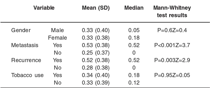
Table 4: Correlation between LNR and gender, metastasis, recurrence and tobacco use in patients with colorectal
adenocarcinoma
Variable Mean (SD) Median Mann-Whitney test results
Gender Male 0.33 (0.40) 0.05 P=0.6Z=0.4
Female 0.33 (0.38) 0.18
Metastasis Yes 0.53 (0.38) 0.52 P<0.001Z=3.7
No 0.25 (0.37) 0
Recurrence Yes 0.52 (0.38) 0.52 P=0.003Z=2.9
No 0.28 (0.38) 0
Tobacco use Yes 0.34 (0.40) 0.18 P=0.95Z=0.05
No 0.33 (0.39) 0.12
RESULTS
Out of the whole 150 patients, 74 (%49.3) people were male and 76 (%50.7) were female (the ratio of female to male patients was 1.02); the overall mean (SD) of age was 56.2 (14.8) (females: 54.4 (14.4); males: 58 (15.07)); the age range was 22-87 years; %37 of the patients were under 50 years old; the range of resected LNs was 0-42 (an
average of 7.16); in 119 (%79.3) patients, the location of tumor was rectosigmoid colon; in 18 (%12) patients, the location of tumor was ascending colon; in 8 (%5.3) patients, the location of tumor was transverse colon; in 5 (%3.3) patients, the location of tumor was descending colon; 4 (%2.7) patients had stage A cancer, 74 (%49.3) patients had stage B cancer; 67 (%44.7) patients had stage C cancer; 5 (%3.3) patients had stage D cancer; 83 (%55.3) patients had a good differentiation degree (grade I); 54 (%36) had an average differentiation degree (grade II); 13 (%7.8) patients had a weak differentiation degree (grade III); visceral metastasis was detected in 47 (%31.3) patients; in 103 (%68.6) patients, no metastasis was observed; and in 36 (%24 patients recurrence was observed.
Table 5: Correlation between LNR and cancer stage, tumor grade and tumor location in patients with colorectal adenocarcinoma
Variable Mean (SD) Median Kruskal-Wallis test results
Stage A 0 (0) 0 Chi2=65.18P<0.001
B 0.1 (0.28) 0
C 0.52 (0.35) 0.5
D 0.94 (0.07) 1
Grade I 0.23 (0.36) 0 Chi2=16.43P<0.001
II 0.39 (0.39) 0.29
III 0.72 (0.34) 0.92
Tumor location Ascending colon 0.55 (0.45) 0.62 Chi2=5.51P=0.138
Transverse colon 0.36 (0.34) 0.33 Descending colon 0.32 (0.46) 0 Rectosigmoid colon 0.30 (0.37) 0.05
Disease Free
80 60 40 20 0
Su
rv
iv
a
l
1.2
1.0
.8
.6
.4
.2
0.0
Inf iltrated Node
pos etive
negative
Fig. 3: Correlation between disease-free survival and total involved lymph nodes in
patients
Table 6: Percentage of positive node patients in terms of resected LNs
Number of Percentage of CI95%
resected LNs positive node patients
≥5 4/41 46-35
≥7 2/43 48-37
≥9 7/47 51-42
≥11 50 54-45
≥13 9/48 52-43
≥15 1/47 51-42
≥17 2/48 50-43
≥19 49 53-44
However, Chi-square test results indicated significant differences in the presence of metastasis,recurrence and tobacco use (p=0.04 , 0.07 and 0.001respectively,higher in male)
T-test results showed no significant relationship between the number of resected LNs and gender, metastasis, recurrence and tobacco use (table 1).
Anova results showed no relationship between the number of resected LN and tumoral location,stage and grade(table 2).
Table 7: Correlation between disease-free survival and gender and tobacco use variable Mean and Log-rank
SD of test results survival rate
Gender Male 34.7±4.15 P=0.03
Female 50.3 ±3.82
Tobacco use Yes 32.37 ±6.7 P=0.4 No 44.9 ±3.26
Fig. 4: Correlation between disease-free survival and gender
Di sease Free
80 60
40 20
0
Su
rv
iv
a
l
1.2
1.0
.8
.6
.4
.2
0.0
-.2
SEX
female
male
Table 8: Correlation between disease-free survival and clinicopathological criteria
Variable Mean (SD) Long-rank of survival rate test results
Age 40 e” 37.6 ±6.8 P=0.62
40< 44.46 ±3.21
Stage A 22.5 ±0.35 P<0.001
B 53.4 ±4.02
C 36.95 ±4.15
D 4 ±0.89
Grade I 48.93 ±3.71 P<0.001
II 40.39 ±4.8
III 11.89 ±2.23
Tumor location Ascending colon 24.49±7.74 P=0.01 Transverse colon 38.5±7.06
Descending colon 27.6±5.49 Rectosigmoid colon 45.61±3.31
Number of resected LNs 11dH 49.92±3.28 P=0.64
11< 45.1±6.21
LNR 0.05> 56.69±4 P<0.001
0.05-0.19 36.78±3.98
0.2-0.39 33.19±6.20
0.4-1 30.62±4.46
CEA 5d” 50.6±3.17 P<0.001
5< 18.4±3.36
negative correlation between CEA and the number of resected LNs (r=-0.16; p=0.04). ANOVA test results showed no significant relationship between the number of resected LNs and tumor location, cancer stage and tumor grade (table 3).
According to Spearman correlation test, the correlation between the number of resected LNs and the number of involved LNs was less than 0.001.
Spearman correlation test results also showed a negative correlation between the LNR and disease-free survival and a positive correlation between the LNR and CEA level (table 3).
Fig. 5: Correlation between disease-free survival and cancer stage Disease Free
80 60
40 20
0
Su
rv
iv
a
l
1.2
1.0
.8
.6
.4
.2
0.0
-.2
STAGE
D
C
B
A
Disease Free
80 60
40 20
0
Su
rv
iv
a
l
1.2
1.0
.8
.6
.4
.2
0.0
GRADE
3.00
2.00
1.00
Fig. 6: correlation between disease-free survival and tumor grade
Disease Free
80 60 40 20 0
Su
rv
iv
a
l
1.2
1.0
.8
.6
.4
.2
0.0
-.2
Location
rectosigmoi d
Descending col on
transvers e col on
asc endi ng colon
Fig. 7: Correlation between disease-free survival and tumoral location disease-free survival decreased Accordingly, the
longest disease-free survival can be seen in LNR=0 and the shortest disease-free survival in LNR=1.
The positive correlation between LNR and CEA is presented in figure (2). Mann-Whitney test results showed a significant relationship between LNR and metastasis and recurrence (table 4).
Kruskal-Wallis test results indicated significant relationships between LNR and cancer stage and tumor grade (table 5).
Based on disease-free survival, the survival rates of node positive and node negative patients were compared. The average survival rates in node negative and node positive patients were 51.75 ±3.96 and 35.39 ±4.07 months respectively (Log-rank test, p=0.002) (figure 3). Thus, survival rate was lower in node positive patients.
In the present study, when a maximum number of 11 LNs was examined, %50 of the patients with <11 LNs were positive (CI: %95,
%45-54). This ratio did not significantly increase when LNs’ numbers increased more than 11 (table 6).
Log-rank test results showed significant differences in survival rate and disease-free survival between male and female patients (table 7).
As shown in figure (4), the survival rate was higher in female patients compared to male ones.
Lon-rank test results also indicated significant relationships between survival rate (based on disease-free survival) and cancer stage, tumor grade, tumor location, LNR and CEA (table 8).
As shown in figure (5), the survival rate was lower in stage D cancer compared to other stages.
As shown in figure (6), patients’ survival rate decreased with the increase of tumor’s grade. As shown in figure (7), the survival rate was lower in patients whose tumors were located in ascending colon.
As shown in figure (8), the survival rate decreased when the LNR approached 1.
Mann-Whitney test results showed that higher levels of CEA were associated with higher incidence of recurrence and metastasis (P<0.001 and P=0.001 respectively)
As shown in figure (9), the survival rate decreased when CEA level>5ng/ml.
DISCUSSION AND CONCLUSIONS In the present study, the examined clinicopathologic criteria included the number of resected and involved LNs, age, gender, cancer stage, tumor grade, tumor location and disease-free survival.The survival rates were determined based on the disease-free survival. The incidence of CRC is similar in men and women [1]. In the present study, the ratio of female to male patients was almost equal (1.02) which was in line with the ratio obtained by Baxter [17]. However, in a study conducted by Lagoudianakis [5], the ratio of male to female patients was 1.5. The incidence of CRC increases after age 50 (the peak incidence is between 60 to 70 years of age); however, %20 of CRC cases are under 50 years old [1]. In this study, the average age of CRC was 56.21 ±14.8 (%37 under 50 years old and %16 under 40 years old). In two other Iranian studies, Jalali [22] and Moghimi [23] have reported the average ages of 51 and 53.5 years for CRC. In other parts of the world, the average ages of 66 years (Byrne [9]), 71 years (Lagoudianakis [5]) and 77 years (Baxter [17] & Moor [24]) have also been reported. It can be concluded that the average age of CRC is lower among Iranian people indicating the importance of
identification of CRC risk factors and screening at ages under 50 years old.
Given that lower survival rates and poorer prognosis are associated with younger people’s CRC [2], the examined patients in this study were divided to two age groups of <40 and >40 years and then compared. However, the results indicated no significant difference in survival rate between the two age groups. This finding was in line with the results of another Iranian study conducted by Jalali [22]. In a study conducted by Chou [24], under 40 years patients had higher cancer stage and poorer prognosis which might be due to genetic and environmental factors.
Most patients in the present study had a well differentiated grade (%53.3) and stage B CRC (%49.3). These findings were consistent with results of previous studies done by Lagoudianakis [5], Baxter [17], Jalali [22] and Moghimi [23]. The higher incidence of stage B CRC can be due to the emergence of disease symptom at this stage or lower stage estimation for lower number of resected LNs. Accordingly, more accurate staging is expected by resecting more LNs.
In the present study, the initial location of tumor was mostly in rectosigmoid colon (%79.3) which was in line with Jalali’s study [22]. The incidence of right side cancer is higher in low risk countries and the incidence of left side cancer is higher in high risk countries [22]. Iran is among the low risk countries for cancer [25].
Disease Free
80 60 40 20 0
Su
rv
iv
a
l
1.2
1.0
.8
.6
.4
.2
0.0
LNR Group
0.4-1
0.2-0.39
0.05-0.19
<0.05
Fig. 8: correlation between disease-free survival and LNR groups
Disease Free
80 60
40 20
0
Su
rv
iv
a
l
1.2
1.0
.8
.6
.4
.2
0.0
-.2
CEA
>5
<=5
In this study, patients were initially compared in terms of gender. The results showed no significant difference in tumor grade, degree of differentiation, age, and tumor location between male and female patients. Jalali [22] found no significant difference in the average age of CRC between male and female patients. Nonetheless, Zisman [20] reported lower age of onset for CRC in male patients. Moreover, in Zisman’s study, tumor location was mostly in distal colon. The prognosis of CRC is usually better in female patients [2].
In this study, the disease-free survival, defined in terms of the presence or absence of metastasis or recurrence, was 12.8 ±2.13 months. The rates of metastasis and recurrence were higher in male patients; thus, the length of disease-free survival was shorter and the survival rate was lower in male patients as well.
Hemoglobin level and rate of tobacco use were higher in male patients; but, no significant difference was observed in CEA level between male and female patients. Jalali [22] also found a significant relationship between being male and tobacco use. In some studies, including a study conducted by Zhao [12], it has been stated that tobacco use can affect the onset of CRC and patients’ survival rate. However, tobacco use did not significantly affect the patients’ survival rates in the present study, which was in line with the results of similar studies (Brian [26], Peppone [27] & Moghimi [23]). In Chen’s study [29], no significant difference was found in CEA level between male and female patients.
In this study, the range of resected LNs was 0-42 (an average of 7.16). The average numbers of resected LNs in node positive and node negative patients were 21.8 and 13.6 respectively. The results showed no significant difference in the average number of resected LNs between the two mentioned groups. The higher number of resected LNs in node positive patients was similar to the findings of Tepper [19] and Wong [31]. The higher number of resected LNs in node positive patients might be due to the following issues:
• The size of LNs might be smaller in node negative patients and their identification be harder;
• There is a linear relationship between the number of resected LNs and the number of involved LNs; thus, an increase in the number of resected LNs leads to an increase in the number of involved LNs;
In the present study, the average number of resected LNs was 7 which was similar to what Jha [16] (8 LNs) found and completely different from what Lagoudianakis [5] (13 LNs) reported.
In this study, the number of resected LNs was not significantly related to age, gender, tumor location, cancer stage, tumor grade and tobacco use.
Similar to the results of this study, the number of resected LNs was not significantly related to age, gender (Gelos [15]), tumor location (Bamboat [4]) and tumor grade (Lagoudianakis [5]) in previous studies. Conversely, Lagoudianakis [5] and Baxter [17] found a negative correlation between age and the number of resected LNs. They also reported a positive correlation between the number of resected LNs and tumor grade.
Gelos [15] found significant relationships between the number of resected LNs and tumor location and tumor grade. Shen [11] also reported a significant relationship between the number of resected LNs and tumor location which was not in line with the present study’s results.
Although the overall survival rates of the patients were not determined in the present study, the effects of various factors on the rate of survival were examined. Accordingly, the rate of survival was somehow (not statistically significant) related to disease-free survival and the number of resected LNs (p=0.07). This finding can be helpful in determining cancer stage and tumor grade more realistically. In other studies conducted by Byrne [9], Cianchi [18] and Tsikitis [13], more resected LNs led to higher survival rates. In Jha study [16], no difference was observed between the rate of survival and <9 resected LNs.
In other words, cancer stage and tumor grade were strongly related to patients’ survival rates and with the increase of stage and grade, survival rate decreased. These findings were in line with Moghimi [23] and Seicean [30] studies.
The role of tumor location in CRC prognosis is in some way controversial. Some studies have reported no significant relationship between them; while, others have focused on better prognosis of the left side cancer [2]. In this study, the rate of survival decreased when tumor was detected in ascending colon that could be due to a delayed diagnosis of CRC in the examined patients. In this study, the increase of CEA level was associated with more LNs involvement, higher recurrence and metastasis rates and lower survival rate. This finding was consistent with the results of Cheng-Jen [28] study.
In the present study, LNR was significantly related to metastasis, recurrence, survival rate, cancer stage, tumor grade and the number of resected LNs. However, it was not related to tumor location, age, gender and hemoglobin level. Similar to the present study’s results, Kim [14] found no significant relationship between LNR, age, gender and tumor location. He did not find any relationship between LNR, tumor grade and CEA level too which was not in line with the present study’s results. In both the present and Kim’s [14] studies, LNR was significantly related to tumor grade and the number of resected LNs. The results of other studies conducted by Shao [3], Byrne [9], Zhao [12] and Moghimi [23] indicated a significant relationship between the number of involved LNs and the survival rate. For that reason, many scholars have
considered the number of involved LNs as an independent major prognostic factor in patients with CRC. To reliably deter mine the absence of metastasis in CRC patients, many scientists believe that the minimum number of resected LNs must be 12; however, they have not systematically found an absolute cut off point yet. In this study, when at least 11 LNs were examined, %50 of the patients had at least one positive node. In other words, for having one positive node, at least 11 LNs were required. In this regard, the minimum optimal numbers of 14 (Tepper [29] & Wong [44]) and 9 (Jha [16]) have also been repor ted. These different numbers indicate that the determination of a minimum optimal number of LNs depends on many factors and cannot be stated with certainty.
Similar to Jha [16] and Kim [14] studies, a linear relationship was found in this study between the number of resected LNs and the number of involved LNs. This finding indicated that resecting the maximum number of LNS can be a good choice because it will lead to a better and more realistic staging of CRC.
The number of resected LNS and removal of tumors used to be considered as indicators for adequacy of surgical procedure. Previous scholars believed that at least 12 LNs are required for an appropriate staging of CRC and that resecting more LNs is associated with a better prognosis of CRC. However, some recent researchers have questioned this approach as they have not found any significant relationship between the number of resected LNs and cancer staging or survival rate. Some scholars believe that considering the number of negative LNs or the ratio of positive LNs to the total number of resected LNs may improve cancer staging.
REFERENCES
1. Anderson, D. K., Billiar, T. R., Dunn, D. L., Hunter, J. G., Matthews, J. B., Pollock, R. E., by Brunicardi, F. Schwartz’ Principles of surgery. Translated by Serati Nouri, et al. Tehran (2010).
2. RosaiJ. Large Bowel. In: Rosai and
Ackermans. Surgical pathology. 9nd ed. Mosby; p. 810-825 (2004).
Colorectal cancer. Zhonghua weichang waikezuzhi.141(4):249-530 (2011). 4. Bamboat ZM, Deperaltia D, Dursum A,
Berger DL, Boreianou L. Factors affecting lymph node yield from patient undergoing colectomy for cancer.tj Colorectal Dis. 26(9):1163-80 (2011).
5. Lagoudianakis E , Pappas A , Koronakis N, Tsekouras D, Dallianoudis J, Kontogianni P, ‘ etal “. Lymph node resecting in colorectal carcinoma specimens.Tumori; 97: 74-78 (2011).
6. Stocchi L, Fazio V, Lavery I, HammelJ. Individualsurgeon,pathologist,and other factors affecting lymph node resect in stage II colon carcinoma.Is a minimum of 12 examined lymph nodes sufficient?. Annals of Surgical Oncology. 18(2)405 (2011 ). 7. Oh TY, Moon SM, Shin US, Lee HR, Park
SH.Impact on prognosis of lymph node micrometastasis and isolated tumor cells in stage II colorecalcancer. J Korean SocColoproctal. 27(2):71-7 (2011). 8. Downing S, Cadogan K, Ortega G, Jaji Z,
Bolorunduro O, Oyetunji T, ‘ etal “.The number of lymph nodes examined debate in colon cancer: How much is enough? Journal of Surgical Research. 264-269 (2010).
9. Byrne LH, Janku F, Bird BR, Keeffee J, Murchu E, Callaghan L, ‘ etal “. The correlation between the number of lymph nodes identified and treatment outcome in colorectal cancer patient:Single-institutionexperience. JClin oncol; 28 (2010). 10. Marks JH, Valsdottir EB, Rather AA, Nweze IC, Newman DA, Chernick MR. Fewer than 12 lymph nodes can be expected in surgical specimen after high-dose radiation therapy for rectal cancer. Dis Colon Rectom; 53(7):1023-9 (2010 ).
11. Shen S, Haupt B, Ro J, Zhu J, Bailey R, Schwartz M. Number of lymph nodes examined and associated clinicopathologic factors in colorectal carcinoma. Archives of Pathology & Laboratory Medicine. 133(5):781-786 (2009).
12. Zhao Y, Li DC, Lou RC, Chen GP, Fan YT. Prognostic significance of metastatic lymph node ratio in colorectal cancer.
ZhonghuaZhong Liu ZaZhi.; 31(10):764-8 (2009).
13. Tsikitis VL, Larson DL, Wolff BG, Kennedy G, Diehl N, Qin R , ‘etal”. Survival in stage III colon cancer is independent of the total number of lymph nodes retrieved. J Am Coll Surg. 208(1):42-7 (2009 ).
14. Kim YS, Kim JH, Yoon SM, Choi EK, Ahn SD, Lee SW ‘etal”. Lymph noderatio as a prognostic factor in patients with stage III rectal cancer treated with total mesorectal excision followed by chemoradiotherapy. Int J RadiatOncolBiol Phys.74(3):796-802 (2009).
15. Gelos M, Gelhaus J, Mehnert P, Bonhag G, Sand M, Philippou S ‘etal”. Factors influencing lymph node resect in ccolorectal surgery. Int J Colorectal Dis. 23(1):53-9 (2008 ). 16. Jha M, Corbet W, Wilson R, Koreli A,
PapaqriqoriadisS. Variance of surgeon versus pathologists in staging of colorectal cancer. Minervachirurgica; 61(5):385-91 (2006).
17. Baxter N, Virnig B, Rothenberger D, Morris A, Jessurun J, Vir ing B. Lymph node evaluation in colorectal cancer patients: A population – based study .Oxford Journals; 97(3):219-225 (2004).
18. Cianchi F, Palomba A, Boddi V, Messerini L, Pucciani F, Perigli G, Bechi P, Cortesini C. Lymph node recovery from colorectal tumor specimens: recommendation for a minimum number of lymph nodes to be examined. World J Surg.; 26(3):384-9 (2002).
19. Tepper J, Connell M, Niedzwiecki D, Hollis D, Compton C, Benson A .Impact of number of nodes retrieved on outcome in patients with rectal cancer. Journal of Clinical Oncology; 19(1):157-163 (2001).
20. Zisman A, Nickolove A, Brand R, Gorchow A, Roy H. Associations Between the Age at Diagnosis and Location of Colorectal Cancer and the Use of Alcohol and Tobacco Implications for Screening. Archives of internal medicine. 166(6):629-34 (2006 ). 21. Murphy J, Pocard M, Jass JR, O’Sullivan GC,
Colon Rectum. 50(10):1526-34 (2007). 22. Jalali, M., Koradji, A., Jalali, A. (2004).
Epidemiological features of colorectal cancer in a 20-year period (1979-1999) in patients referred to Tehran’s Imam Khomeini Hospital. Iran University of Medical Sciences Journal. 11th year, (43), 723-730.
23. Moghimi, B., Safaei, A., Zali, M. Evaluation of survival rates and prognostic factors in patients with colorectal cancer. Ilam University of Medical Sciences Journal. Volume XVI, (1), 33-42 (2008).
24. Moore J, Hyman N, Callus P, Littenberg B. Staging error does not explain the relationship between the number of lymph node in a colon cancer specimen and survival. Surgery.147(3):358-365 (2010). 25. Ansari R, Mahdavinia M, Sajadi A, Nouraie
M, Kamangar F, Bishehsari F, ‘ etal “.Incidence and age,distribution of colorectal cancer in Iran: Results of a population based cancer registry. Cancer letters. 240(1):143-147 (2006).
26. Brian C, Lynch C, Cerhan J, Cantor K.Cigarette smoking and risk of Bladder, Pancreas,Kidney. 11(1):28-37.
27. Peppone L, Hyland A, Moysich K, Reid M, Piazza K, Purnell J, ‘etal”.Examining the
association between cigarette smoking and colorectal cancer using historical case-control data. Cancer Epidemiology.; 33(3-4):182-188 (2009 ).
28. Cheng-Jen M, Jan-Sing H, Wen-Ming W, Yu-Chung SU, Che-Jen H, Tsung-Jen H‘etal”. Multivariate Analysis of Prognostic Determinants for Colorectal Cancer Patients with High Preoperative Serum CEA Levels: Prognostic Value of Postoperative Serum CEA Levels. The Kaohsiung Journal of Medical Sciences.22(12):604-609 (2006). 29. ChenZK, Ouyang ZT. Relationship between
carcinoembryonic antigen and cyclooxygenase 2 expression and colorectal cancer. Ai Zheng. 22(2):164-7 (2003). 30. Seicean R, Funariu G, Seicean A, Mocan T,
Ciuce C. Results and prognostic factors in rectal cancer surgically treated with curative intent—experience of a single tertiary center.Chirurgia(Bucure). 106(3):333-40 (2011 ).