* To whom all correspondence should be addressed. E-mail: [email protected]
Watershed Algorithm and Adaptive Threshold Canny Edge
Detection Based Automatic Segmentation of Tibio Femoral
Cartilage from MRI Images
Punit Kumar Singh1*, Garima Sharma2 and Prashant Kumar Pandey3
1Department of Biomedical Engineering, BBDNITM, Lucknow, India. 2Department of Biomedical Engineering, Bundelkhand University, Jhansi, India.
3Department of Electronics and Communication Engineering, Ansal Technical Campus, Lucknow, India.
http://dx.doi.org/10.13005/bbra/2517
(Received: 25 March 2017; accepted: 17 April 2017)
Restorative Analysis of the femoral ligament by thickness estimation utilizing the strategies of picture division has demonstrated very viable device for joint ailment determination. There is a lot of work has been done in different procedures of division of MRI pictures of ligament. In this review we have created Watershed calculation for programmed division of knee ligament pictures along with application of versatile shrewd edge discovery which has made it more compelling. The space amongst tibia and femur in knee joint is portioned by Watershed Algorithm. Extra separating through morphological operations-picture widening and disintegration with double picture opening capacities is then connected to expel uproarious locales to precisely segmentthe required area from the generally portioned district. Edges in the harsh district were removed out by the versatile limit watchful edge identifier. At last the precise division was accomplished by extricating the picture information in the highlighted area.
Keywords: Automatic Segmentation, MR Images, Watershed Algorithm, Edge Canny Detection.
T h e q u a n t i t a t i v e e x a m i n a t i o n o f ligament morphometricproperties from attractive reverberation imaging (MRI) is turning into the standard strategy these days for appraisal of joint ailment, for example, rheumatoid joint inflammation, osteoarthritis et cetera. These are extremely significant devices for assessment of joint illness movement and observing of treatment1,2,3.MR imaging is the most favored
strategy for ligament imaging as a result of its non - intrusiveness, high delicate tissue difference and its multi-planar and three-dimensional information procurement2,3. However in MRI the tissues with
close dim level, for example, ligaments may show up as various powers. Likewise the utilization of just a solitary heartbeat grouping for ligament division confines the measure of automatization accomplished. Besides, it has been demonstrated that a solitary heartbeat grouping is not adequate for the precise assessment of the phase of osteoarthritis in knee joint4,5,8.
PC illustrations has permitted the formation of capable altering apparatuses that has streamlined the intelligent division required by semi-robotized approaches like the one displayed in this work. In this manner goal of the present review is to outline a quick and precise articular ligament division system which is the most imperative essential for exact ligament thickness estimation. The achievement of any ligament ensuring and restoring convention relies on precisely deciding the ligament thickness which is the crucial markers of joint sicknesses 8,9.
LiteratureReview
The watershed transform has been widely used in many fields of picture handling, including medicinal picture division. Due to the number of favorable circumstances that it has: it is a straightforward, natural and quick technique that can be parallelized (a practically direct speedup was accounted for various processors up to 645), and it creates an entire division of the picture in isolated areas regardless of the possibility that the difference is poor, along these lines staying away from the requirement for any sort of form joining. Moreover, a few specialists have proposed strategies to implant the watershed change in a multi scale system, in this manner giving the upsides of these portrayals8.
“In a review performed by Ghosh et al. (2000), a low pass channel based concealing was connected to make up for the inhomogeneous loop gathering profile. Inundation based watershed calculation driven by variety in pixel power, spatial nearness and location of territorial minima and bunching - was connected to each veiled picture volume to play out the essential division errand8. A lexible” circle based, iterative separation
minimization calculation was executed for ligament thickness dissemination. For intra/entomb gather correlation, volumes of the patellar, tibia1 and femoral ligament were standardized by the epicondylar separate (ED), a parameter invariant to OA.Edges can be identified utilizing different edge finders. These incorporate Sobel, Prewitt, Roberts, Laplacian of Gaussian (LoG), Zero intersections and Canny. In this paper we have utilized LoG to recognize the edge of the picture. The watershed change is an intense morphological instrument for picture division. Tragically, the watershed division procedure prompts an over division issue10, 11.
Beucher and Lantuejoul were the first to apply the idea of watershed to computerized picture division issues. A decent number of works has as of now been done on watershed division and these are accessible in the distributed or online writing12, 13. The Laplacian of Gaussian edge recognition
administrator is utilized with the watershed calculation to produce the last division comes about with less over division15. The slope greatness of a
picture is considered as a topographic surface for the watershed change. Watershed lines can be found by various ways. The entire division of the picture through watershed change depends for the most part on a decent estimation of picture angles. The consequence of the watershed change is corrupted by the foundation clamor and creates the over-division. Additionally, under division is created by low-differentiate edges produce little size angles, making particular areas be mistakenly combined14.
A watershed change as a way to isolating covering objects. We can consider the picture as a scene or topographic help where the dim level of every pixel is deciphered as its height in the alleviation. Drenching the scene in a lake with gaps penetrated in nearby minima, catchment bowls will top off with water beginning at these neighborhood minima. At focuses where water originating from various bowls would meet, dams are fabricated. This procedure closes when the water level has achieved the most astounding top in the scene. Therefore, the scene is apportioned into locales or bowls isolated by dams, called watershed lines or just watersheds16. The watershed is connected
to the picture slope and the watershed lines isolate homogeneous districts, giving the coveted division result. The slope picture for the change is regularly discovered utilizing the morphological angle. Be that as it may, clamor in the inclination picture brings about over-division which can have a critical antagonistic effect on the nature of the division comes about. The nature of the inclination assess impacts them division execution17. So the
multiscale division. Because of number of focal points watershed change has been generally utilized as a part of many fields of picture preparing. This strategy is the morphological based picture division. This strategy create a total division of the picture in isolated locale regardless of the possibility that the differentiation is poor, along these lines maintaining a strategic distance from the requirement for any sort of form joining18.
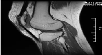
Human Knee joint is composed of femoral head femur and tibia. The surface between femoral head and the inner surface of tibia are covered with a layer of cartilage tissue, with the synovial fluid filled between two layers of cartilage as lubricant. Usually in the MR images, it’s difficult to distinguish layers of cartilage because they integrate closely. By traction technology to stretch knee joint, the femoral head and femoral cartilage can be seen clearly in MR images. Figure 1 shows MR projective image of a knee joint after traction, and articular cartilage in the figure is displayed as a high-brightness[Fig-1]. Addition to the femoral head and femora cartilage, some of surrounding soft tissue and noise are also shown as a high brightness.
The upsides of the watershed change are that it is basic, instinctual information, and can be parallelized. The primary disadvantage of this strategy is the over-division because of the nearness of numerous nearby minima.
The disadvantage is here remunerated by joining this change with a checking performed.
The inside marker is resolved naturally by joining systems including Canny edge location, thresholding and morphological operation.
The principle goal of this paper is to discover the crevices in existing writing. The diverse division systems are looked into and found that marker based is best in the greater part of cases since it denote the areas then section them. In any case, advancing the checking locales is as yet a region of research.
METHODOLOGY
Image Acquisition and Pre Processing
MR pictures were taken from knee joint of elderly females (>50 years) utilizing quick ruined inclination reverberate arrangement to projective picture in the sagittal and coronal bearing of the
knee joint utilizing 1.5T HDE MR framework (Manufactured by GE). Projective imaging parameters were as per the following: redundancy time(TR)/resound time(TE)= 10.4/4.8 ms; flip point = 15º; field of view(FOV)= 160 mm x 160 mm; lattice = 256 x 256; segment thickness = 1.6 mm; number of signs gained = 2; obtaining time = 4 min 38 s.
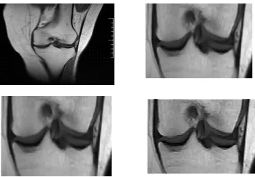
P i c t u r e p r e - p r e p a r i n g i n c l u d e d introduction, smoothing and upgrade. The picture insertion was executed to upgrade the precision of picture division, which enhanced the determination from 256 x 256 to 399 x 376 for sagittal and 368x361 for coronal picture and making the measure of each picture pixel change from 0.625mm x 0.625mm to 0.3125mm x 0.3125mm. Figure 2 and 3 demonstrates the sagittal and coronal pictures of knee joint after interjection.
In this work types of images coronal and sagittal planes are used and the process applied to both the images are 100% similar in spite of some changes of about 5% due to the orientation of the Knee in sagittal and coronal planes and morphological operation applied to them. The images were taken in MATLAB and converted from type RGB to Grayscale so that it is comfortable to work with the single pixel values instead of three.
Images pre-processing was carried out so as to make it easy to work with.For this, 1.Filtering: The image is filtered from unwanted noise by using 2-D median filtering operator. 2.Cropping:The images are cropped in order to focus the process on the area of interest by conventional cropping function (imcrop by size-116x174 approx.) 3. Resizing or Interpolated: The images were interpolated (Bi-cubic Interpolation) by imresize function to have 1.5 times bigger (174x261 approx. acc. to cropped image size) version of image and finally 4. Sharpening: The images were sharpened with the imfilter function using appropriate inbuilt filters or convolution kernel.
Image Segmentation for Area of Interest Harsh Segmentation Using Watershed Algorithm
Fig.1. MR Image of a Human Knee Joint Fig. 2. Sagittal Image of Knee Joint
Fig. 3. Coronal Image of Knee Joint Fig. 4. Watershed Images
two catchment basins. Watershed algorithms then find the catchment basins and the ridge lines in an image.
The algorithm works as follows: Suppose a hole is punched at each regional local minimum and the entire topography is flooded from below by letting the water rise through the holes at a uniform rate. Pixels below the water level at a given time are marked as flooded. When we raise the water level incrementally, the flooded regions will grow in size. Eventually the water will rise to level where two flooded regions from separate catchment basins will merge. When this occurs, the algorithm constructs a one-pixel thick dam that separates the two regions. The flooding continues until the entire image is segmented into separate catchment basins divided by watershed ridge lines. Figure 4 shows how watershed segmentation algorithm works on image [Fig-4].
Removing Noise in Rough Segmentation by Morphological Operations
The areas in the binary image which are not desired are considered as the noise areas and these are to be removed from the binary image. These areas are of two types: smaller than the desired area (in sagittal and coronal both) and larger
than the desired (in sagittal image). So these noisy areas are removed by using the process of dilation and erosion with the morphologically opening binary image using area open function (removing small object)-[5].The larger noise areas are first to be detected and then to be removed separately from binary image13. After removing these entire
noisy regions desired region is produced11.
Accurate Edge Extraction and Segmentation
The Canny edge indicator is the principal subordinate of a Gaussian and nearly approximates the administrator that improves the result of flag to-commotion proportion and confinement. The Canny edge location calculation is condensed by the accompanying documentation.
Let I[i, j] denote the image. The result from convolving the image with a Gaussian smoothing filter using separable filtering is an array of smoothed data,
S[i, j] = G[i, j;á ]* I[i, j],
Fig, 5. (A) Noise sifted picture of sagittal plane, (B) Cropped Image of sagittal plane, (c) Resize of edited picture 1.5 times in sagittal plane and (D) Sharpened picture of sagittal plane
Fig. 7. (a) Binary image of sagittal plane based on pixel properties, (b) Dilated version of the binary image extracted previously
Fig. 8. (a) Binary image of coronal plane based on pixel properties, (b) Dilated version of the binary image extracted previously (c) 1's complement of the binary image obtained previously
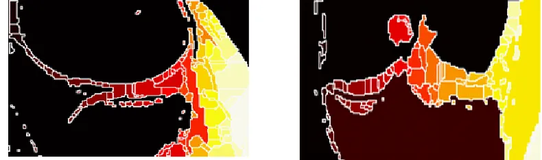
Fig. 9. Watershed segmentation of previous image in colourmap‘HOT’(a) sagittal plane (b) coronal plane
P[i,j] ~ (S[i,j + 1]- S[i,j] + S[i + 1,j + 1]- S[i + 1,j])/2 Q[i,j] ~ (S[i,j] – S[i + 1,j] + S[i,j + 1]- S[i + 1,j + 1])/2.
The limited contrasts are found the middle value of over the 2x2 square so that the x and y fractional subsidiaries are processed at a similar point in the picture. The greatness and introduction of the inclination can be registered from the standard recipes for rectangular-to-polar transformation:
M[i,j]=
Q[i,j]=arctan(Q[i,j],P[i,j])
Where the arctan work takes two contentions and creates an edge over the whole
hover of conceivable headings. These capacities must be figured effectively, ideally without utilizing drifting point math. It is conceivable to register the inclination size and introduction from the incomplete subsidiaries by table query. The arctangent can be registered utilizing for the most part settled point arithmetic2 with a couple of basic drifting point figurings performed in programming utilizing number and settled point math. Sedgwick gives a calculation to a whole number estimation to the inclination edge that might be adequate for some applications.
Fig.10. Watershed segmentation on original grayscale image (a) sagittal plane (b) coronal plane
Fig. 11. Binary image of sagittal plane after removing (a) small noisy regions (b) Large noisy regions (c) all the noise
Fig. 12. Binary image of coronal plane after removing (a) small noisy regions (b) Large noisy regions (c) all the noise
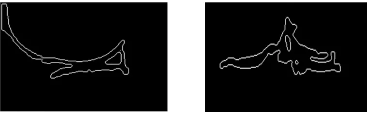
Fig.13. Canny edge detection of the binary image obtained previously (a) sagittal plane (b) coronal plane
of the segmented object in the binary image3.
Now applying these boundaries in the original image by superimposing the canny edge boundary image on the original grayscale image by proper pixel manipulation in these images1. At last final
segmented Region is highlighted by showing the corresponding pixels of the object (in binary image) on the actual grayscale image and making the other pixels of single same non-highlighted value18. The
Fig.14. Outlining the boundary of the desired segmented Region (a) sagittal plane (b) coronal plane
Fig.15. Desired segmented Region between Femur and Tibia (a) sagittal plane (b) coronal plane
RESULT AND DISCUSSION
The proposed systems were produced in MATLAB R2010a conditions. The trials were performed on a PC (I3 Processor: 3 GHz; RAM: 2 GBytes; HD:220GBytes). The execution of the created procedure was assessed on 20 real MRI Images of Knee joints and just aftereffects of agent pictures are exhibited in this review.
The results of the study were compared with the actual result of the same knees and found the similarity was above 85%.The thickness of the cartilage is calculated by obtaining the pixel distance from tibia and femur and then calibrating it with proper value.Comparison is made with the diagnostic reports and images.
We actually apply the two process of segmentation here one after the other that is the watershed algorithm segmentation [Fig-9,10] process and the little amount of the morphological operations with the canny edge detector [Fig -13,14,15] at the final segmentation process. If morphological operations [Fig- 7,8] in this segmentation are considered they may prone to
some variation with the original image they can change the pixel values from 5-15% which lead to the error result of about 5-10%. Here in the operations of Erosion and dilation there are some pixel disturbances from that of original. This can be calculated by calculating the number of pixels diversions from the original or desired area of the thickness and then this value can be considered as a parameter to judge the errors in the image or may be represented as percentage by a ratio of val(no. of pixel diverted/no. of pixels in desired area to be segmented).
the positive side of the segmentation work it will gives the 90-95% accurate results under the given circumstances prescribed in the work.
We tentatively demonstrated that the proposed strategy within the sight of the accessible informational indexes indicated viable in 93.5% of the slices. The disappointment of the calculation in the rest of the cuts was for the most part because of poor imaging condition, inadequate footing, and inconsistency in the round state of hard and ligament tissues. In such cases, even the judgment of a specialist in deciding the correct area of the articular space was dubious. We have to assess the calculations with more informational collections, coordinate the proposed procedure in a product bundle for ligament division, ligament thickness delineate and relating 3D perceptions.
Later on we will research how the power in homogeneities in the MRI information can be managed. We are as of now analyzing the utilization of a consolidated dynamic shape model of the femur and tibia to expand the power and exactness of the bone division. At present, our outcomes show that some manual post-preparing is as yet required to draw level with human-rater execution. Notwithstanding, we are sure that with our proposed system and the recommended upgrades we will have the capacity to build the quantity of situations where manual post processing will end up plainly old.
CONCLUSION
To accomplish programmed femoral ligament with space amongst femur and tibia division on MR picture, a programmed division strategy in light of Watershed Algorithm and versatile shrewd edge discovery has been proposed. Addition, smoothing and upgrade were actualized in the picture pre-preparing to enhance the picture quality. The territory of intrigue was chosen and the objective region for the harsh division was made sense of by considering the anatomical structure imperative (Pixel qualities) of Knee joint. Watershed Algorithm was utilized to generally fragment the picture for the hole in the middle of femur and tibia. Extra separating through Morphological operations-picture expansion and disintegration with parallel picture opening capacities is then connected to evacuate boisterous
areas to precisely fragment the required locale from the generally sectioned district. Picture marked and sifted in a custom one by one way to evacuate the uproarious areas ones and procure the correct internal and external edges of the space amongst femur and tibia. Edges in the unpleasant area were separated out by the versatile limit shrewd edge locator. As indicated by the properties of the pixel on distinguished edge, these edges and paired picture were then used to highlight the real fragmented locale in the real grayscale. At last the exact division was accomplished by extricating the picture information in the highlighted Region. Division explore approves the method. The entire process did not require manual mediation and the effectiveness of division process was progressed. This programmed division strategy is effectively connected for both the plane i.e. sagittal and coronal.
ACKNOWLEDGEMENTS
This research was supported by the Sarkar Diagnostic and Hind Hospital, Lucknow. We thank them for technical aid and providing data for this research.
REFERENCES
1. “Knee Cartilage Extraction and Bone-Cartilage Interface Analysis from 3D MRI Data Sets” Jos´e G. Tamez-Pen˜a, Monica Barbu-McInnis and SaaraTottermanVirtualScopics, 350 Linden Oaks, Rochester NY, 14625, USA,2004. 2. “Automatic Segmentation of Femoral Cartilage
from MR Image Based on Hough Transform and Adaptive Canny Detection” Yu Cao1, Xia Liu1, Yang Cao1 and Yu-nan Liu2*. International Journal of Signal Processing, Image Processing and Pattern Recognition, 2013; 6(4).
3. “Automated Segmentation of the Articular Space in MR Images of the hip joint” M. Khanmohammadi, R. A. Zoroofi, Y. Sato, T. Nishiif, K. Nakanishi”, H. Tanaka’. N. Sugano”, H. Yoshikawa”, H. Nakamurat, S. Tamura Visual Information Engineering,IET International Conference, 2006.
5. J. Carballido-Gamio1, K. Lee1, E. Ozhinsky1, S. Majumdar, “MRI cartilage of the knee: segmentation, analysis, and visualization” United State. Proc. Intl. Soc. Mag. Reson. Med. 2004; 11.
6. HeikoSeim, Dagmar Kainmueller, Hans Lamecker, Matthias Bindernagel, Jana Malinowski, and Stefan Zachow, “Model-based Auto-Segmentation of Knee Bones and Cartilage in MRI Data”, 14195 Berlin, Germany,2010. 7. S. Martelli, V. Pinskerova,”The shapes of the
tibial and femoral articular surfaces in relation to tibio femoral movement” rom the Rizzoli Orthopaedic Institute, Bologna, Italy and the Orthopaedic Clinic, Prague, Czech Republi,J Bone Joint Surg [Br], 84-B:607-13,2002. 8. S. Ghosh, Beuf, D. C. Newitt, M. Ries, N. Lan
and S. Majumda.”Watershed Segmentation of High Resolution Articular Cartilage Images for Assessment of OsteoArthritis”,Proceedings of the 22nd Annual International Conference of the IEEE Engineering in Medicine and Biology Society (Cat. No.00CH37143), Chicago, IL, 2000; 4: 3174-3176.
9. “Improved Watershed Transform for Medical Image Segmentation Using Prior Information” V. Grau*, A. U. J. Mewes, M. Alcañiz, Member, IEEE, R. Kikinis, and S. K. Warfield, Member, IEEE Transactions on Medical Imaging, 2004; 23(4) .
10. S. Thilagamani, N.Shanthi, “A Novel Recursive Clustering Algorithm for Image Oversegmentation”, European Journal of
Scientific Research, 2011; 52(3): pp.430-436. 11. P e t e r E g g l e s t o n , “ U n d e r s t a n d i n g
Oversegmentation and Region Merging”, Vision
Systems Design, December 1, 1998.
12. M. W. Hansen and W. E. Higgins, “Watershed-based maximum-homogeneity filtering,” IEEE Trans. Image Process., 1999; 8(7): 982-988. 13. K. Parvati, B. S. PrakasaRao and M. Mariya
Das, “Image Segmentation Using Gray-Scale Morphology and Marker-ControlledWatershed Transformation”, Discrete Dynamics in Nature and Society, 2008: 1-8.
14. Dr.(Mrs)S.N Geethalakshmi and T.Jothi, “ S e g m e n t a t i o n B a s e d O n E n h a n c e d Morphological Watershed Algorithm”, Journal of Global Research in Computer Science, 2010; 1(4).
15. P.P.Acharjya1, A. Sinha2, S.Sarkar3, S.Dey4, S.Ghosh5 Department of CSE, Bengal Institute of Technology and Management, Santiniketan, India “A New Approach Of Watershed Algorithm Using Distance Transform Applied To Image Segmentation” International Journal of Innovative Research in Computer and Communication Engineering, 2013; 1(2). 16. Amanpreetkaur, AshishVerma, Ssiet, Derabassi
(Pb.) India “The Marker-Based Watershed Segmentation- A Review” International Journal of Engineering and Innovative Technology (IJEIT), 2013; 3(3).
17. AnjuBala “An Improved Watershed Image Segmentation Technique using MATLAB”
International Journal of Scientific & Engineering
Research, 2012; 3(6).