Int. J. Nanosci. Nanotechnol., Vol. 11, No. 4, Dec. 2015, pp. 249-256
Short Communication
Investigation on Influences of Synthesis
Methods on the Magnetic Properties of
Trimetallic Nanoparticles of
Iron-Cobalt-Manganese Supported by Magnesium Oxide
A. Dahmardeh and A. M. Davarpanah*
Department of Physics, Faculty of Science, University of Sistan and Baluchestan, Zahedan, I. R. Iran
(*) Corresponding author: a.m.davarpanah@phys.usb.ac.ir
(Received: 16 May 2015 and Accepted: 06 Sep 2015)
Abstract
Using Fe(NO3)3.9H2O, Co(NO3)2.6H2O and Mn(NO3)2.4H2O the magnetic properties of nanoparticles
trimetalic Iron - Cobalt - Manganese, with supported Magnesium oxide have been prepared by Co-precipitation and Solvothermal methods. The prepared samples are characterized by Scanning Electron Microscope (SEM), X-Ray Diffraction (XRD), Vibrating Sample Magnetometer (VSM) and Brunaer-Emmett-Teller (BET) surface area measurements. Data from SEM showed spherical and nearly uniform spherical shape particles for the samples which were synthesized with support by co-precipitation and by solvothermal methods, respectively. According to the patterns of XRD, the crystal size of the nanoparticles prepared by co-precipitation method with support are in the range of 25 to 30 nm and the size of the particles are in the range of 45 to 50 nm using solvothermal method. The results of the BET from all of the nanoparticles synthesized show that precursors have highest surface area of the calcinated samples. According to the VSM results taken at room temperature (RT), the samples with support after calcination were placed in the category of soft ferromagnetism.
Keywords: Catalyst, Co-precipitation, Nanoparticles, Soft ferromagnetism, Solvothermal.
1. INRODUCTION
In recent years, Nano science and Nano technology, have become especially to a branch of special research with specific applications [1]. The properties and applications of nanoparticles are subjects that have been much researched about them because they have unique physical and chemical properties [2, 3]. The magnetic properties of materials in Nano-scale are used in high-density recording, image color, high-frequency devices and so on [4]. Nanoparticles of tri-metallic alloys have especially applications in the petrochemical and synthetic fuels. Synthetic fuel and Fischer–Tropsch reaction are important issues for scientific research with economical advantage in the twenty-first century. In general, Fischer–
Tropsch reaction includes CO
hydrogenation on heterogeneous catalysts which is an economical process to convert synthesis gas (a mixture of Hydrogen and Carbon monoxide) to a wide range of Hydrocarbon products. Although, several metals have been known as catalysts for this reaction but the Fischer–Tropsch reactions of synthesis have industrial and commercial aspects only on catalysts based on Iron and Cobalt. Amongst the transition metals [5-6], the most common metals as a catalyst in the synthesis Fischer–Tropsch are VIII metals such as Rh, Ru, Ni, Co, Fe [7].
metal-based catalysts. However, the controllable synthesis of trimetallic nanomaterials as well as the exact role played by the addition of a third metal in their composition over catalytic performances remains unclear. Trimetallic nanoparticles have shown improved catalytic
performances relative to their mono- and bimetallic counterparts for a variety of reactions that include Cyclohexene and Glucose oxidation, the electro oxidation of formic acid, and C–C coupling [8-10]. Despite these very attractive features, studies on the synthesis of trimetallic noble metal nanomaterials are still limited, and the role of the third metal over the performances, relative to their bimetallic systems, remains unclear [11]. Trimetallic nanoparticles may possess an even greater degree of catalytic activity and selectivity than the bimetallic ones because more variables (such as tuning) are available, however, there have been only a few report of trimetallic nanoparticles in literatures thus far [12-13]. Selection a method of syntheses of Nano-particles is very important and depends on the chemical composition, application, and laboratory facilities, economic cost, type of initial reaction and so on, which should be considered when selecting a method of making Nano-materials. In general, the methods of making Nano materials are divided into three categories:
1- Physical methods 2- Chemical methods 3- Mechanical methods [7]. The purpose of this study is investigation of influence of synthesis methods, such as co-precipitation and solvothermal methods, on the magnetic properties of the Iron, Cobalt, and Manganese nanoparticles by adding support of Magnesium Oxide.
2. PREPARATION PROCEDURES
2.1 Preparation of the samples by co-precipitation method
In the co-precipitation method for preparing nanoparticles of metallic Iron, Cobalt, Manganese, were prepared two
molar solution of nitrate salts Co(NO3)2.6H2O (2 M) (99% Merck), Fe(NO3)3.9H2O (2 M) (99% Merck) and Mn(NO3)2.4H2O (2 M) (99% Merck) with the same molar ratios were premixed and then the 10 wt.% of MgO (based on the total catalyst weight) was added to mixed solution of Cobalt, Iron and Manganese Nitrates. The resulting solution was heated to 70°C in a round bottom flask fitted with a condenser. Aqueous Na2CO3 (2 M) (99% Merck) was added drop wise to the mixed nitrate solution with stirring while the temperature was maintained at 70°C until pH = 9.7 ± 0.1 was achieved. The resulting precipitate was then left in this medium for 2 hrs. The aged suspension was filtered and then washed several times with warm distilled water until no further Na+ was observed in the washings [12], as tested by flame atomic adsorption. The precipitate material was then dried in an oven at 120°C for 12 hrs to get a material denoted as the catalyst precursor, which was subsequently calcinated in static air in a furnace at 600 °C for 6 hrs.
2.2 Preparation of the samples by solvothermal method
for 6 hrs and the sample has been powdered in mortar.
3. RESULTS and DISCUSSIONS 3.1 Results of Scanning Electron Microscope
The shape and size of the sample were determined using SEM system, model MIRA II LMU/TESCAN located in the Iranian Research Organization for Science and Technology. Figures 1 and 2 show SEM images of trimetallic nanoparticles with the support MgO prepared by co-precipitation method before and after calcination respectively. In the Figure 1, the pre-calcinated particles have stucked together, but after calcination (Figure 2), most of particles have became nearly spherical and average size of particles were about 30 nm (using ruler and MATLAB softwares).
Figure1: SEM image of nanoparticles
prepared by co-precipitation with MgO support before calcination.
Figures 3 and 4 show nanoparticles that produced by solvothermal, which represent better uniformity. In this method, we can see from Figure 3 that the particles have been stucked together before calcination, but after calcination (Figure 4), most of the particles have been found uniformly and
spherical in average size 45 nm. So, we can say that the changing method could affect size and distribution of nanoparticles.
The results of the SEM images show that the produced samples by co-precipitation
method are more agglomerated
nanoparticles respect to the ones which produced by solvothermal method.
Figure2: SEM image of nanoparticles
prepared by co-precipitation with MgO after calcination.
Using solvothermal method is available to achieve better nanoparticles with spherical morphology and particle size distribution. It should be noted that other important factors can show effects on the performance of the nanoparticles such as phases in the sample, the distribution of particles in the tissue sample and the sample structure.
3.2 Results of X-Ray Diffraction
intensity of the samples that prepared by co-precipitation method.
Table1: Results of XRD for the samples
that produced by co-precipitation method
Peak No
Crystalline Phases 2θ
1 CoMnO4 15.78
2 Fe2O3 35.60
3 CoMnO4 38.31
4 C3N4 45.80
5 CoFe2O4 62.81
6 CoFe2O4 66.09
The average size of the particles is 27 nm and a maximum intensity is observed at an angle of 35.60°. The MgO supported samples that prepared by co-precipitation method have phases such as: Fe2O3 (Rhombohedral), CoFe2O4 (cubic), CoMnO3 (Cubic).
Figure3: SEM image of nanoparticles
prepared by solvothermal with MgO support before calcination.
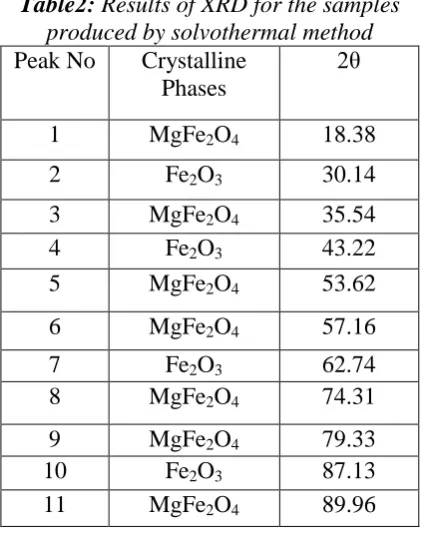
Table2 shows crystal phases and the intensities of peak points from the XRD spectra of the samples that prepared by solvothermal method. The average particle
size is 47 nm and a maximum intensity is observed at an angle of 35.54°. Also Table 2 shows that the sample prepared in this method, involves MgFe2O4 (Cubic) and Fe2O3 (Rhombohedral) Phases. Figure 6 shows XRD spectrum of the trimetallic Iron-Cobalt-Manganese supported MgO prepared by solvothermal.
Figure4: SEM image of nanoparticles
prepared by solvothermal with MgO support after calcination.
3.3 Results of BET
increases surface area and decreases size of particles that prepared by solvothermal. It can be said, because the MgO support with Oleylamine as reductive factor was used in solvothermal method, causes an increase in concentration and reduces the volume of solvent in autoclave and resulting in decreased surface area respect to co-precipitation method.
Figure 5: XRD spectrum of the trimetallic
Iron-Cobalt-Manganese with MgO prepared by co-precipitation.
Table2: Results of XRD for the samples
produced by solvothermal method
Peak No Crystalline Phases
2θ
1 MgFe2O4 18.38
2 Fe2O3 30.14
3 MgFe2O4 35.54
4 Fe2O3 43.22
5 MgFe2O4 53.62
6 MgFe2O4 57.16
7 Fe2O3 62.74
8 MgFe2O4 74.31
9 MgFe2O4 79.33
10 Fe2O3 87.13
11 MgFe2O4 89.96
4.3 Results of VSM
All of magnetic measurements have been done by VSM system where is located in the Nano Magnetic Laboratory, in the University of Sistan and Baluchestan, Zahedan, Iran, The system is made of
Meghnatis Daghighe Kavir, (MDK), Company, Kashan I. R. Iran [14]. Figure 7 shows magnetization curve in terms of the intensity of the magnetic field for the samples of trimetallic with Magnesium oxide support that have prepared by co-precipitation method before calcination. The sample has a saturation magnetization 0.65104 emu/gr and residual magnetization 0.01353 emu/gr and the coercivity of 100 Oe. Because the coercivity is 100 Oe, so this material is ferromagnetic, and has not reached to the saturation level.
Figure6: XRD spectrum of the trimetallic
Iron-Cobalt-Manganese supported MgO prepared by solvothermal.
-10000 -8000 -4000 0 4000 8000 10000
-0.7 -0.6 -0.4 -0.2 0.0 0.2 0.4 0.6 0.7
Ma
g
n
e
tiza
tio
n
(e
mu
/g
r)
Applied Magnetic Field (Oe)
Fe-Co-Mn with MgO
Figure7: Curve of M-H for the sample of
trimetallic Fe-Co-Mn with MgO support before calcination prepared by
co-precipitation.
2. field from ±2000 to ±10000 Oe where the sample has not saturated.
Figure 8 shows magnetization curve in terms of applied external magnetic field for trimetallic sample with magnesium oxide support which is prepared by co-precipitation method after calcination. The sample has a saturation magnetization of 6.09519 emu/gr and residual magnetization of 0.31191 emu/gr. If we have a look at the range between – 4000 to +4000 Oe according to the area under the curve, hysteresis loop is very low, so it can be said that the sample is in soft ferromagnetic materials’ category.
-10000 -8000 -4000 0 4000 8000 10000
-7 -6 -4 -2 0 2 4 6 7 Ma n e tiza tio n (e mu /g r)
Applied Magnetic Field (Oe)
Fe-Co-Mn with MgO
Figure8: M-H curve for the trimetallic
Iron-Cobalt-Manganese with MgO support after calcination prepared by
co-precipitation.
-10000 -8000 -4000 0 4000 8000 10000
-14 -10 -5 0 5 10 Ma g n e tiza tio n (e mu /g r)
Applied Magnetic Field (Oe) Fe-Co-Mn with MgO
Figure9: Curve of M-H for the sample of
trimetallic Iron-Cobalt-Manganese with MgO support before calcination that
prepared by solvothermal.
Figure 9 shows magnetization curve in terms of the external applied magnetic field for the sample of trimetallic with
magnesium oxide support that is prepared by solvothermal method before calcination. The sample has a saturation magnetization of 10.7545 emu/gr and residual magnetization of 0.1511 emu/gr and the coercivity of zero Oe. We can consider the sample as superparamagnetic since the coercivity is zero, and because of the phases of production, Fe3O4 and Fe2O3 pre-calcinated.
-10000 -8000 -4000 0 4000 8000 10000
-25 -20 -10 0 10 20 25 Ma g n e tiza tio n (e mu /g r)
Applied Magnetic Field (Oe)
Fe-Co-Mn with MgO
Figure10: curve of M-H for the trimetallic
Iron-Cobalt-Manganese with MgO support after calcination that prepared by
solvothermal.
Figure 10 shows magnetization curve in terms of magnetic field for the sample of trimetallic with magnesium oxide support that is prepared by solvothermal method after calcination. The sample has a saturation magnetization of 6.09519 emu/gr and residual magnetization of 0.31191 emu/gr and the coercivity of 100 Oe and according to the information obtained from this curve, it is placed in the category of ferromagnetism.
tetrahedral states, it may change the interaction and exchange in the two situation. Therefore it creates different effects on the magnetic properties of the sample. Several experimental studies of the relations between coercivity and particle size have been reported [15-18].
4. CONCLUSION
In this study, trimetalic nanoparticles, Iron – Cobalt, Manganese, with supported Magnesium oxide have been prepared by co-precipitation and solvothermal methods. The prepared samples are characterized by Scanning Electron Microscope (SEM), X-ray Diffraction (XRD), Brunaer-Emmett-Teller (BET) surface area measurements and Vibrating Sample Magnetometer (VSM). The results of SEM images showed that the particles were spherical in co-precipitation method with MgO as support. However, in solvothermal method, the synthesized samples had distribution and uniformity better than co-precipitation method and they were almost spherical. The XRD results showed that in co-precipitation
method, particle sizes were between 25 and 30 nm and in solvothermal method, particle sizes of approximately were 45 to 50 nm. The results of VSM at room temperature show that, the sample which was prepared by co-precipitation with
MgO support, pre-calcined had
diamagnetic properties, but after calcination at 600°C and the removal of some phase, it became soft ferromagnetic. Moreover, the sample that was prepared with MgO support after calcination at the same temperature, because of having small area under the curve, it is categorized as soft ferromagnetic materials.
ACKNOWLEDGEMENTS
The authors appreciate Prof. A. A. Mirzaie and Dr M. Arsalanfar, for their unwavering scientific advices. The authors thank to Ms Nickparsa and Ms Taherzadeh for their help on synthesis of the samples. The authors are thankful to the University of Sistan and Baluchestan, I.R. of IR, for financial support.
REFERENCES
1. Koch, C. C. (2007). Nano structured Materials processing, properties and potential application, by Noyes publications/William Andrew Publishing, Norwich, New York, USA, 1-30.
2. Strevens, A. E., Drury, A., Lipson, S. M., Kroll, M., Blau, W. J., Horhold, H. H., (2005). Appl. Phys. Lett. 86: p 143503
3. Endo, M., Muramatsu, H., Hayashi, T., Kim, Y. A., Terrones, M., Dresselhaus, M. S. (2005). “Pure and Clean Double-Walled Carbon Nanotubes” Nature, 433, 476.
4. Dorman, J. L., Fiorani, D. (1992). Magnetic Properties of Fine Particles, North Holland, Amsterdam.
5. Arsalanfar, M. (2012). “The effect of preparation procedures and operational conditions over the catalytic
performance of Fe-Co-Mn catalysts on the Fischer-Tropsch synthesis process and study of kinetic and
mechanism reaction” PhD thesis, University of Sistan and Baluchestan.. 50-79 and 130-160.
6. Penaoshea, De. La., Alvarez-Galvan, V. A., Campos-Martin, M. C., Fierro, J. M., Gaul, J. L. (2007). “Fisher-Tropsch synthesis on mono- and bimetallic Co and Fe catalysts in fixed bed and slurry reactors” Appl. Catal.
A: Gen,:. 326, 65-73
7. Wang, C., Xu, L., Wang, Q. (2003) “Review of direct producing light olefins via CO hydrogenation” J. Nat.
Gas. Chem, : 12, 10-16
8. Zhang, H., Toshima, N. (2013). “Glucose oxidation using Au-containing bimetallic and trimetallic nanoparticles” Catal. Sci Technol : 3, 268–278
doi: 10.1039/C2CY20345F.
9. Ghiaci, M., Aghabarari, B., Botelho, Do., Rego, A. M. (2011). “Efficient allylic oxidation of cyclohexene catalyzed by trimetallic hybrid nano-mixed oxide (Ru/Co/Ce)” Appl. Catal. A, : 393. 225–230
10. Jiang, K., Cai, W. B. (2014). “Carbon supported Pd–Pt–Cu nanocatalysts for formic acid electrooxidation: synthetic screening and componential functions” Appl. Catal. B, :. 147. 185–192.
11. Thenner S., Rodrigues, Anderson G. M. da Silva; Alexandra Macedo, Bruna W. Farini, Rafael da S. Alves;
Pedro H. C. Camargo. (2015). “Probing the catalytic activity of bimetallic versus trimetallic Nanoshells” J.
12. Arsalanfar, M., Mirzaei, A. A., Bozorgzadeh, H. R. (2012). “Effect of calcination conditions on the structure and catalytic performance of MgO supported Fe-Co-Mn catalyst for CO hydrogenation” Journal of Natural
Gas Science and Engineering, :. 6, 1-13
13. Marabet, D., Abassi, A., Cherizol, R., Trong-On, Do. (2012). “One-pot solvothermal synthesis of mixed Cu-Ce-Ox Nano catalysts and their catalytic activity for low temperature CO oxidation” Applied Catalysis A:
General 447– 448,. 60-64.
14. http://www.mdk-magnetics.com
15. Chernavskii, P. A., Dalmon, J. A., Perov N. S. and Khodakov, A. Y. (2009). “Magnetic Characterization of Fischer-Tropsch Catalysts” Oil & Gas Science and Technology Rev. IFP, :. 64, 1, 25-48
16. Sort, J., Surinach, S., Munoz, J. S., Baro, M. D., Wojcik, M., Jedryka, E., Nadolski, S., Sheludko, N., Nogues, J. (2003). “Role of stacking faults in the structural and magnetic properties of ball-milled cobalt”
Phys. Rev. B 68, 014421, 7.
17. Stoner, E. C., Wohlfarth, E. P. (1948). “A Mechanism of Magnetic Hysteresis in Heterogeneous Alloys”
Philos. T. Roy. Soc. A, 240, 599-642.
18. Chen, C., Kitakami, O., Shimada, Y. J. (1998), “Particle size effects and surface anisotropy in Fe-based