Int. J. Nanosci. Nanotechnol., Vol. 14, No. 2, June. 2018, pp. 153-158
Short Communication
Theoretical Analysis of the Optical Properties
of Gold Nanoparticles Using DDA
Approximation
H. Akherat Doost, M. H. Majles Ara*and E. Koshki
Department of Physics, Kharazmi University, Mofateh Ave, Tehran, Iran.
(*) Corresponding author: [email protected] (Received: 09 November 2015 and Accepted: 07 August 2016)
Abstract
This article describes a study, using numerical simulation, of the optical properties of nano particles as a function of their size. Many methods introduced to simulate and calculate the interaction of light and particle, such as Mie analysis, boundary element and finite element methods. The Discrete Dipole Approximation (DDA), wherein a target geometry is modeled as a finite array of polarizable elements and DDSCAT simulation, are employed for determining extinction, absorption and scattering cross sections by gold nano particles with 15, 35, 55, 75, 100, 125 and 150 nanometers in diameter in different wavelengths. In this method, the particle is represented as a cubic array of electric dipoles. The results show that Plasmon resonance dependent on nano particles size. In other project, the angular distribution of different sizes of gold nano particles are calculated and scattering of these particles are compared. This study can be helpful in designing appropriately shaped nano particles for in-vivo applications like photo-thermal cancer treatment and optical sensors.
Keywords: Absorption and scattering of gold nanoparticles, Discrete Dipole Approximation (DDA), DDSCAT simulation.
1. INTRODUCTION
Geometry of nano particles and efficient modeling of light scattered from small features and particles on surfaces are primary factor and important facility for detecting and analyzing how incident light is scattered and absorbed by particles. Recently physical properties of different sizes of metallic nanoparticles and optical models of nano- metrology systems based on laser are theoretically analyzed [1, 2]. Prediction of evanescent wave scattering by particles on a surface is a challenging problem and knowledge of nanoparticle-light interactions and their cross sections calculation are very helpful in many studies such as soot agglomerates in combustion systems, interstellar dust grains in astrophysics, biological cells in biomedical applications and aerosols in
purposes, the forward model should be able to accommodate arbitrarily shaped objects on a surface. Numerical approaches such as the null-field method with discrete sources and the finite-difference time-domain method [12] could be employed for that purpose. We have used the discrete-dipole approximation (DDA) method to compute all cross sections of
nanoparticles. Discrete dipole
approximation (DDA) is a well-established and widely accepted method of solving light scattering by particles from direct propagating illumination. DDA was proposed by Purcell and Pennypacker (PP) [14], who replaced the scatterer by a set of targets, these dipoles interact with each other and the incident field and each of which attains a dipole moment due to both the incident beam and the other oscillating dipoles within the target. The DDA is a popular method in the light-scattering community and it has been reviewed by several authors. The DDA was further developed by Draine and coworkers [3, 4, 5, 6, 7], who describes the current state of the DDA and its historical development. It also explains the equivalence of the DDA and methods based on the volume integral equation formulation and popularized the method by developing a publicly available computer code DDSCAT [6]. Later it was shown that the DDA also can be derived from the integral equation for the electric field. This derivation was apparently first performed by Goedecke and O'Brien [9] and further developed by others (see, for instance, [10, 13, 15, 24] and certain DDA applications are concerned with very prolate or oblate particles [16, 17, 22]. In this paper, at first, the extinction, scattering and absorption cross sections for different sizes of gold nanoparticles were determined using DDA technique and then a comparison of plasmon resonance peaks for all nanoparticles was made.
2. DIPOLE DISCRETE
APPROXIMATION METHOD
In this method, the particle is represented as a cubic array of electric dipoles. Assume the number of dipoles is N. The jth dipole has a polarizability j, and the total
electric field at the dipole is Ej.Therefore,
the polarization Pjin response to the total
electromagnetic field can be described by
, .
j j j inc j jk k
k j
P E E A P
(1)
where Ejis the local electric field, which
is the sum of the incident field and the retarded radiation fields from the other dipoles, The incident field is assumed to be a plane wave and is in the form of
.
, 0
j
ik r inc j
E E e where k is the wave
vector. A is the dipole-dipole interaction matrix,
2 3 2 2 exp 1 3 . jk kjk jk k jk
jk
jk jk k jk jk k jk
k r r P
ikr
ikr A P
r r P r r P
r (2)
Here, k is the wave number, and
jk j k
r r r . Let Ajj j1 so that we can obtain:
inc
A P E (3)
P andEinc are 3N-dimensional vectors, and A is a 3N × 3N matrix. Equation 3 can be solved iteratively by means of the complex conjugate gradient (CCG) algorithm in combination with the fast Fourier transform technique. When the polarization of each dipole is known, the extinction, absorption and scattering cross-sections can be computed using
4 . , 2 Im kinc j j ext
k j inc
C E P
E
(4)* 1 2 2 1 3 0 . Im 4 2 3
N j j j
abs j j P P k C
E k P
(5)
sca ext abs
3. RESULTS AND DISCUSSIONS
It is possible to calculate scattering, absorption and extinction cross-sections of different sizes of gold nanoparticles by using DDA (Fig.1, 3-4). All the simulation results for particles of arbitrary geometries and different sizes reported in this work were obtained. Extinction is the sum of scattering and absorption which is a measure of the light that would not be received by the detector that is collinear with the nanoparticle and source.
Figure 1. Comparison extinction spectra of 15,35,55,75,100,125 and 150 nm diameters for Au nanoparticles.
The extinction peak for Au nanoparticles occurs at the surface plasmon resonance (SPR) frequency. At this frequency, conduction electrons oscillate coherently. SPR frequencies of gold nanoparticles occur in the visible part of the spectrum, which is useful for nanoantens, bistable devices and solar cell applications. Figure 1 shows that the extinction efficiency (i.e. extinction coefficient per unit cross sectional area of the nanoparticle) increases linearly with particle size up to a size of 150 nm. For particle sizes lower than 55 nm, the peak wavelength remains nearly constant (~520 nm) but for particle sizes higher than 55 nm, shifts to higher wavelengths. Peak wavelength values for spheres with size of 15 nm, 75 nm and 125 nm are 495 nm, 500 nm and 540 nm, respectively.
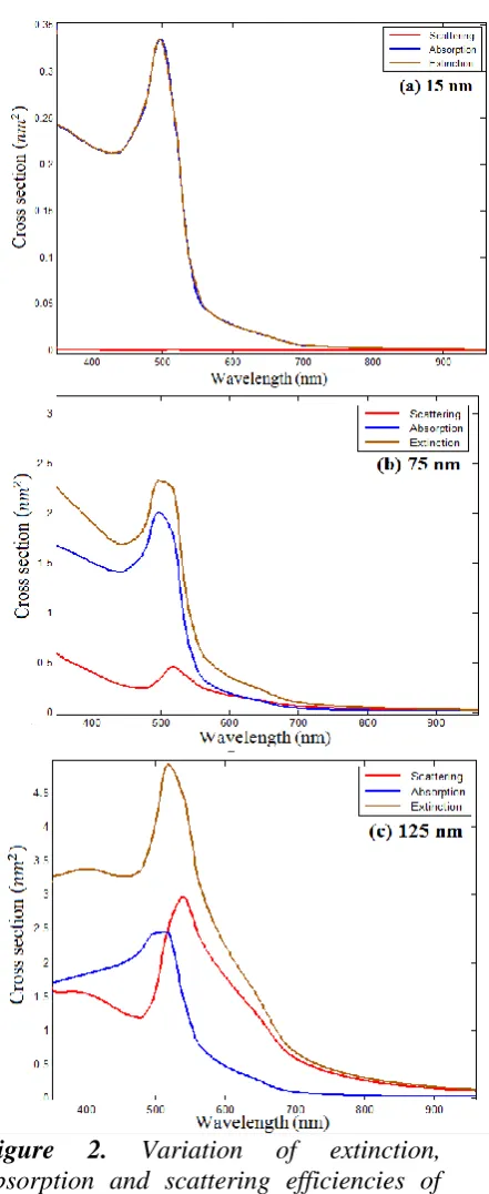
Figure 2. Variation of extinction, absorption and scattering efficiencies of gold nanoparticles of diameter (a) 15nm, (b) 75nm and (c) 125 nm.
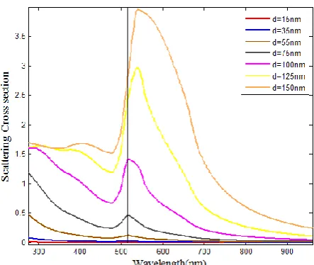
spherical gold nanoparticles. It is seen that for smaller nanoparticles, the extinction efficiency is predominantly governed by absorption efficiency. As the size of nanoparticle increases, scattering efficiency becomes more dominant. As Well As comparison of different sizes of gold nanoparticles absorption and scattering cross sections were shown in figures 3 and 4, respectively. It is clear that noble metal nanoparticles such as gold exhibit a strong optical extinction peak in the visible range of spectrum.
Figure 3. Comparison absorption spectra of
15,35,55,75,100,125 and 150 nm diameters for Au nanoparticles.
Figure 4. Comparison scattering spectra of
15,35,55,75,100,125 and 150 nm diameters for Au nanoparticles.
The scattered far field in spherical coordinates (𝐸𝑠𝜃,𝐸𝑠∅) for a unit amplitude
incident field is given by
𝐸𝑠𝜃 = 𝑒
𝑖𝑘𝑟
−𝑖𝑘𝑟𝑐𝑜𝑠∅. 𝑆2(𝑐𝑜𝑠𝜃) 𝐸𝑠∅ =𝑒
𝑖𝑘𝑟
𝑖𝑘𝑟 𝑠𝑖𝑛∅. 𝑆1(𝑐𝑜𝑠𝜃)
𝐸𝑠𝜃 is the scattered far-field component in the scattering plane, defined by the incident and scattered directions, and 𝐸𝑠∅
is the orthogonal component. The angle ∅
is the angle between the incident electric field and the scattering plane.
With respect to the application in a device, placing nanoparticles at an interface is a more realistic configuration. This also plays an important role when scattering efficiencies are judging. In the following, we compute scattering intensities to display results as a polar diagram of θ in both the upper half circle (0<θ<π) and lower half circle (π<θ<2 π).
(a)
(b)
Figure 5. Angular scattering diagram of upper and lower half circle for 55, 100, 150 diameters of Ag nanoparticles. It can be observed that scattering in the backward hemisphere is exiguity larger than that in the forward hemisphere.
4. CONCLUSION
In this paper, the optical spectra of different sizes of gold nanoparticles have been simulated using DDASCAT code. Results showed that with increasing in particle size, Coulomb's force in electrons motion was smaller and electrons oscillated with low energy. It is clear that for smaller nano spheres, only absorption efficiency contributes significantly towards extinction efficiency, but with an increase
in size the contribution from scattering dominates, which was seen in Figure 2, and a linear relationship was also seen between the extinction efficiency and particle size for the smaller nano particles. Comparison between nano particles spectra showed that with increasing in size, SPR peaks had red shift and this has extent application in photonic such as nanoantens and optical bistability devices. Finally angular distributions of Au nano particles in 55, 100, 150 nm diameters were computed that can be helpful in designing appropriate target nano particle geometry for in-vivo applications like photo-thermal cancer treatment, optical limiting devices and optical sensors.
REFERENCES
1. Salehian, M., Mohammadian, A., Vaezzadeh, M., Saeidi, M., (2017). “Influence of size on the melting temperature of metallic nanoparticles”, Int. J. Nanosci. Nanotechnol., 13: 91-95.
2. Olyaee, S., Dashtban, Z., (2014). “Analysis of Frequency Leakage in Different Optical Paths of Nano-Metrology Systems Based on Frequency-Path Models”, Int. J. Nanosci. Nanotechnol., 10: 79-96.
3. Draine, B. T., (1988). “The discrete-dipole approximation and its application to interstellar graphitgrains”, Astrophysics Journal., 333: 848-872.
4. Draine, B. T., (1993). “Goodman J. J. Beyond clausius-mossotti - wave-propagation on a polarizablepoint lattice and the discrete dipole approximation”, Astrophysics Journal., 405: 685-697.
5. Draine, B. T., Flatau, P. J., (1994). “Discrete-dipole approximation for scattering calculations”, Journal of Optical Society of America A., 11: 1491-1499.
6. Draine, BT., (2000), “The discrete dipole approximation for light scattering by irregular targets”, Academic Press, NewYork.
7. Draine, B. T., Flatau, P. J., (2004). “User guide for the discrete dipole approximation code 6.1”, http://xxx.arxiv.org/abs/astroph/0409262.
8. Francoeur, M., Venkata, P. G., Mengu, M. P., (2007). “Sensitivity analysis for characterization of gold nanoparticles and agglomerates via surface Plasmon scattering patterns”, Journal of Quantitative Spectroscopy and Radiative Transfer., 106: 44-55.
9. Goedecke, G. H., O'Brien, S. G., (1988). “Scattering by irregular inhomogeneous particles via thedigitized Green's function algorithm”, Applied Optic Journal., 27: 2431-2438.
10.Lakhtakia, A., (1992). “Strong and weak forms of the method of moments and the coupled dipolemethod for scattering of time-harmonic electromagnetic-fields”, International Journal of Modoern Physics C., 3: 583-603.
11.Mackowski, D. W., (2008). “Exact solution for the scattering and absorption properties of sphere clusters on a plane surface”, Journal of Quantitative Spectroscopy and Radiative Transfer., 109: 770-788.
12.Muller, J., Parent, G., Jeandel, G., Lacroix, D., (2011). “Finite-difference time-domain and near-field-to-far-field transformation in the spectral domain: Application to scattering objects with complex shapes in the vicinity of a semi-infinite dielectric medium”, Journal of the Optical Society of America A., 28: 868-878. 13.Piller, N. B., (1999). “Coupled-dipole approximation for high permittivity materials”, Optical
Communication Journal., 160: 10-14.
14.Purcell, E. M., Pennypacker, C. R., (1973). “Scattering and adsorption of light by nonspherical dielectric grains”, Astrophysics Journal., 186:705–14.
15.Rahola, J., (1996). “Solution of dense systems of linear equations in the discrete-dipole approximation SIAM”, Journal Science Computer., 17: 78-89.
17.Sokolowska, A., Rudnicki, J., Kostecki, M., Wojtkiewicz, S., Sawosz, P., Chodun, R., (2014). “Electric field used as the substitute for ultra sounds in the liquid exfoliation of hexagonal boron nitride”, Journal of Micro electron Engineering., 126: 124-8.
18.Venkata, P. G., Aslan, M. M., Mengu, M. P., Videen, G., (2007). “Surface plasmon scattering patterns of gold nanoparticles and 2D agglomerates ASME”, Journal of Heat Transfer., 129: 60-70.
19.Videen, G., (1991). “Light scattering from a sphere on or near a surface”, Journal of the Optical Society of America A., 8: 483-499.
20.Videen, G., Turner, M. G., Iafelice, V. J., Bickel, W. S., Wolfe, W. L., (1993). “Scattering from a sphere near a surface”, Journal of the Optical Society of America A., 10: 118-126.
21.Videen, G., Aslan, M.M., Mengu, M.P., (2005). “Characterization of metallic nano-particles via surface wave scattering: A. Theoretical framework and formulation”, Journal of Quantitative Spectroscopy and Radiative Transfer., 93: 195-206.
22.Wojtkiewicz, S., Sawosz, P., Kostecki M., Sokolowska, A., (2013). “Optical method for characterization of nanoplates in lyosol”, Journal of Micro electron Engineering., 108: 121-6.
23.Aslan, M. M., Mengu, M. P., Videen, G., (2004). “Characterization of metallic nano-particles via surface wave scattering: B. Physical concept and numerical experiments”, Journal of Quantitative Spectroscopy and Radiative Transfer., 93: 207-217.