Study the Cytotoxic and Pro-Apoptotic Activity of
Artemisia
Annua
Extracts
Seyed Ahmad Emami 1, Shahrzad Zamani Taghizadeh Rabe 2, Ali Ahi 3, Mahmoud Mahmoudi 4∗, -afiseh Tabasi 5
1
Associate Professor of Pharmacogneosy, Department of Pharmacogneosy, School of Pharmacy, Mashhad University of Medical Sciences, Mashhad, Iran
2
MSc in Immunology, Immunology Research Center, Bu-Ali Research Institute, Mashhad University of Medical Sciences, Mashhad, Iran
3
BSc in Plant Culture, Department of Pharmacogneosy, School of Pharmacy, Mashhad University of Medical Sciences, Mashhad, Iran
4
Professor of Immunology, Immunology Research Center, Bu-Ali Research Institute, Mashhad University of Medical Sciences, Mashhad, Iran
5
MSc in Physiology of Animal sciences, Immunology Research Center, Bu-Ali Research Institute, Mashhad University of Medical Sciences, Mashhad, Iran
Summary
Growth inhibitory activity of Artemisia species on gastric cancer cells have been reported. This study focused on growth inhibitory activity of different extracts from Artemisia annua on gastric cancer cells. Methanol, ethylacetate, dichloromethane and hexan extracts were prepared from aerial parts of Artemisia annua by step to step procedure. Cultivated gastric cancer cell line (AGS) and normal fibroblast cell line (L929) were incubated with different concentrations of extracts for 24 hours and cell growth inhibition was determined using MTT assay. Induction of apoptotic and/or necrotic death in AGS cells was evaluated using Annexin V and propidium iodine staining method. Cytotoxic activity of extracts was shown as growth inhibition percent of treated cells compared to untreated cells. Prepared extracts from Artemisia annua showed significant inhibitory effect on AGS cancer cells mainly by induction of apoptosis. They showed less inhibitory effect on L929 normal cells. Methanol extract showed the highest growth inhibitory effect (IC50: 500 µg/ml) on
AGS cells and it induced apoptosis more than others. So, evaluation of Artemisia annua in prevention or treatment of gastric cancer is recommended. Also, isolation of effective compound/s from this extracts and determination of their mechanisms of actions is suggested.
Keywords: Artemisiaannua, Gastric cancer cell line, Fibroblast cell line, Growth inhibitory activity, Cell death pattern
∗ ∗∗ ∗
Author of correspondence: Professor Mahmoud Mahmoudi
Immunology Research Center, Bu-Ali Research Institute, Mashhad University of Medical Sciences, Mashhad, Iran
Introduction
Prevention of cancer is one of the most important public health and medicinal purposes in this century. In past two decades many natural products have been studied for their chemopreventive effects. Many plants with different pharmacological properties are known to full of chemical compounds that may be potential for the prevention or treatment of malignancies (1,2). Apoptosis has been characterized as a fundamental cellular activity to maintain the physiological balance of the body. It is also plays a necessary role as a protective mechanism against carcinogenesis by eliminating damaged cells or abnormal excess cells proliferated (3,4). Emerging evidences have demonstrated that the anticancer activities of certain chemotherapeutic agents are involved in the induction of apoptosis, which is regarded as the preferred way to manage cancers (3,4).
The genus Artemisia L. is one of the largest and most widely distributed of the Astraceae (Compositae). This genus is a large and heterogeneous genus, numbering over 400 species distributed mainly in the temperate zone of Europe, Asia and North America. Some chemical classes of the genus include monoterpenes, sesquiterpenes, sesquiterpene lactones, flavonoides, coumarins, sterols, polyacetylenes etc (5,6).
Different species of Artemisia have a vast range of biological effects including antimalarial (6), cytotoxic (7), antibacterial, antifungal (6) and antioxidant (8) activities.
With the aim to identify the anti-proliferative principles presents in Artemisia annua, we prepared methanol, ethylacetate, dichloromethane and hexan extracts from it. The human gastric cancer cell line (AGS) was studied as models of tumor. We demonstrated here that these extracts inhibited AGS cancer cell growth and induced apoptosis in treated cells.
Materials and Methods
Plant material
Aerial parts of Artemisiaannua (persian name, Gandwash) collected from Islamabad near Maraveh tapeh-Shahrabad road, North Khorasan province, height, 945 m. Dr. V. Mozaffarian, Research Institute of Forest and Rangelands, Ministry of Jahad Keshavarzi, Iran, was identified this plant. Voucher specimen of the plant has been deposited in the Herbarium of School of Pharmacy, Mashhad University of Medical Sciences (MUMS), Mashhad, Razavi Khorasan province, Iran. The collected materials were dried under shade and stored in a cool place until analysis.
Extraction Procedure
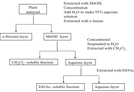
The shade dried aerial part of plant sample (100 g) was chopped in small pieces and then crushed into powder by a blinder. Plant sample was macerated in pure methanol for 24 hours. The sample then extracted using a percolator. The extracted solution was concentrated at 50 °C under reduced pressure to dryness. A small amount of water was added to MeOH to obtain a 95%-aqueous methanol solution. This solution was added to the concentrated extract was then extracted with an equal volume of n-hexane (3 times) to give an extract containing non-polar compounds. The methanol layer was evaporated to dryness and then suspensioned in water. The suspension was partitioned between CH2Cl2 and ETOAC, successively. Each obtained extract was concentrated at
Figure 1. Portioning scheme using immiscible solvent.
Plant material
Extracted with MeOH, Concentration
Add H2O to make 95% aqueous
solution
Extracted with n-hexan
MeOH layer
n-Hexane layer
CH2Cl2-soluble fraction
Concentrated Suspended in H2O
Extracted with CH2Cl2
Aqueous layer
EtOAc- soluble fraction
Extracted with EtOAc
Aqueous layer
Cell culture and treatment
AGS (human gastric carcinoma cell line) and L929 (mouse fibroblast cell line) were obtained from Natural Cell Bank of Iran (NCBI), Tehran, Iran. Cell lines were cultured as a monolayer culture in Eagle’s medium, modified according to Dulbecco (DMEM from Gibco Co., USA) and supplemented with 10% fetal bovine serum (Gibco Co., USA), 1% penicillin/streptomycin (100 IU ml-1 and 100 µgml-1, respectively from Gibco Co., USA) in a 5% CO2 humidified atmosphere at
37°C. For the experiments, cells were removed from the flasks using a 0.25% Trypsin-EDTA solution.
Growth inhibition assay
Growth inhibition of tumor and normal cells by obtained extracts was measured by a rapid colorimetric assay, using 3-(4,5-dimethylthiazol-2-yl)-2,5-diphenyl tetrazolium bromide (MTT) and compared with untreated controls (9). The assay is based on the metabolic reduction of soluble MTT by mitochondrial enzyme activity of viable tumor cells into an insoluble colored formazan product, which can be measured spectrophotometrically after dissolving in dimethyl sulfoxide (DMSO). Briefly, cells (1×10⁴ cells per each well) were seeded into 96-well microculture plates and allowed to adhere for 24 h before treatment. Then, each cell line was exposed to extracts at 250, 500, 1000, 1500, 2000, 2500, 3000 and 4000 µg/ml concentrations for 24 h. The first column of each microplate was assumed as negative control (containing no extracts). All different treatments done by setting up three independent experiments on separate days, each time with three or more wells for each concentration in 96-well plates. To assay the cell growth, 25 µl of MTT solution (5 mg ml-1 in phosphate buffer solution) was added to each well and the plate was incubated for 3 h at 37°C. Then, 200 µl of DMSO was replaced and pipetted to dissolve any formazan crystals. The optical density (OD) was read on an Elisa reader (Microplate reader MR 600, Dynatech, USA) at a wavelength of 570 nm. The inhibitory rate of cell proliferation was calculated by the following formula:
Growth inhibition (%) = (OD control - OD treated)/OD control × 100%.
Also, growth inhibitory activity of extracts against cell lines expressed as IC50 values (the extract
concentration reducing 50% in the absorbance of treated cells, with respect to untreated cells) were determined from dose response curves.
Determination of cell death pattern
In brief, AGS cells were cultured overnight in 24-well culture plate (2×10⁵ cells) and then treated with IC50 values of different extract from Artemisia annua for 24 h. After incubation, cells were
harvested with 0.25% trypsin-EDTA solution, washed with PBS and then resuspended in Annexin V-FITC binding buffer and incubated at room temperature for 5 min in the dark with 5 µl of Annexin-V FITC and 5 µl of PI. Cells were analyzed (10,000 events) by flow cytomety (Ex. = 488nm; Em. = 530nm) using a FACScan flow cytometer (Becton Dickinson) within 1 h (10). The results has shown as the induction of early apoptosis (Annexin V+/PI-) versus late apoptosis/necrosis (Annexin V+/PI+ or Annexin V-/PI+) in treated cells.
Statistical analysis
Data were expressed as mean ± SD. The significance of difference was evaluated by ANOVA test. A probability level of P < 0.05 was considered statistically significant.
Results
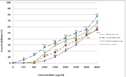
As the first step of our investigation, we assessed the effect of methanol, ethylacetate, dichloromethane and hexan extracts of Artemisia annua on the growth of gastric cancer cell line (AGS) after 24 h exposure (Figure 2). All different concentrations of extracts caused significant decreases in cancer cell growth (p<0.01). Low concentration (250 µg/ml) of ethylacetate extract had inhibitory effect (p≤0.05) and higher concentrations showed more inhibitory effect (p<0.01) on AGS cancer cells. Concentration of 250 µg/ml of dichloromethan extract had no significant (p=0.28) toxicity on AGS cells. But it had significant inhibitory activity in higher concentrations. Growth of AGS cells after treatment with 250 µg/ml and 500 µg/ml of hexan extract didn’t significantly (p=0.13, p=0.64) changed. Comparing of growth inhibitory activity of extracts revealed that different concentrations of methanol extracts had the highest and hexan extract had the weakest inhibitory effect on AGS cell growth (Figure 1). IC50 values of methanol, ethylacetate,
dichloromethane and hexan extracts were 500, 1259, 1167 and 2250 µg/ml, respectively.
Also, we evaluated the growth inhibitory activity of methanol, ethylacetate, dichloromethane and hexan extracts of Artemisia annua on fibroblast cell line (L929) after 24 h exposure (Figure 3). All concentrations of methanol extract had less toxic effect on L929 cells rather than AGS cells. Concentration of 250 µg/ml and 500 µg/ml of ethylacetate extract had no toxic effect on L929 cells. But, higher concentrations showed significant inhibitory (p<0.01) effect. Dichloromethan extract at 250 µg/ml had no toxic effect on L929 cells and at 500 µg/ml concentration didn’t show significant (p=0.23) inhibitory effect on L929 cells. Concentration of 250 µg/ml and 500 µg/ml of hexan extract didn’t show any toxicity on L929 cells. Concentration of 1000 µg/ml from hexan extract inhibited the growth of L929 cells (p≤0.05). All of the extracts had less inhibitory effect on L929 normal cells rather than AGS cancer cells.
Figure 3. Growth inhibition effect of different extracts from Artemisia annua on L929 fibroblast cells was shown as mean ± SD. The asterisks are indicator of statistical differences obtained separately at different time points compared to their controls shown in figure as *P < 0.05, **P < 0.01 and ***P < 0.001.
FACS analysis was used to quantify apoptotic and or necrotic death after treatment with IC50 values
Figure 4. Cell death pattern induced by IC50 values of different extract from Artemisia annua in
AGS cells after 24 h. Cells were stained with Annexin V-FITC and propidium iodide (PI) to determine quantitative evidences for increased apoptosis and/or necrosis in treated cancer cells.
Discussion
Cancer causes significant morbidity and mortality and is a major public health problem worldwide (11). Since the majority of cancer chemotherapeutants severely affect the hosts' normal cells, the use of natural products has now exceptional value in the control of cancers (12, 13). One of the approaches is the selection of a plant based on the prior information on its folk medicinal usages (14, 15).
Artemisia annua is a good candidate to look for chemopreventive agents because of its antitumor activity on different cancer cells. Essential oil of Artemisia annua could induce apoptosis of cultured SMMC-7721 cells (16). Artemisinin, a compound isolated from the sweet wormwood (A. annua), has shown to have selective toxicity towards hepatocarcinoma cell line in vitro. Also, it given orally to retard breast cancer development in DMBA-treated rats (17). The cytotoxic activity of nine terpenoids and flavonoids isolated from Artemisia annua were showed significant cytotoxicity against P-388, A-549, HT-29, MCF-7, and KB tumor cells (7). Aqueous and methanol herbal extracts from Artemisia annua showed inhibitory activity against human digestive tumor cell
line. Concentration of 1000 µg/ml of its ethanol extract caused a 51% until 70% decrease in the growth of human liver carcinoma cell line (SMMC-7721), human gastric cancer cell line (BGC-823), human colon adenocarcinoma cell lines (LoVo and SW-116) and esophagus adenocarcinoma cell line (CaEs-17). Aqueous extract showed little cytotoxicity on cancer cells. So, it has been concluded that ethanol extract contains main anticancer constituents (18).
These extracts mainly inhibited the proliferation of gastric cancer AGS cells and not normal human fibroblast-like cells that is shows specificity of these extracts towards neoplastic cells. Thus, our data confirmed previous study that showed cytotoxicity of Artemisiaannua extracts on cancer cell lines (16-18). Also, we showed that the inhibition of the proliferation of AGS cells by these extracts was the result of apoptosis induction that was evidenced by FACS analyses. In which, treated AGS cells showed high percentage of Annexin V positive cells. Methanol, ethylacetate, dichloromethane and hexan extracts caused significant induction of early apoptosis at 60%, 50%, 51% and 51%, respectively. Also, treatment with these extracts induced increase in late apoptosis/necrosis at 5%, 13%, 6% and 15%, respectively.
In brief, we prepared different extracts from Artemisia annua that had promising antitumor activities. These extracts inhibited AGS gastric cancer cell growth by the inhibition of proliferation and inducing apoptosis. These extracts selectively showed anti-proliferative effects on AGS cancer cells and had less toxicity on normal human fibroblast-like cells.
In comparison with our results, methanol extract prepared in our study showed high antitumor effect too. Also, we suggested that main anticancer constituents of Artemisia annua are in its alcohol extract. Because of high cytotoxicity of other extracts of Artemisia annua, we recommend to more experiments on non-polar extract of Artemisia annua in order to isolation of new anticancer compounds.
So, Artemisia annua could be considered as a promising chemotherapeutic agent in cancer treatment. Future work will focus on isolation and elucidation of antitumor compounds from tested extracts and assessment of their antitumor effect on gastric cancer AGS cells.
Acknowledgements
The authors would like to thank the authorities in research council of Mashhad University of Medical Sciences (MUMS).
References
1- Rao CV, Newmark HL, Reddy BS. Chemopreventive effect of farnesol and lanosterol on colon carcinogenesis. Cancer Detect Prev 2002; 26: 419-425.
2- Zhang H, Spitz MR, Tomlinson GE, Schabath MB, Minna JD, Wu X. Modification of lung cancer susceptibility by green tea extract as measured by the comet assay. Cancer Detect Prev 2002; 26: 414–418.
3- Hengartner MO. The biochemistry of apoptosis. Nature 2000; 407: 770-776.
4- Brown JM, Wouters BG. Apoptosis, p53, and tumor cell sensitivity to anticancer agents. Cancer Res 1999; 59: 1391-1399.
5- Mucciaralli M, Maffel M. Introduction of the genus In: Wright CW, eds. Artemisia. Taylor and Francis, London, 2002: 1-50.
6- Tan RV, Zheng WF, Tang HQ. Biologically active substances from genus Artemisia. Planta Med 1998; 64: 295-302.
7- Zheng GQ. Cytotoxic terpenoides and flavonoides from Artemisia annua. Planta Med 1994; 60: 54-57.
8- Cakir A, Kilic H, Kodali S, Mavi A, Yildirim A. Screening of chemical composition and antifungal and antioxidant activities of the essential oils from three Turkish Artemisia species. J Agric Food Chem 2005; 53: 1408-1416.
9- Mosmann T. Rapid colorimetric assay for cellular growth and survival: application to proliferation and cytotoxicity assays. J Immunol Methods 1983; 983:55-63.
11- Smith-Warner SA, Elmer PJ, Tharp TM, Fosdick L, Randall B, Gross M, Wood J, Potter JD. Increasing vegetable and fruit intake: randomized intervention and monitoring in an at-risk population. Cancer Epidemiol Biomar Prev 2000; 9 (3): 307–317.
12- Mascarenhas M. Structure–activity characterization, a quick method to screen mushrooms for the presence of antitumor glucans. Mushroom Research 1994; 3: 77–80.
13- Suffness M, Pezzuto JM. Assays related to cancer drug discovery In: Hostettmann K, eds. Methods in Plant Biochemistry: Assays for Bioactivity, vol. 6, Academic Press, London, 1990: 71– 133.
14- Chapuis J, Sordat B, Hostettmann K. Screening for cytotoxic activity of plants used in traditional medicine. Journal of Ethnopharmacology 1988; 23: 273–284.
15- Cordell GA, Beecher CCW, Pezzuto JM. Can ethnopharmacology contribute to the development of new anticancer drugs? Journal of Ethnopharmacology 1991; 32: 117–133.
16- Li Y, Li MY, Wang L, Jiang ZH, Li WY, Li H. Induction of apoptosis of cultured hepatocarcinoma cell by essential oil of Artemisia Annul L. Sichuan Da Xue Xue Bao Yi Xue Ban 2004; 35: 337-9.
17- Henry L, Narendra P. Singh Oral artemisinin prevents and delays the development of 7,12-dimethylbenz[a]anthracene (DMBA)-induced breast cancer in the rat. Cancer Lett 2006; 231: 43– 48.