University of Pennsylvania
ScholarlyCommons
Publicly Accessible Penn Dissertations
1-1-2012
Chemical Genetic Approaches for Elucidating
Protease Function and Drug-Target Potential in
Plasmodium Falciparum
Michael Harbut
University of Pennsylvania, [email protected]
Follow this and additional works at:http://repository.upenn.edu/edissertations
Part of theChemistry Commons,Parasitology Commons, and thePharmacology Commons
This paper is posted at ScholarlyCommons.http://repository.upenn.edu/edissertations/518
For more information, please [email protected].
Recommended Citation
Harbut, Michael, "Chemical Genetic Approaches for Elucidating Protease Function and Drug-Target Potential in Plasmodium Falciparum" (2012).Publicly Accessible Penn Dissertations. 518.
Chemical Genetic Approaches for Elucidating Protease Function and
Drug-Target Potential in Plasmodium Falciparum
Abstract
Plasmodium falciparum is a protozoan parasite and the causative agent of malaria, which kills upwards of 1 million people annually. With the increasing prevalence of drug-resistant parasites, considerable interest now exists in the identification of new biological targets for the development of new malaria chemotherapeutics. However, given the genetic intractability inherent in studying P. falciparum, it is imperative that novel approaches be developed if we are to understand the role of essential enzymes. My work presented here focuses on the development and use of chemical tools to study malarial proteases, a class of enzymes that have been shown to play essential roles throughout the parasite lifecycle, but the majority of which though are still uncharacterized. In Chapter 2 I develop a novel set of activity-based probes (ABPs) based on the natural product metallo-aminopeptidase (MAP) inhibitor bestatin. I show the bestatin-based ABP allows the functional characterization of MAP activity within a complex proteome. In Chapter 3, I utilize an extended library of bestatin-based ABPs to define the function of two essential malarial MAPs, PfA-M1 and Pf-LAP. I find that PfA-M1 is necessary in the proteolysis of hemoglobin and that lethal inhibition starves parasites of amino acids. I also show that Pf-LAP has a role other than hemoglobin digestion, as parasites are susceptible to its inhibition prior to the onset of this process. In Chapter 4, I use a suite of specific small molecules to validate the P. falciparum signal peptide peptidase (PfSPP) as a drug target. This work shows that PfSPP is a druggable enzyme and that parasites are extremely vulnerable to its inhibition. Evidence is also presented that suggests this enzyme may play an important role in the parasite's endoplasmic reticulum stress-response.
Degree Type Dissertation
Degree Name
Doctor of Philosophy (PhD)
Graduate Group Pharmacology
First Advisor
Doron C. Greenbaum
Keywords
Activity-based probe, Drug design, Protease, Small molecule
Subject Categories
CHEMICAL GENETIC APPROACHES FOR ELUCIDATING PROTEASE
FUNCTION AND DRUG-TARGET POTENTIAL IN PLASMODIUM
FALCIPARUM
Michael B. Harbut
A DISSERTATION
in
PHARMACOLOGY
Presented to the Faculties of the University of Pennsylvania
in
Partial Fulfillment of the Requirements for the
Degree of Doctor of Philosophy
2012
Supervisor of Dissertation:
Doron C. Greenbaum, PhD Assistant Professor of Pharmacology
Graduate Group Chairperson:
Vladimir R. Muzykantov, PhD Professor of Pharmacology
Dissertation Committee:
John B. Hogenesch, PhD, Associate Professor of Pharmacology (Chair)
Scott L. Diamond, PhD, Arthur E. Humphrey Professor of Chemical and Biomolecular Engineering
David S. Roos, PhD, E. Otis Kendall Professor of Biology
Acknowledgements
Presented herein is work that could only have come to fruition with the
support, both scientifically and otherwise, of numerous individuals. My most
immediate gratitude is dedicated towards my mentor, Doron Greenbaum. His
support facilitated this work in a variety of ways. Just as importantly, he fostered
an intellectually rigorous and enthusiastic scientific environment, the
consequences of which I will appreciate long after I’ve left the lab. I am also
grateful to the various members of the Greenbaum lab over the past five years
who always made the lab lively and enjoyable to be in.
I am appreciative of the time and guidance provided by my committee,
Drs. Scott Diamond, John Hogenesch, David Roos, and David Speicher. I am
grateful for support and funding provided by the Pharmacology Graduate Group
as well as the Parasitology Training Grant. This work was produced in
collaboration with many scientists, both at Penn and elsewhere, whose efforts,
advice, and outstanding science I am thankful for.
I have numerous friends who have provided a cathartic outlet from the
stress of lab for which I am extremely grateful. My family has always provided
support but never pushed me in any one direction, and for this deft touch, I am
and will always be extremely thankful. Finally, Laura has been the one variable
that has remained constant during the last five years, for which I am more
Abstract
CHEMICAL GENETIC APPROACHES FOR ELUCIDATING PROTEASE
FUNCTION AND DRUG-TARGET POTENTIAL IN PLASMODIUM
FALCIPARUM
Michael B. Harbut
Advisor: Doron C. Greenbaum, PhD
Table of Contents
TITLE………i
ACKNOWLEDGEMENTS………ii
ABSTRACT………..……….…iii
TABLE OF CONTENTS.……….…iv
LIST OF TABLES...….………...vii
LIST OF FIGURES.………...…viii
LIST OF SCHEMES……….x
CHAPTER 1: INTRODUCTION………..………1
1.1 Malaria 1
1.2 The Plasmodium falciparum life cycle 4
1.3 Proteolytic enzymes and their roles during the erythrocytic stage of P. falciparum 7
1.4 Understanding protein function in P. falciparum: a chemical-genetics approach 11
1.5 Using chemical genetics to study protease function in P. falciparum 13
1.6 Proteases as drug targets in P. falciparum 16
CHAPTER 2: DEVELOPMENT OF BESTATIN-BASED ACTIVITY-BASED PROBES FOR METALLO-AMINOPEPTIDASES...21
2.1 Introduction 20
2.2 Results and Discussion 25
2.3 Experimental procedures 36
3.2 Results 41
3.3 Discussion 64
3.4 Experimental procedures 68
CHAPTER 4: CHEMICAL VALIDATION OF SIGNAL PEPTIDE PEPTIDASES AS POTENTIAL ANTI-PROTOZOAN DRUG TARGET...78
4.1 Introduction 78
4.2 Results 81
4.3 Discussion 98
4.4 Experimental procedures 101
CHAPTER 5: CONCLUSIONS AND FUTURE DIRECTONS...105
5.1 A bestatin-based chemical biology strategy reveals distinct roles for M1 and M17 family aminopeptidases 105
5.2 Chemical validation of signal peptide peptidases as potential anti-protozoan targets 109
REFERENCES...116
List of Tables
List of Figures
Figure 1.1: The malaria parasite erythrocytic pathway………...6
Figure 2.1: General mode of binding of bestatin………...24
Figure 2.2: Anatomy of ABPs and labeling of aminopeptidase N………...28
Figure 2.3: Aminopeptidase N ABP labeling by fluorophore-containing bestatin.30
Figure 2.4: “Click” chemistry-based ABP labeling of aminopeptidase N………...33
Figure 2.5: ABP labeling of the malarial M1 metallo-aminopeptidase in cell
lysates……….34
Figure 3.1: Identification of PfA-M1 and Pf-LAP as the targets of the anti-parasitic
MAP inhibitor bestatin………...………44
Figure 3.2: Bestatin-based ABP libraries reveal distinct chemotypes produced by
ABPs with increased specificity for either PfA-M1 or Pf-LAP……...48
Figure 3:3: Biochemical and structural characterization of BTA and PNAP
specificity against PfA-M1 and Pf-LAP…...………53
Figure 3:4: DV-localised endoproteases are not inhibited by BTA……….54
Figure 3.5: Inhibition of PfA-M1 kills parasites via disruption of Hb digestion
whereas Pf-LAP kills via a distinct mechanism……….58
Figure 3.6: Inhibition of PfA-M1 causes DV swelling but does not prevent
proteoytic cleavage of full length Hb…..………61
Figure 3.7: Inhibition of PfA-M1 blocks proteolysis of specific Hb oligopeptides.62
Figure 3.8: Small dipeptide species accumulate in BTA-treated parasites……...63
Figure 4.1:SPP/PS Inhibitor pharmacology against protozoan parasites and
Figure 4.2: Yeast activity-based SPP assay………..87
Figure 4.3: Generation of parasites resistant to potent PfSPP inhibitors………..91
Figure 4.4: PfSPP localizes to the parasite ER and is important during the
trophozoite stage...………94
Figure 4.5: Inhibition of PfSPP synergizes with ER stress in P. falciparum and
potentiates the antimalarial effect of an inhibitor of the ERAD
List of Schemes
Scheme 2.1: Synthesis of bestatin-based ABPs………..……….27
Chapter 1: Introduction
1.1 The burden of malaria
Plasmodium is the etiological agent of malaria, a devastating global public
health burden that each year leaves over 400 million people infected and up to 1
million dead, over 75% of them African children [1]. The disease manifests itself
as a serious illness characterized by recurrent fevers, metabolic acidosis,
respiratory distress, and anemia, and fatal cases occur disproportionately in
young children [2]. Four species of Plasmodium infect humans and all are
transmitted through the bite of the female Anopheles mosquito. The species
Plasmodium falciparum is the most severe, accounting for the vast majority of
malaria-associated deaths. This is largely the result of its propensity to produce
surface changes upon the infected erythrocyte that cause cytoadherence and
disruption of the microvasculature in organs such as the lungs, kidneys, and
brain. [3]. While over 50% of the world’s population is exposed to malaria, the
African continent shows the greatest burden from this disease and accounts for
more than 90% of all malaria deaths. This, along with the compounding burdens
of tuberculosis and HIV/AIDS within these populations, has had catastrophic
socio-economic consequences for sub-Saharan South Africa [4].
While malaria is today perceived as a tropical disease, it is only in the last
century that malaria has been eliminated from most developed Western nations.
Elimination of malaria from North Africa and Europe was achieved primarily
through the systematic control or elimination of the mosquito vector, such as
breeding habitats [5]. The World Health Organization launched the Global
Malaria Eradication Program in 1955, was key to this accomplishment in Europe,
but similar success was not shared in sub-Saharan Arica [6]. In the second half
of the nineteenth century eradication efforts were largely abandoned due to
diminishing resources, strife, concern about DDT, insecticide resistance, and the
growing thought that eradication in Africa was virtually impossible.
Today, where it is endemic, malaria is managed by two primary methods:
1) vector control (indoor residual spraying, insecticide treated nets) and 2)
treatment. While a significant effort has been put towards the development of a
malaria vaccine, the most advanced candidate (RTS,S) has shown only modest
success in clinical trials and is not likely to see widespread use until 2015 [7]. In
addition, almost complete vector coverage will still necessitate the use of drug
administration to help eliminate malaria in high transmission areas.
Quinine, derived from the bark of the tree Cinchona calisaya, has been
used to treat malaria since the 17th Century. Due to its low cost and ease of use,
the quinine-related drug chloroquine remained a constant in the control of acute
uncomplicated malaria for over 40 years since the discovery of its antimalarial
properties in 1946 [8]. Chloroquine inhibits the parasite’s mechanism for heme
detoxification, likely resulting in lethal lysis of the lysosome-like organelle where
this occurs. Other frequently used antimalarials target parasite-essential
metabolic pathways, such as the tetrahydrofolic acid synthesis
Unfortunately, P. falciparum resistance to chloroquine arose within 20
years of its introduction and by the 1980s widespread resistance was reported in
most high-transmission areas, virtually nullifying the effectiveness this drug.
Resistance to drugs with specific enzyme targets such as
sufadoxine-pyrimethamine arose at an even quicker pace [9].
Modern malaria chemotherapy relies on the active ingredient of the
Qinghao plant, a sesquiterpene lactone called artemisinin, and its use in
combinations with other antimalarials. The mechanism of action of artemisinin
and artemisinin derivatives is still debated, but its properties have facilitated its
use as a frontline defense, including rapid activity against existing drug-resistant
strains and blood stages of the parasite, in addition to mature sexual stage
gametocytes, which helps reduce transmission. By using artemisinin in
combination therapy (ACT), that is, two medicines with different mechanisms of
action, the probability for the emergence of parasite resistance is drastically
reduced. Indeed, in areas where artemisinin monotherepy has predominated,
reports of resistance are beginning to emerge [10].
The global spread of resistance to current antimalarial standards has
spurred an innovation in malaria research over the last 20 years. The current
antimalarial drug-development pipeline is mostly devoid of newly defined
molecular targets, being dominated by alternative artemisinin-based combination
therapies or new generations of validated inhibitors (www.mmv.org). Thus, it is
pathways and enzymes. This way, a more rational approach to the validation of
novel targets may be achieved to further the development of new antimalarials.
1.2 The Plasmodium falciparum life cycle
Plasmodium spp are obligate intracellular protozoan parasites, and
progress through a complicated life cycle both in a mosquito vector and human
host. The asexual erythrocytic cycle, which in P. falciparum is synchronous and
lasts 48 hrs, is the cause of morbidity and mortality associated with malaria [11].
Transmission occurs via the bite of an Anopheles mosquito, which injects
sporozoite forms that migrate to the liver. After multiple rounds of replication in
hepatocytes, merozoites are released into the blood stream and the erythrocytic
life cycle of the parasite begins. This cycle is initiated by the invasion of host red
blood cells by the merozoites and the formation of a parasitophorous vacuole
inside the erythrocyte within which the new ring stage parasite resides. Parasites
progress through the ring stage for approximately 20 hrs. This is followed by a
highly metabolically active growth stage, the trophozoite stage, which is
accompanied by the uptake and degradation of host hemoglobin. At about 36 hrs
post invasion, parasites begin a process of asexual reproduction leading to the
formation of 16-32 daughter merozoites. Finally, the merozoite-containing red
blood cells are lysed around 48 hrs post infection, releasing the parasites, and
Catabolic processes are known to mediate a number of events during the
parasite’s life cycle. Essential to those processes are protease enzymes, both
Figure 1.1: The malaria parasite erythrocytic pathway. Merozoite parasites invade the erythrocyte (RBC) and develop into ring stage parasites within the parasitophorous vacuole (PV). The peak of the parasite’s metabolic activity occurs during the growing trophozoite stage, during which host cell cytoplasm is endocytosed and large-scale hemoglobin proteolysis in carried out within the digestive vacuole (DV). In addition, the parasite sets up a complex secretory pathway during this stage for delivering proteins to the PV and host cell. During the schizont stage multiple rounds of nuclear division occur, which results in the formation of daughter merozoites. At roughly 48 hrs post infection (for P. falciparum), the parasite ruptures from the host cell.
D V
DV
DV DV
1.3 Proteolytic enzymes and their roles during the erythrocytic stage of P. falciparum
Proteolytic enzymes, or proteases, catalyze the degradation of proteins or
peptides and have wide-ranging and important roles from single-cell prokaryotes
to metazoans. There are approximately 100 predicted proteases in the
Plasmodium genome that span all classes and several proteases are thought to
participate in critical pathways during the life cycle of P. falciparum [12]. As such,
proteases and the biological events they mediate have been considered potential
anti-malarial drug targets. Specifically, three events during the erythrocytic life
cycle have been extensively studied so as to uncover the proteolytic events
necessary: 1) merozoite invasion of the erythrocyte, 2) hemoglobin degradation,
and 3) egress from the erythrocyte [13-15].
Early evidence for the importance of proteolysis during invasion was
uncovered using purified P. knowlesi merozoites. Pre-treatment of the
merozoites with a variety of protease inhibitors significantly decreased the
invasion capacity of the parasite [16]. Follow-up work in P. falciparum has
focused on primarily on the role of proteases that process merozoite-associated
proteins. During invasion, sets of adhesin molecules are released from the
parasite apical secretory organelles, the micronemes, which facilitate high affinity
binding of the host cell and parasite. The most abundant of these is a glycosyl
phosphatidylinositol (GPI)-anchored protein named merozoite surface protein-1
and at the end of schizogony is proteolytically processed by a serine protease
called PfSUB1, resulting in a non-covalent complex of several polypeptides. Two
other proteins are also part of this complex (MSP6 and MSP7) and are
processed as well. The importance of this primary processing event is still
unclear, but it is possible that it allows a conformational change that enables the
complex to function on the free merozoite at the time of invasion. During
subsequent invasion events, the N-terminus is shed from the merozoite surface
by a membrane-bound subtilisin-like protease, PfSUB2 [18].
Apical membrane antigen 1 (AMA-1) is another important adhesin whose
function depends on proteolytic processing [19,20]. Like MSP1, AMA-1 is
synthesized in the apical complex of the merozoite and undergoes multiple
rounds of proteolytic maturation prior to circumferential redistribution on the
merozoite surface. In addition, once it is translocated from the apical complex to
the parasite surface it is then shed from the membrane by PfSUB2. Additional
cleavage is carried out by an intramembrane serine protease, rhomboid 1
(PfROM1), the biological relevance of which is still being assessed [21].
Once established inside the erythrocyte, the parasite begins to
endocytose host hemoglobin (Hb) and proteolytically digests it into its constituent
amino acids within a lysosome-like organelle called the digestive vacuole (DV)
[15]. This process peaks during the parasite’s most active metabolic stage,
between 20-36 hours post infection, and is likely carried out to relieve osmotic
pressure for the growing parasite and for utilization of individual amino acids
parasite, and has long been considered an “Achilles heel” of the parasite. Thus
the putative proteases involved have been focus of intense study for their drug
target potential.
The upstream endoproteases that catalyze the initial cleavage of the Hb
molecule are the aspartyl protease plasmepsins (I-IV) and the cysteine protease
falcipains (2, 2’, 3) [24,25]. The roles for cysteine and aspartyl protease activity
were originally identified by biochemical characterization of whole DVs and
purified enzymes, where the hydrolysis of native and denatured hemoglobin was
analyzed under assumed physiologic conditions [26-28]. The discovery of
additional proteases was facilitated by the P. falciparum genome sequencing
project. The active roles of the individual enzymes are still uncertain. Some have
described it as an ordered process, initiated by the plasmepsins at the α
Phe33-Leu34 conserved hinge region of Hb, whereas others have suggested that the
falcipains initially cleave rapidly at multiple sites in intact hemoglobin [15,29]. A
striking functional redundancy exists amongst these proteases, suggesting the
importance of this process to the parasite. For instance, the falcipains appear to
be able to compensate for the loss of all four DV plasmepsins [30]. A
metalloprotease, falcilysin, has also been localized to the DV and is capable of
cleaving small Hb peptides 11-15 residues in length, suggesting it has a role
downstream of the plasmepsins and falcipains [31]. Finally, exopeptidases
release individual amino acids from the N-terminus of Hb, and their roles are
At the end of its erythrocytic life cycle, the parasite disrupts both the
membrane to the parasitophorous vacuole and the host erythrocytes to allow
egress from the cell. That protease inhibitors can block egress tells us that
rupture is indeed an active process and not a passive consequence of parasite
growth [32]. The SERA family of proteins have been a primary focus of
protease-mediated rupture in P. falciparum based on the evidence that the P. berghei
ortholog to SERA8 is essential for egress from the oocyst wall of the mosquito
midgut [33]. The gene products of all the SERA family members have been
detected in the asexual blood stage during late schizogony. In addition, SERA5,
an essential gene product, is proteolytically processed during rupture. Processed
products of SERA5 precursor are found in culture supernatants, and only the
precursor could be detected in unruptured schizonts [34,35]. Mechanical
disruption of parasites releases unprocessed SERA5, suggesting that a specific
pathway of events is necessary during rupture for SERA processing. The
mechanistic importance of this processing has yet to be uncovered. Intriguingly,
the SERA proteins all share a conserved papain-like cysteine protease domain,
suggesting that proteolytic processing of the respective SERAs may represent an
activation event of a non-catalytic zymogen precursor. While a refolded papain
domain of SERA5 has show weak chymotrypsin-like activity, the role of SERAs
during egress remain to be elucidated [36].
Even with our current limited understanding of protease function in P.
falciparum biology, the critical role these enzymes have in the biology of the
novel essential functions, the results of which may lead to putative drug target
candidates. To facilitate these discoveries, tools beyond classical genetic
approaches, which remain burdensome in P. falciparum, must be developed and
utilized.
1.4 Understanding protein function in P. falciparum: a chemical-genetics approach
Significant biochemical and biological work has helped to elucidate
essential protease-mediated events during the P. falciparum life cycle. Yet, of the
approximately 100 proteases encoded by the parasite genome, fewer than 15
have been fully characterized, making this biological system ripe for study.
Unfortunately, the systematic approaches used today to study gene function in
other organisms, such as RNAi, are not available in P. falciparum, leaving
classical gene disruption, via homologous recombination, the main genetic
technique [37]. Additionally, the procedure for targeted gene disruption is
inherently labor-intensive, slow, and inefficient, due to difficulties in the culturing
of the parasite and extremely low transfection rates. These technical limitations
and the fact that the parasite is grown as a haploid renders it currently difficult to
genetically disrupt essential genes. The development of a tetracycline-based
conditional gene expression system in the related apicomplexan organism
Toxoplasma gondii has not been successfully applied for functional studies in P.
falciparum [38].
molecules to modulate the activity of protein families or individual proteins to
provide insight into protein function and validate potential therapeutic targets.
This assemblage of techniques is broadly called “chemical-genetics” [39]. As
opposed to manipulation of DNA, as in classical chemical genetics, small
molecules usually modulate protein function, such as by active site binding or
preventing protein-protein interactions. In addition, small molecules offer greater
temporal control, allowing for the discrimination of effects at restricted stages.
Two approaches to chemical-genetics can be undertaken to discover
small molecules that modify protein function. The first approach is analogous to
classical reverse genetics, in which a gene of interest is permanently or
conditionally disrupted or modified in such a way that protein it codes for ceases
to function. The knockout or knockdown of the gene can give functional insights
as to its role in the system under study. Reverse chemical genetic strategies aim
to identify small molecules that target a specific protein. These are often carried
out through traditional medicinal chemistry efforts with significant underlying
knowledge about the protein of interest. Recently, the Wandless lab developed a
novel reverse chemical genetics strategy, that allows for the regulatable control
of protein stability [40]. The approach relies on FK506-binding protein (FKBP)
mutants that contain a degron that normally destabilizes the protein, but is
stabilized by a small molecule. This degron can be appended to a protein of
interest and will destabilize the protein in the absence of the small molecule.
Application or removal of the small molecule allows for the conditional expression
falciparum to validate the role of a calcium dependent kinase (PfCDPK5) in
egress [41]. Parasites expressing the kinase with the destabilization domain
failed to rupture from the host erythrocyte when the stabilizing small molecule
was removed from culture. This phenotype was rescued upon application of the
small molecule and subsequent stabilization of the kinase.
The second chemical genetics approach is known as forward chemical
genetics. This technique seeks to identify a protein responsible for a phenotype
or pathology under study. This is often done through the use of small molecule
libraries in phenotypic screens. Upon the small molecule-mediated production of
a phenotype of interest, the target(s) of the small molecule that produces the
phenotype is identified. High throughput screens in P. falciparum are still
primarily focused on parasite replication, and informative assays such as protein
export to the erythrocyte surface are complex and not as amenable to high
throughput screening. As such, phenotypic screens are rare for P. falciparum.
1.5 Using chemical genetics to study protease function in P. falciparum Small-molecule based approaches have been critical to the understanding
of protease function in P. falciparum. In a study to assess the role of proteases
during invasion, Dluzewski and colleagues uncovered the first evidence for the
proteolytic breakdown of hemoglobin within the digestive vacuole (DV) after
discovering the accumulation of undegraded hemoglobin within the swollen DV of
parasites treated with leupeptin (a general cysteine and serine protease inhibitor)
protease activity by inhibiting Hb degradation in parasites with cysteine protease
inhibitors [28]. Biochemical characterization of Hb digestion from purified DVs
also facilitated the discovery of aspartyl protease activity in this process [26].
These studies, using inhibitors with broad selectivity, could at best suggest a role
for a specific class of proteases involved in Hb degradation. Only with extensive
studies, including expression analysis, immunofluorescence localization,
biochemical purification and characterization, and finally genetic knockout, could
the individual roles of the endoproteases involved in Hb catabolism be ascribed
to particular proteins. Further study of the putative downstream exopeptidases in
the Hb catabolic pathway has been hindered because they are genetically
essential and refractory to knockout [31,43,44].
More recently, much progress has been made in identifying the proteases
required for rupture from the host erythrocyte, using specific inhibitors and both
reverse and forward chemical genetic approaches. The Bogyo lab recently
undertook a forward chemical genetics screen that utilized a facile and rapid
method for the assessment of parasite staging by flow cytometry [45]. This
allowed the authors to screen a library of over 1,200 covalent serine and cysteine
protease inhibitors that blocked parasite egress. Upon identification of a serine
protease inhibitor that blocked egress (JCP-104), they then undertook a
chemical-genetics approach known as activity–based protein profiling (ABPP).
ABPP is a chemical strategy that utilizes mechanism-based, tagged small
molecule inhibitors. Appending a reporter tag such as a biotin or fluorophore to
fluorescent scanning) and the identification of the inhibitor target(s) by
biotin-(strept)avidin chromatography capture, gel separation, and mass spectrometry
analysis. Using JCP-104 as an ABP allowed the identification of the protein
target of the inhibitor, PfSUB1. In addition, it was shown that JCP-104 inhibited
the processing of SERA5, the blocking of which prevented egress.
In this same study, a cysteine protease capable of blocking rupture was
also identified. Again, the suitability of ABPP in P. falciparum was illustrated
when the authors were able to identify the target of the inhibitor, JCP-405, by
utilizing the inhibitor as an ABP, and showed that it targeted two cysteine
proteases, dipeptidyl-aminopeptidases 1 and 3 (DPAP3). DPAP1 was previously
implicated as a hemoglobinase, while the function of DPAP3 was unknown.
Using a forward chemical genetics strategy, the authors screened a focused
library of dipeptide vinylsulfone inhibitors similar to JCP-405 to identify selective
inhibitors of DPAP1 and DPAP3. Upon identification of specific inhibitors, they
showed that selective inhibition of DPA3 resulted in a block of parasite rupture,
prior to PfSUB1 processing of SERA5.
The treatment of late-stage parasites with the cysteine protease inhibitor
E64 results in the accumulation of merozoites locked in the erythrocyte. E64 is a
general cysteine protease inhibitor and thus targets multiple cysteine proteases
[46]. The Greenbaum lab recently utilized chemical genetics combined with
biochemical and cell-biological approaches to reveal a role for the
host-erythrocyte calpain protease in rupture [47]. Using an activity-based probe (ABP)
schizogony and localized that the host cell membrane upon activation. Using an
endogenous inhibitor specific for calpain 1, calpastatin, along with
immunodepletion of calpain 1 the authors showed that human calpain 1 activity
was coopted by the parasite and necessary for degradation of the host cell.
Proteases that mediate invasion have also been uncovered using
chemical genetic approaches. Using a chemical screen, the Bogyo lab showed
that falcipain 1 was the only active parasite cysteine protease during the invasive
merozoite stage [48]. In a reverse chemical genetics approach, they identified a
specific inhibitor of falcipain 1 by screening a small chemical library. Use of this
specific inhibitor showed that inhibition falcipain 1 had no effect on parasite
growth or hemoglobin degradation, but did result in unruptured schizonts,
illustrating that falcipain 1 was indeed necessary for egress.
These studies demonstrate the promise and feasibility of using chemical
tools in forwarding the understanding of protease roles in P. falciparum biology.
Furthermore, they also help to provide proof of principle in the druggability of
protease targets and identify putative starting points for drug development
initiatives.
1.6 Proteases as drug targets in P. falciparum
The validity of targeting proteases to alleviate a pathology as a therapeutic
strategy is illustrated by the clinical success of a number of protease inhibitor
drugs, including inhibitors of HIV aspartyl protease, dipeptidyl peptidase IV
[49]. As the roles of many proteases in P. falciparum are essential for the
parasite’s life cycle, it is not unreasonable to consider the parasite proteases as
potential drug targets. Moreover, extensive biochemical knowledge of protease
mechanism and structure in addition to the existence of focused inhibitor libraries
allows for a rational approach to the design of potential drugs.
Though the sequencing of the P. falciparum genome has led to the
identification of at least 100 putative proteases, viewing proteases of P.
falciparum as drug targets is a strategy that predates that long predates the
genome-sequencing effort [50]. Two of the most significant efforts were directed
towards the hemoglobin-catabolizing proteases, the cysteine protease falcipains
and aspartyl protease plasmepsin. Efforts to identify optimal inhibitors to these
enzymes were facilitated through screening of existing cysteine and aspartyl
protease inhibitor libraries and through the development of novel inhibitors based
on scaffolds known to inhibit the respective protease families [24,51]. Genetic
approaches were also utilized, and numerous labs showed through the knockout
of the individual genes that exceptional functional redundancy existed amongst
all four plasmepsins and also falcipains (only falcipain-3 was shown to be
essential) [30,52,53]. Unfortunately, plasmepsin inhibitors with high potency
towards the recombinant enzyme often showed limited potency against cultured
parasites [24]. In addition, inhibitors for both the falcipains and plasmepsins
showed problems with bioavailability, undermining their effectiveness [54]. The
combination of these issues has recently limited enthusiasm for these enzymes
Multiple other proteases are expected to have a role in hemoglobin
degradation, working downstream on the peptide products of Hb produced by
plasmepsin, and may represent improved drug targets over falcipains or
plasmepsins. These proteases include falcilysin, a metallo-aminopeptidases
(PfA-M1), a proline aminopeptidase (PfAPP), and a cathepsin C-like cysteine
protease DPAP1. All four have been validated as genetically essential during the
erythrocytic stage. DPAP1 and PfA-M1 are the only proteases of the group
shown to be amenable to chemical inhibition. A recent study utilizing ABPs to
characterize DPAP1 activity identified nonpeptidic covalent inhibitors of DPAP1
lethal to parasites at low nanomolar concentrations. Unfortunately, the most
potent inhibitors of DPAP1 were toxic in mice, while a less potent but non-toxic
inhibitor did decrease parasite levels in a mouse model.
Given the parasite-essential nature, proteases involved in rupture and
invasion may also represent attractive drug targets. DPAP3, SERA5, PfSUB1,
and PfSUB2 have each been shown to be genetically essential. In addition,
DPAP3 and PfSUB1 are amenable to chemical inhibition in live parasites.
PfSUB2 has not been targeted by small molecules, but a PfSUB2 propeptide
inhibits PfSUB2 sheddase activity in parasites. SERA5 represents an unusual
cysteine protease in that a cysteine-to-serine replacement has occurred within its
putative canonical catalytic site. Its protease-like domain has been expressed
and, upon renaturing, shown to have weak chymotrypsin-like protease activity
that was reversible with a serine protease inhibitor, although the biological
characterization of SERA5 revealed a number of anomalies in addition to the
non-canonical active site, further complicating any rational drug discovery efforts
centering on SERA5.
Proteases represent a highly druggable class of parasite enzymes, based
both on our present knowledge of their enzymology and keys to inhibition as well
as their importance in numerous biological processes during the P. falciparum life
cycle. With the increase of data from analysis of the P. falciparum genome
project our awareness of putative protease targets is great. At the same time,
translating these potential targets into validated targets requires not just
knowledge of the enzyme’s existence, but a better foundational understanding of
the biology and biochemistry of the protease within the context of the parasite’s
life cycle, which will allow us to fully exploit this class of enzymes. This
understanding will only come about with the development of novel technologies,
such as activity-based protein profiling, coupled with classical cell biological,
biochemical, and genetic techniques. Herein, work is presented that exploits
these and other approaches in an attempt to forward our knowledge of P.
falciparum protease biology while testing potential as drug-target. In Chapter 2 I
present work on the development of a novel activity-based probe based on the
metallo-aminopeptidase inhibitor, bestatin, as published in Harbut et al., (2008).
This novel chemical reagent is then utilized in a study on
metallo-aminopeptidases in P. falciparum, in which we identify the protease parasite
targets of bestatin. Upon identification of bestatin-based specific inhibitors to two
PfA-M1 results in a block of hemoglobin peptide degradation, implicating this PfA-PfA-M1
in this process. Inhibition of Pf-LAP is lethal to parasites prior to the onset of bulk
hemoglobin degradation, suggesting a role beyond the release of Hb amino
acids. The latter observations are presented in Chapter 3 and were published in
Harbut et al., (2011). In Chapter 4, I present work that chemically validates the
malarial signal peptide peptidase (PfSPP) as a drug target along with data that
indicates the P. falciparum endoplasmic-reticulum associated degradation
(ERAD) pathway represents an untapped parasite vulnerability. Parasites are
sensitive to SPP inhibitors, and utilizing a variety of approaches I show that the
PfSPP protein is indeed targetable by small molecules. Data is also presented
that parasites are sensitized to SPP inhibitors under conditions that produce
endoplasmic reticulum stress and that SPP inhibitors combined with inhibitors of
Chapter 2: Development of bestatin-based activity-based
probes for metallo-aminopeptidases
*2.1 Introduction
Metallo-aminopeptidases (MAPs) are exopeptidases that catalyze the
hydrolysis and cleavage of a single N-terminal amino acid from a peptide or
protein substrate. The families of MAPs are a large and diverse set of peptidases
and comprise the M1, M17, and M18 families, which in humans totals 16
potential enzymes (www.merops.org). MAPs are widely distributed in organisms
from bacteria to humans and play essential roles in protein maturation and
regulation of the metabolism of bioactive peptides [55-57]. In addition, MAPs
have been linked to several pathophysiological states including cancer and
hypertension [58,59].
A major challenge in elucidating the function of peptidases during normal
or pathological processes lies in their complex post-translational regulation.
Petidase activity is usually tightly controlled post-translationally, with mRNA
levels frequently showing little correlation to active protein levels. In addition,
peptidases can be localized to any part of a cell and are frequently found in
multiple locations with different functions. A targeted proteomics approach
whereby only active proteins are enriched by the use of activity-based probes
(ABPs) can address these complicating issues and provide complementary data
to more traditional genetic and biochemical approaches. These ABPs typically
possess three main structural components: (i) a mechanism-based inhibitor
scaffold to covalently or non-covalently target catalytic residues or the active site
of peptidases, (ii) a linker, and (iii) a reporter tag, such as a fluorophore or biotin,
for the visualization and characterization of labeling events, and eventual affinity
purification of target proteins. The mechanism-based inhibitor scaffold ensures
that the ABP binds to the peptidase in an activity-dependent manner. Therefore
these ABPs can identify peptidases with differential levels of activity during a
biological process and potentially identify candidate enzymes that regulate the
specific phenotype under study. In addition, ABPs allow for screening of small
molecule libraries and identification of specific inhibitors that can be used to
validate the role of the target enzyme. To date, ABPs have been developed for
more than a dozen enzyme classes including: peptidases, kinases,
phosphatases, glycosidases and oxidoreductases [60-64].
Although the MAP superfamily is quite large and divergent, MAPs utilize a
common catalytic mechanism by the coordination of one or two Zn atoms in the
active site to activate water for nucleophilic attack of a peptide or protein
substrate. To exploit this mechanism several classic inhibitor scaffolds have been
developed to target the MAP family including, most prominently, phosphinic
acids, hydroxamic acids and the bestatin family [65-69].Both phosphinic acids
and hydroxamic acids have the capacity to inhibit metallo-endopeptidases and
peptide deformylase while the bestatin scaffold appears specific for MAPs.
We have thus chosen to explore the bestatin scaffold to develop ABPs to
Actinomycetes that inhibits most MAP families, including the M1, M17, and M18
families. Bestatin was originally found to be a potent aminopeptidase B and
leucine aminopeptidase inhibitor and has been crystallized with leucine
aminopeptidase, leukotriene A4 hydrolase, and aminopeptidase N [70-72].
Bestatin is thought to modulate many biological pathways, including the
induction of apoptosis in tumors [73,74]. It is also known to possess
anti-inflammatory properties [75]. Therefore a MAP-specific ABP would be a powerful
tool to tease apart the functions of multiple MAP pathways. In the present study,
we set out to develop the first MAP-specific ABP, exploiting bestatin for use as
the inhibitor scaffold.
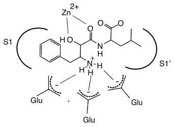
Bestatin resembles a Phe-Leu dipeptide substrate. However, the first
residue contains a α-hydroxy group that, along with the neighboring carbonyl,
co-ordinates the catalytic zinc atom resulting in a competitive active site-directed
inhibitor (Fig. 2.1). In addition, the free amine of bestatin is coordinated by one or
more glutamate residues in the MAP active site [72]. Bestatin family members
are slow- and tight-binding inhibitorswith well-defined interactions with the S1 and
S1’ active site pockets [69]. Thus bestatin represents an ideal candidate for ABP
development for MAPs due to its family specificity, potency in the low nanomolar
to micromolar range, synthetic tractability and potential for expansion through
Figure 2.1: General model of interactions of bestatin in the active site of metallo-aminopeptidases.
S1
S1!
Zn2+
N H
O O
H O O
N H
Glu
Glu
Glu
-
-+
H H
2.2 Results and Discussion
To design an ABP family for MAPs, we chose to derivatize the core
bestatin inhibitor scaffold using a solid-phase synthetic strategy (Scheme 2.1).
Bestatin has a free carboxyl group available for functionalization and previous
x-ray crystal structures of MAP-bestatin complexes indicated that extension at this
carboxylate was unlikely to perturb inhibitor binding [72]. Thus we attached the
inhibitor scaffold to solid-phase resin at the carboxyl end of the molecule. Our
first attempt to develop a MAP-directed ABP probe involved the addition of a
spacer, a UV crosslinker, and a biotin affinity tag to the C-terminus of the core
bestatin scaffold (Scheme 2.1). We included a benzophenone UV crosslinker
since this is a non-covalent, reversible inhibitor scaffold. Synthesis was
accomplished on solid-phase using Rink amide resin and, to our knowledge,
represents the first reported solid-phase synthesis of this class of molecules.
We initially explored the placement of the spacer and UV crosslinker
relative to the core bestatin scaffold (MH01 and MH02, Fig. 2.2) and found that
there was little difference in labeling efficiency of a model enzyme, purified
porcine aminopeptidase N (Fig. 2.2A). We then assessed the ability of MH01 to
label the model aminopeptidase in an activity-dependent manner (Fig. 2.2B). Our
results show that, indeed, the bestatin-based probe is an activity-dependent
probe of MAPs. Firstly, competition with the unbiotinylated parental compound,
competitive and therefore binding at the active site (Fig. 2.2C, lane 2). Preheating
of the sample, a process that denatures all protein targets, abrogated labeling
(Fig. 2.2C, lane 3), indicating that this labeling was dependent on properly folded,
active enzyme. Lastly, UV exposure was necessary for labeling owing to the fact
Figure 2.2: Anatomy of ABPs and labeling of aminopeptidase N. (a) Structure of ABPs MH01 and MH02. (b) Aminopeptidase N (0.13 U) was treated with 10, 1, or 0.5 µM of either MH01 or MH02 for 1 hr in 50mM Tris-HCl, pH 7.8, 0.5 µM ZnCl2 (buffer A). Certain reaction mixtures were UV
While a biotin tag is ideal for affinity tagging purposes, its use is not
optimal for higher throughput activity-based profiling due to the time and labor
involved in producing western blots. Additionally, many cells and tissues contain
endogenously biotinylated proteins that complicate analysis of biotinylated
probe-based western blots. To circumvent these shortcomings we synthesized a
fluorophore-tagged version of the bestatin-based probe, which would allow for
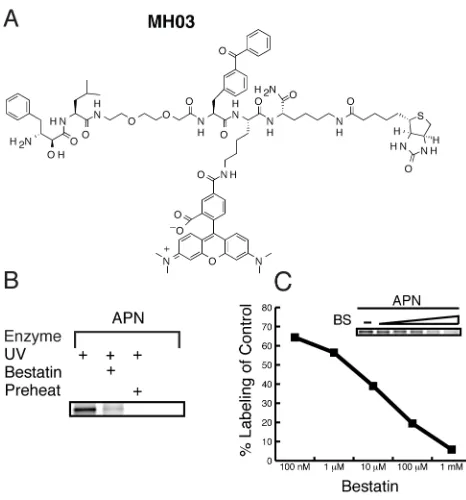
direct detection of labeled targets in a gel-based read-out. For the fluorescent
bestatin-based probe we added a TAMRA group in addition to the biotin, creating
a dual function ABP, MH03, making it suitable for both affinity purification and
fluorescence applications (Fig. 2.3). Labeling of purified porcine aminopeptidase
N with the dual label probe was performed under the same conditions as with the
original biotinylated MH01 (Fig. 2.3B). Labeled proteins were analyzed by
SDS-PAGE coupled with in-gel fluorescent scanning (Typhoon, GE). MH03 showed
similar activity-dependent labeling as MH01 (Fig. 3B) demonstrating the
robustness of this ABP scaffold. Some residual labeling was observed (Fig 2.3B,
lane 2), which is normal in ABP labeling experiments, although this may have
Next, we wanted to demonstrate that utility of the fluorescent MH03 for
relative quantification of enzyme labeling. To do this, porcine aminopeptidase N
was preincubated with increasing concentrations of bestatin followed by the
addition of a single concentration of the fluorescent MH03. The densities of each
labeled band, representing active enzyme, were quantified using a Typhoon
flatbed fluorescent scanner (GE). In-gel fluorescent scanning of the labeled
peptidase band showed a decrease in aminopeptidase N labeling by MH03
relative to increasing concentration of bestatin preincubation (insert in Fig. 2.3C).
Percent competition values were calculated by dividing the density of the bestatin
preincubated aminopeptidase N band relative to the untreated band in lane 1
(Fig. 2.3C). We demonstrate a dose dependent relationship that is amenable to
relative quantification with a dynamic range of several orders of magnitude and
will allow future screening efforts of derivative libraries.
One of the technical challenges facing the development and use of ABPs
with biotin or fluorophore tags is limited or biased uptake by live cells. In some
cases, ABPs have been used to label enzymes in living cells, but a more
universal system for labeling would employ a small, hydrophobic tag. We
therefore chose to add a small alkyne tag to our MAP probe in order to utilize the
popular “click” bio-orthogonal chemistry, which employs a 1,3-dipolar
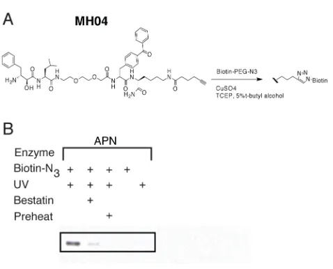
azide/alkyne cycloaddition [76,77]. We thus replaced the biotin of MH01 with a
C-terminal alkyne resulting in a click probe, MH04 (Scheme 2.2 and Fig. 2.4). We
observed facile “click” addition of a biotinylated azide following labeling of porcine
enzyme was not affected by this procedure, as illustrated by bestatin
Figure 2.4: “Click” chemistry-based ABP labeling of aminopeptidase N. (a) Structure of MH04 probe. (b) Aminopeptidase N labeling was performed as described in Figure 2.2C
using MH04. The ligation of the biotin-azide reporter tag was performed by adding 100 µM of the biotin-azide tag, followed by 1 mM TCEP (tris(2-carboxyethyl) phosphine hydrochloride), 100 mM ligand (tris[(1-benzyl-1H-1,2,3-triazol-4-yl)methyl]amine) (17x stock in DMSO:t-butanol 1:4), and 1 mM CuSO4. Reactions were allowed to proceed for 1 h at
room temperature, then quenched with equal volume of SDS-PAGE loading buffer. Labeled protein was visualized as in Fig. 2.2B.
Finally, to validate the utility of the probe as an ABP for MAPs in the
context of a complex proteome, we assessed labeling of an aminopeptidase N
homolog from the malarial parasite (PfA-M1) from whole cell lysates. To facilitate
the identification of PfA-M1 from P. falciparum we utilized a parasite line that
expresses the endogenous PfA-M1 as a YFP C-terminal fusion. Homogenized
whole cell lysates from P. falciparum were labeled with MH01. The resulting
proteome was separated by SDS-PAGE and western blot analysis was
performed to first visualize biotinylated proteins. A 150 kD protein was labeled by
MH01, which corresponds to the approximate weight of the PfA-M1-YFP fusion
protein (Fig. 2.5A). Preincubation of the protein lysate with unbiotinylated
bestatin (Fig. 2.5A, lane 2) resulted in loss of labeling of the 150 kD band
illustrating that bestatin was competitive with MH01 for this protein target.
Additionally, the target was labeled in an activity-dependent manner, as
preheating of the proteome prior to labeling abrogated any labeling (Fig. 2.5A,
lane 3). Finally, to identify this target the blot was stripped and reprobed using an
anti-YFP antibody (Fig. 2.5B). The anti-YFP revealed the presence of the
expected 150kD fusion protein in all lanes and this band overlaid the exact
position where the biotinylated protein appeared. It should be noted that
PFA-M1-YFP fusion appeared in all lanes with the anti-YFP antibody, whereas the
biotinylated species only appeared when the active peptidase was labeled. This
data thus confirmed the specific activity-based labeling of the PFA-M1
In conclusion, we have developed a novel activity-based probe class, with
specificity for MAPs, based on the bestatin inhibitor scaffold. The use of a biotin,
fluorophore, or alkyne moiety did not alter the activity-dependent labeling profile
for the scaffold and, thus, the suite of ABPs presented in this manuscript should
allow for a variety of labeling methodologies. We therefore believe that this ABP
may prove to be a valuable tool for future characterization of MAP activity in a
wide variety of biological systems. We are now currently pursuing the expansion
and application of these probes for use against the malarial parasite.
2.3 Experimental Procedures
General method for phase peptide synthesis of ABPs: Standard
solid-phase peptide synthesis was performed on Rink amide resin, using
HBTU/HOBt/DIEA in an equimolar ratio in DMF for 30 min at RT. Coupling of the
α-hydroxy-β-amino acid required HATU for 1 hr. Each amino acid was double
coupled. Fmoc protecting groups were removed with 20% piperidine/DMF for
30 min. To cleave products from resin, a solution of 95%TFA:2.5%TIS:2.5%H2O
was added to the resin. After standing for 2 hrs, the cleavage mixture was
collected, and the resin was washed with fresh cleavage solution. The combined
fractions were evaporated to dryness and the product was purified by reverse
phase-HPLC. Fractions containing product were pooled and lyophilized. Reverse
phase HPLC was conducted on a C18 column using an Agilent 1200 HPLC.
with a concentration gradient 0-70% of acetonitrile (0.1% Formic acid). LC/MS
data were acquired using LC/MSD SL system (Agilent). Solid-phase peptide
chemistry was conducted in polypropylene cartridges, with 2-way Nylon
stopcocks (Biotage, VA). The cartridges were connected to a 20 port vacuum
manifold (Biotage, VA) that was used to drain excess solvent and reagents from
the cartridge. MH01: C62H90N10O14S, predicted mass 1230.64, observed [M+H]
1231.4. MH02: C57H81N9O12S, predicted mass 1115.57, observed [M+H] 1116.3.
The synthesis of the dual function ABP MH03 was accomplished using
standard solid phase peptide synthesis as detailed above, However, the TAMRA
was added after product cleavage from resin due to the instability of TAMRA to
TFA. This was accomplished using a Boc protected lysine and addition of
TAMRA using HBTU/HOBt/DIEA in DMF after resin cleavage for 3 h. MH03:
C85H107N13O16S, predicted mass 1597.77, observed [M+2H] 799.8.
Synthesis of MH04 was accomplished as depicted in Scheme 2.2. The
deprotection of the Aloc group was conducted under a positive flow of argon.
The resin was solvated with dichloromethane for 5 min. The solvent was
drained, and PhSiH3 (24 eq.) in CH2Cl2 was added to the resin followed by
Pd(PPh3)4 (0.25 eq.) in CH2Cl2. After agitating the resin for 1 hr by bubbling with
argon, the solution was drained, and the resin was washed with CH2Cl2 (3x).
Synthesis of the biotin-azide was accomplished using standard solid-phase
synthesis using a bromo-acetic acid as the final group. The replacement of the
bromo group by the azide was achieved by heating with NaN3 at 60oC. MH04:
Chapter 3: A bestatin-based chemical biology strategy reveals
distinct roles for malarial M1 and M17 family aminopeptidases
†3.1 Introduction
Malaria is a global disease causing at least 500 million clinical cases and
more than 1 million deaths each year [78]. While significant efforts to control
malaria via insect vector elimination have been pursued, chemotherapy remains
the principal means of malaria control. Moreover, the emergence of drug
resistance in Plasmodium falciparum, the causative agent of most
malaria-associated deaths,necessitates the discovery of novel antimalarials.
P. falciparum has a complex life cycle involving mosquito and human
hosts. Thislife cycle involves both sexual and asexual stages of growth, wherein
the human asexual erythrocytic phase (blood stage) is the cause of
malaria-associated pathology. The erythrocytic stage begins when extracellular parasites,
initially released from the liver, invade red blood cells. Once established in a
specialized vacuole inside the host erythrocyte, parasites grow from the initial
ring stage to the trophozoite stage, wherein much of the host hemoglobin (Hb) is
proteolyzed. Parasites then replicate during the schizont stage to produce
expanded populations of invasive merozoites that then rupture from the host cell
approximately 48 hours post-invasion and go on to recapitulate the life cycle [79].
Peptidases are critical to parasite development throughout the life cycle and
therefore are considered to be potential antimalarial drug targets [45,80,81].
One proteolytic pathway that has received significant attention is the
multi-step degradation of host Hb [15]. While residing inside the host red blood cell,
malaria parasites endocytose and proteolytically digest host Hb in a specialized
lysosomal-like digestive vacuole (DV). This process liberates amino acids that
the parasite can utilize for protein synthesis and general metabolism [22] and
may reduce pressure on the host cell produced by the growing parasite [23].
Multiple endoproteases make initial cuts in full-length Hb; however, genetic
knockout studies of these enzymes, plasmepsins 1-4 and falcipain 2/2’, have
revealed that all are non-essential and functionally redundant [52,53,82]. While
falcipain 3 has not been shown to be dispensable, it is expressed later in the
parasite lifecycle and may have roles beyond Hb degradation. On the other hand,
several exopeptidases, some of which may have roles in Hb degradation, are
likely genetically essential [43,44]. Among these non-redundant enzymes are
cysteine dipeptidyl aminopeptidase 1 (DPAP1) and three
metallo-aminopeptidases (MAPs): aminopeptidase N (PfA-M1), aminopeptidase P
(PfAPP), and leucyl aminopeptidase, (Pf-LAP) [44].
MAPs have been postulated to be important for the parasite life cycle.
Early studies suggested that MAP activities were absent from the DV lumen,
leading to the proposal that Hb peptides are exported to the cytosol for further
degradation by MAPs [83-85]. However, more recent localization and
while Pf-LAP is located in the parasite cytosol and has been proposed to act on
exported globin peptides [86-88]. A cytosolic aspartyl MAP, PfDAP, has been
shown to hydrolyze substrates with an amino-terminal Asp or Glu residue, but its
contribution to blood-stage peptide catabolism appears to be dispensable as
genetic disruption causes no overt phenotype [44]. Importantly, none of these
studies provide any biological evidence, most importantly in live parasites, for a
direct role for MAPs during Hb peptide catabolism.
Unfortunately in P. falciparum, genetic approaches to study essential
genes are limited and thus direct evidence for the biological roles of these MAPs
is still lacking [37]. As a complementary approach, we have developed a
MAP-specific chemical genetics platform that utilizes activity-based protein profiling
(ABPP) based on the natural product inhibitor bestatin to study the roles of MAPs
[89]. ABPP is a chemical strategy that utilizes mechanism-based, tagged small
molecule inhibitors to discover new enzymes, profile their activity state in
complex proteomes, and identify potential functions for these enzymes during a
specific biological process [90]. ABPP has been utilized for a variety of enzyme
classes, including serine hydrolases, peptidases, histone deacetylases and
kinases [60,61,91,92].
(-)-Bestatin is a natural product dipeptide analog of actinomycetes that
potently inhibits multiple families of MAPs including the M1 and M17 families
[70-72,93] (Fig. 3.1A). Importantly, bestatin has been shown to inhibit growth of P.
falciparum parasites in culture and in mouse models of malaria [94-96]. In
catabolism showing that parasites treated with bestatin had decreased levels of
hemazoin formation, the detoxified biomineral byproduct of Hb digestion [97];
likewise, isoleucine uptake was decreased in these bestatin treated parasites.
However, the MAP(s) targeted by bestatin, which are responsible for these
processes, were not identified.
Herein, we report on a multidisciplinary effort combining bestatin-based,
small molecule ABPs with biochemical and peptidomic approaches to functionally
analyze two essential aminopeptidases, PfA-M1 and Pf-LAP.
3.2 Results
Identification of bestatin targets in P. falciparum
Because of the paucity of genetic tools for analysis of essential proteins in
P. falciparum and a lack of highly specific inhibitors with which to probe the
individual roles of MAPs, we chose to study the functions of these essential
parasite MAPs through the development and application of a MAP specific ABPP
platform. ABPP utilizes tagged mechanism-based inhibitors, or activity-based
probes (ABPs), to characterize families or individual active peptidases within
complex proteomes. ABPs typically possess two main structural components that
contribute to their target specificity: (i) a mechanism-based inhibitor scaffold to
covalently or noncovalently target catalytic residues or the active site of
peptidases and (ii) a reporter tag, such as a fluorophore or biotin, for the
of target proteins. The mechanism-based inhibitor scaffold ensures that ABPs
bind to the enzyme(s) in an activity-dependent manner.
We decided to use the natural product, bestatin, as the scaffold for the
development of ABPs for MAPs since it is a general MAP inhibitor, kills
Plasmodium parasites, and is synthetically tractable using both solution and
solid-phase chemistry [69,89,98]. To identify the target(s) of bestatin in P.
falciparum, we utilized a previously published bestatin-based ABP, MH01
(Fig.3.1B) [89]. MH01 contains a biotin moiety to allow for monitoring of protein
binding and affinity purification for target identification and also utilizes a
benzophenone for irreversible UV crosslinking to protein targets, as the inhibition
of MAPs with bestatin is noncovalent [89]. Asynchronous cultures of 3D7
parasites were treated with saponin to lyse the erythrocyte and parasitophorous
vacuole membranes and isolated whole parasites were harvested by
centrifugation. Crude parasite lysates, including both soluble and membrane
proteins, were treated with 5 µM MH01, exposed to UV light, and analyzed by
western blot using streptavidin-HRP to detect biotinylated proteins. The observed
labeling pattern consisted of four bands at approximately 100 kDa, 70 kDa, 55
kDa, and 25 kDa (Fig. 3.1C). Western blot analysis with PfA-M1 or Pf-LAP
antibodies of parasite lysates labeled with MH01 showed that all four labeled
species could be accounted for with these two antibodies: three bands
corresponded to different species of PfA-M1 and one to Pf-LAP (Fig. 3.1C).
PfA-M1 is known to be proteolytically processed from a 120 kDa proform to a 115