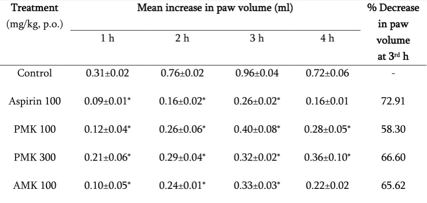
ANTI-INFLAMMATORY ACTIVITY OF ALKALOIDS FROM MURRAYA KOENIGII LEAVES IN ANIMAL MODELS
Full text
Figure

Related documents
Die in dieser Arbeit gefundenen inhibitorischen Effekte von α -Tocopherol auf die Expression von Schlüsselrezeptoren der Cholesterin-Homöostase in Makrophagen wie den
the occurrence of these symptoms and their association with measures of COPD burden for effective COPD management. Findings from this study indicate that symptoms during the NT and
The status of designed and operated heavy ion accelerators and colliders all over the world in the kinetic energy range from 1 to 50 GeV/u in lab system is as follows: 1)
The pollution load index (PLI), contamina- tion factor (CF), enrichment factor (EF) and geo- accumulation index (I geo ) were used to determine the metal contamination in
The Aigaleon bell-krater is also decorated with a wreath or swag in the handle zone, although its technique (gilded clay?), form, and even exact position differ from
Application of Magnetic Dicationic Ionic Liquid Phase Transfer Catalyst in Nuclophilic Substitution Ractions of Benzyl Halides..
In this study, we wish to examine whether a psychological group intervention targeting people with poorly controlled type 1 diabetes can be helpful in augmenting quality of life
ATR: Acori Tatarinowii Rhizoma; AGR: Acori Graminei Rhizoma; ACR: Acori Calami Rhizoma; AAR: Anemones Altaicae Rhizoma; CM: Chinese medicine; DAD: diode‑array detector;
