Digital Impression System- A Virtual New Generation A Study
When Compared with Conventional Impression System
Yash Desai
Yerala Medical Trust Dental College and Hospital, Navi Mumbai, Maharashtra, India
ABSTRACT
Digital impression has been introduced into dentistry since 1980’s. To overcome the difficulties occurred by conventional impression techniques to the patient and the practitioner, digital impressions with (intraoral scanner) IOS and CAD/CAM (Computer-aided design and manufacturing) technologies have been introduced in today’s dental practice. In such technologies different types of lights are used to capture images. Diode lasers have been used which produce invisible light at near infrared wavelength ranging from 805nm to1,064nm which are used for capturing soft tissues only. After the image has been captured, the final image is either stored in the system and used later on for fabrication at chairside or the image is sent to milling unit for fabrication of prosthesis or it is digitally transmitted to a laboratory. However different technologies have their different system for recording centric relation. Patient comfort and time saving procedure are the advantages of this digital impression technique.
Statistical analysis: The analysis was performed with ‘Wilcoxon Rank Test’ where p<0.05.
Results: There were remarkable differences between conventional and digital impression techniques (p<0.05) in terms of total working time & processing steps.
Conclusion: The overall study of this article is to make the practitioners understand the importance of clinical application of digital impression technique as well as the patients preferred digital impression technique more than conventional impression technique.
Keywords : Digital Scanner, Software, Camera
I.
INTRODUCTION
Impression in life has its own meaning but in dentistry impression is nothing but a negative replica of teeth or tissues of the oral cavity. Impression has a lot of importance in dentistry especially in prosthodontics for making crowns, dentures, inlays, onlays, veneers using different impression materials and techniques in daily routine dental practice. In olden times impression plaster were the first material which were used to take impressions, but with time changes and technologies impression plaster were replaced with reversible and irreversible hydrocolloids and elastomers. However with all such
conventional techniques inaccuracy and error were still creating a problem so to overcome all these hurdles digital impression technique was introduced using scanners and milling machine. The first digital impression scanner was introduced in 1980’s by a group of engineers for fabrications of dental restorations. These systems are capable of capturing three dimensional (3D) virtual images of anatomic landmarks or tooth preparations from which restorations are directly fabricated.
3. iTero system.
Figure 1. The LAVA™ chairside oral scanner.
Figure 2. The CEREC system.
Figure 3. The iTERO system.
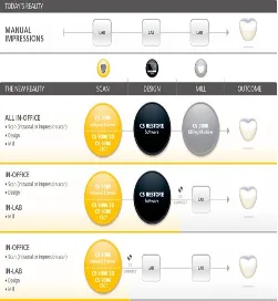
Steps required in digital imaging are as follows:- 1. Digital scanner
2. Software to create using CAD/CAM system. 3. Connection to export the image.
4. Milling unit to fabricate the prosthesis.
Figure 4. steps required in digital imaging.
II.
METHODS
STUDY AND PATIENT SELECTION
INCLUSION/EXCLUSION CRITERIA:
Twenty subjects were selected aged between 19-21 years for the study which fulfilled the inclusion criteria who had no experience of conventional or digital impression technique and on examination of the subjects it showed no history of poor oral hygiene, no history of poor general health, no periodontal disease, and no poor mental health. Exclusion criteria for the study included no experience of conventional or digital impression system, previous history about any orthodontic treatment or appliances, any previous history of space maintainers during mixed dentition period.
CLINICAL SCENARIO:
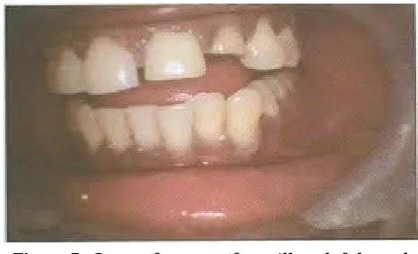
The subjects were shown informational video depicting the restorative steps of the clinical scenario having ‘crown fracture of maxillary left lateral incisor’. However the impression system technique was not shown from the video.
Figure 5. Crown fracture of maxillary left lateral incisor.
CONVENTIONAL IMPRESSION TECHNIQUE: The operator selected metallic perforated stock trays for both maxillary and mandibular arches for the subject and the material used was alginate impression material. The impression was taken by selective pressure technique. The interocclusal relationship was taken by a bite registration material and the materials were used according to the manufactures guidelines and were performed by the operator on the subjects.
DIGITAL IMPRESSION TECHNIQUE:
For digital impression system the appointment was re-scheduled again after 1-2 weeks following the conventional impression technique. The digital impression technique was performed along the chairside by CAD/CAM system. This technique consists of electronic data of the virtual models for both maxillary and mandibular arches and bite registrations by scanning were also recorded. The digital scanning procedure were also carried out according to the manufactures guideline’s by the same operator who had carried out conventional impression technique. After the cessation of digital impression technique the treatment time were again noted down in seconds and the clinical steps were also listed down by the same operator. The same standardized questionnaires were asked to the subjects after the completion of this digital impression technique.
STANDARDIZED QUESTIONNAIRES WHICH WERE ASKED TO THE SUBJECTS ARE AS FOLLOWS:-
1. Which was the preferred impression technique ? 2. Which impression technique was more
comfortable ?
3. Which impression technique was more time saving ?
4. Which impression technique caused more irritation to you ?
Which impression technique did you find cost effective ?
STATISTICAL ANALYSIS
III.
RESULTS
EFFICIENCY CONVENTIONAL IMPRESSION TECHNIQUE
DIGITAL IMPRESSION TECHNIQUE
P-VALUE
Tray selection/patient information
18.43±2.12 18.71±3.20 >0.05
Adhesive application/lab presentation
27.62±3.15 13.50±1.89 <0.001*
Upper impression/upper scanning
240.75±16.38 102.19±17.77 <0.001*
Lower impression/lower scanning
218.15±10.41 90.84±10.16 <0.001*
Bite registration/bite scanning
90.86±10.74 13.86±3.82 <0.001*
Total treatment time 595.81±42.80 239.1±21.59 <0.001*
For all the following steps the treatment time was recorded in seconds. The time for both the techniques were recorded by taking their mean value ( SD±)
SUMMARY OF CONVENTIONAL IMPRESSION TECHNIQUE
The mean overall treatment time for conventional impression technique was 595.81±42.80. The mean treatment time for individual steps were as follows:- for tray selection the mean treatment time came up to 18.43±2.12, the mean time for adhesive application came upto 27.62±3.15, the mean time for upper arch impression came upto 240.75±16.38, the mean time for lower arch impression came upto 218±218.15±10.41, the mean time for bite registration came upto 90.86±10.74.
SUMMARY OF DIGITAL IMPRESSION TECHNIQUE
The mean overall treatment time for digital impression technique was 239.10±21.59. the mean treatment time for individual steps were as follows:- the mean time for patient information in digital impression technique came upto 18.71±3.20, the mean
time for lab presentation came upto 13.50±1.89, the mean time for upper arch impression scanning came upto 102.19±17.77, the mean time for lower arch impression scanning came upto 90.84±10.16, the mean time for bite scanning interocclusally came upto 13.86±3.82.
IV.
CONCLUSION
With the following results recorded the conclusion of this study is drawn as follows:-
1. The digital impression technique was more comfortable to the subjects than the conventional impression technique.
3. The digital impression technique was more preferred, accepted and effective than convention impression technique.
V.
REFERENCES
[1]. Sjogren G, Molin M, Van Dijken JW. A 10-year prospective evaluation of CAD/CAM-manufactured (CEREC) ceramic inlays cemented with a chemically cured or dual-cured resin composite. Int J Prosthodont. 2004;17(2):241-245
[2]. Lee H, Ercoli C, Funkenbusch PD, Feng C. Effect of subgingival depth of implant placement on the dimensional accuracy of the implant impression: an in vitro study. J Prosthet Dent. 2008;99(2):107-113. doi: 10.1016/S0022-3913(08)60026-8
[3]. Brosky ME, Pesun IJ, Lowder PD, Delong R, Hodges JS. Laser digitization of casts to determine the effect of tray selection and cast formation technique on accuracy. J Prosthet Dent. 2002;87(2):204-209
[4]. Burns J, Palmer R, Howe L, Wilson R. Accuracy of open tray implant impressions: an in vitro comparison of stock versus custom trays. J Prosthet Dent. 2003;89(3):250-255
[5]. Birnbaum N, Aaronson HB, Stevens C, Cohen B. 3D digital scanners: a high-tech approach to more accurate dental impressions. Inside Dentistry. 2009;5.
[6]. Powers J. In: Craig’s Restorative Dental Materials. Powers J, editor. St Louis: Mosby; 2006. Impression materials; pp. 275-312.
[7]. Christensen GJ. Impressions are changing: deciding on conventional, digital or digital plus in-office milling. JADA. 2009;140.
[8]. Henkel GL. A comparison of fixed prostheses generated from conventional vs digitally scanned dental impressions. Compend Contin Educ Dent. 2007;28:422-424.
[9]. De La Cruz JE, Funkenbusch PD, Ercoli C, Moss ME, Graser GN, Tallents RH. Verification jig for implant supported prosthesis: a comparison of standard impressions with verification jigs made of different materials. J Prosthet Dent.:330-335. [10]. Syrek A, Reich G, Ranftl D, Klein C, Cerny B,
Brodesser J. Clinical evaluation of all-ceramic crowns fabricated from intraoral digital impressions based on the principle of active wavefront sampling. J Dent. 2010;38:554-559. [11]. Brawek PK, Wolfart S, Endres L, Kirsten A,
Reich S. The clinical accuracy of single crowns exclusively fabricated by digital workflow the comparison of two systems. Clin Oral Investig. 2013;17(9).
[12]. Karl M, Shubinski P, Taylor T. Effect of intraoral scanning on the passivity of fit of implant-supported fixed partial prostheses. Quintessence Int. 2012;43:555-562.
[13]. Polido WD. Digital impressions and handling of digital models: the future of dentistry. Dental Press J Orthod. 2010;15(5):18-22.
[14]. Christensen GJ: Impressions are changing: deciding on conventional, digital or digital plus in-office milling. J Am Dent Assoc 2009;140:1301-1304.
[15]. Christensen GJ: Will digital impressions