R E S E A R C H A R T I C L E
Open Access
Protein signature characterizing
Helicobacter pylori
strains of patients with
autoimmune atrophic gastritis, duodenal
ulcer and gastric cancer
Valli De Re
1*†, Ombretta Repetto
1†, Stefania Zanussi
2, Mariateresa Casarotto
2, Laura Caggiari
1,
Vincenzo Canzonieri
3and Renato Cannizzaro
1,2,3,4Abstract
Background:Helicobacter pylori(H. pylori) represents a key factor in the etiology of autoimmune atrophic gastritis (AAG), duodenal ulcer (DU) and gastric cancer (GC). The aim of this study was to characterize the differential protein expression ofH. pyloriisolated from gastric biopsies of patients affected by either AAG, DU or GC.
Methods:TheH. pyloristrains were isolated from endoscopic biopsies from the stomach of patients with gastric disease. Protein profiles ofH. pyloriwere compared by two-dimensional difference in gel electrophoresis (2D-DIGE) coupled with mass spectrometry (MS) for the identification of significantly different spots (Studentt-test,p< 0.05). Results:A total of 47 differentially expressed spots were found betweenH. pyloriisolated from patients with either DU or AAG diseases and those isolated from patients with GC (Anova < 0.05, log fold change >1.5). These spots corresponded to 35 unique proteins. The identity of 7 protein spots was validated after one-dimensional electrophoresis and MS/MS analyses of excised gel portions. InH. pyloriisolated from DU-patients a significant increase in proteins with antioxidant activity emerged (AroQ, AspA, FldA, Icd, OorA and ScoB), together with a higher content of proteins counteracting the high acid environment (KatA and NapA). In H. pylori isolated from AAG-patients proteins neutralizing hydrogen concentrations through organic substance metabolic processes decreased (GroL, TrxB and Tuf). In addition, a reduction of bacterial motility (FlhA) was found to be associated with
AAG-H. pylori isolates. In GC-H. pylori strains it was found an increase in nucleic acid-binding proteins (e.g.
DnaG, Tuf, RpoA, RplU) which may be involved in a higher demand of DNA- and protein-related processes. Conclusion: Our data suggest the presence of specific protein signatures discriminating amongH. pyloriisolated from either AAG, DU or GC. Changes in protein expression profiles evaluated by DIGE succeeded in deciphering part of the molecular scenarios associated with the differentH. pylori-related gastric diseases.
Keywords:Adenocarcinoma, Autoimmune atrophic gastritis, Comparative proteomics, DIGE, Duodenal ulcer, Gastric cancer,Helicobacter pylori
* Correspondence:vdere@cro.it †Equal contributors
1Facility of Bio-Proteomics, Immunopathology and Cancer Biomarkers, IRCCS CRO National Cancer Institute, Via F. Gallini 2, 33081 Aviano, Italy
Full list of author information is available at the end of the article
Background
Helicobacter pylori (H. pylori) is a class I bacterial
pathogen (IARC) colonizing approximately 50% of the
world’s population. The infection increases the risk of
extragastric and gastric diseases, including duodenal ulcer (DU), autoimmune atrophic gastritis (AAG) and
gastric cancer (GC) [1–4]. It is estimated that about
3%H. pylori-infected individuals will develop a GC
with an increased risk of 3-6-fold compared with non-infected population [5, 6].
Many virulent H. pylori genes have been reported to
have a role in clinical outcomes of infection, with a
pre-dominant involvement of the plasticity region and cag
pathogenicity island genes in GC development [7–10].
However, the precise mechanisms for GC development
by H. pylori infection are still not completely
under-stood. Analysis of the H. pylori proteome offered valid
tools to delineate post-translational modifications and the complexity of gene expression and regulation
char-acterizing H. pylori protein profiles associated with a
particular clinical outcome [11–13]. The aim of this
study was to investigate the H. pylori proteome profile
by two-dimensional difference in gel electrophoresis (2D-DIGE) coupled with mass spectrometry (MS) and
bioinformatics in order to correlate some differential H.
pylori proteins to the clinical outcomes of gastric
dis-eases in an Italian population.
Methods
Bacterial strains and culture conditions
The study was approved by the Internal review board and ethical committee of the IRCCS CRO, and Italian
National Cancer Institute (IRB-14-2013). The H. pylori
strains were isolated from endoscopic bioptic samples from the stomach (corpus and/or antrum), as previ-ously reported [14]. Briefly, the biopsies were cultured
in H. pylori Selective Medium (Bio-Mèrieux, Rome,
Italy), and incubated at 37 °C in a microaerophilic en-vironment (Campygen Oxoid, Ltd., Basingstoke,
Hamp-shire, England) until growth evidence for at least 13–14
days. Several sweeps of colonies, considered
representa-tive of the wholeH. pyloripopulation, were subcultured
in agar-blood plates, and after 3 days of incubation
were collected and stored at −80 °C in a microbial
storage medium (Microbank; Pro-Lab Diagnostics, Richmond Hill, Canada). Strains were revitalized after a
median of 9 months (range of 2–98 months) in H.
pylori Selective Medium, expanded in Columbia sheep
blood agar, and then used for proteome extraction. Bac-terial DNA extraction and PCR on the virulence factor
CagA gene were performed inH. pylori strains isolated
from patients accordingly to Repetto et al. [14] and Fas-ciana et al. [15].
Patient characteristics
Fresh human gastric biopsies were obtained after patient
informed consent. Patients were considered H.
pylori-infected if results from cultures and histologic examin-ation of the biopsy stained by Giemsa and/or serology
forH. pylori(H. pyloriIgG ELISA kit, BIOHIT
Health-Care, Helsinki, Finland) were positive. According to
confirmed histological patient diagnosis,H. pylori
posi-tive isolates were divided into DU-H. pylori (n= 11);
AAG-H. pylori (n= 5), and GC-H. pylori(n= 25). Tissue
biopsies were further grouped based on their anatomic gastric localization (A = antrum and C = corpus). Data of
patients from whomH. pylorihad been isolated are
sum-marized in Table 1 and Additional file 2: Table S1.
Protein labeling and DIGE
Proteins from frozenH. pyloricultures were extracted in
methanol/chloroform, quantified and labeled as previ-ously reported [14]. Prior to co-resolution on the same immobilized pH gradient (IPG) dry strip and two
dimensional electrophoresis (2DE) gel, 25 μg of two
bacterial lysates from two different strains was differen-tially labeled with 100 pmol cyanine fluorescent dyes (Cy3 and Cy5, GE Healthcare) and mixed with the Cy2-labeled internal standard, as described previously [16]. Internal standard included equal amounts of all the samples (nr = 41) within the experiment for a total of 21 gels. A dye swapping strategy was adopted to avoid a dye labeling bias. First dimensional isoelectric focusing (IEF) was carried out on 11-cm IPG strips (IPG pH 3 to 10 Bio-Rad, Milan, Italy) with Protean® IEF unit. The second dimension was performed using
pre-cast 12% gels on Criterion™ Cells (Bio-Rad, Milan,
Italy). For preparative gels, 300 μg of unlabelled protein
pooled from equal amounts of samples was used, and stained with the ProteoStain solution (Proteomics Consult, Kampenhout, Belgium). Proteome maps were imaged
using a Typhoon 940™ laser scanner (GE Healthcare,
Uppsala, Sweden) and analysed using the DeCyder soft-ware version 6.5 (GE Healthcare). The EDA module was used for multivariate analysis of protein expression data, derived from BVA, and it allowed getting information
about the ‘principal component analysis, PCA’ and the
pattern analysis. Student’s t test was performed to assess
the statistical significance of differentially expressed pro-teins based on average spot volume ratio. Based on average spot volume ratio, spots for which relative expression changed at least 1.5-fold (increase or decrease) at 95%
con-fidence level (Student t-test; p< 0.05) were considered to
be significant.
Protein identification by mass spectrometry
LC-MS/MS. MALDI-TOF MS was performed on a Voyager-DE PRO Biospectrometry Workstation mass spectrometer (AB Sciex). While LC-MS/MS was per-formed using a LTQ XL-Orbitrap ETD equipped with a NanoEasy-HPLC (PROXEON, Thermo Fisher Scientific).
Matched spots of interest were excised from the Coomas-sie Blue preparative gel, destained, trypsin-digested, and tryptic peptides were extracted by trifluoroacetic acid (TFA). In case of MALDI-TOF analyses, peptides were subjected to Zip Tip cleanup (Millipore, Milan, Italy),
mixed with α-Cyano-4-hydroxycinnamic acid matrix
solution (1:1, v:v) (LaserBio Labs, Sophia-Antipolis Cedex, France), and spotted on the MALDI target. The collected MALDI mass spectra were then processed by peptide mass fingerprinting (PMF) using Data Explorer (AB Sciex). Database searches were done with the MASCOT search engine version 2.3 (Matrix Science, London, UK), limiting the searches to bacterial proteins. Fig. 1 shows an
example of a characteristics 2D gel map of an H.
pylori-isolated strain with the indication of some of the identified proteins. To get an overview of the regulated proteins and their possible functional connections, the
identi-fied H. pylori-regulated proteins were analysed using
the STRING tool (version 10; http://string-db.org) [17], after converting the protein accession numbers
into ‘Kyoto Encyclopedia of Genes and Genomes,
KEGG’gene entries (http://www.genome.jp/kegg/). For
each protein, KEGG pathways, biological processes and molecular functions were analysed according to the Gene Ontology (GO) description.
Validation of the protein identified by using LC-MS/MS analysis
The correct identification of some proteins of interest was confirmed by searching them in gel portions of the
corresponding MW after 12% 1DE. PooledH. pylori
pro-tein extracts (15 μg per lane) were separated by 1DE,
and images of ProteoStain-stained gel were acquired
with the Typhoon Trio 9400™laser scanner. Gel portions
corresponding to the MW of around 52 kDa (Fig. 1, nr1, nr2), 22 kDa (Fig. 1, nr3) 15 kDa (Fig. 1, nr4), 12 kDa (Fig. 1, nr5, nr6), and 10 kDa (Fig. 1, nr7, nr8), were cut, reduced by incubation with 10 mM dithiothreitol (1 h at 57 °C), and alkylated with 55 mM iodoacetamide (45 min at room temperature). Samples were further
washed with NH4HCO3, dehydrated, trypsin digested
and processed for LC-MS/MS analyses.
Results
Proteomics analysis ofH. pyloristrains
H. pyloristrains isolated from gastric biopsies of patients
affected by either AAG, DU or GC were analyzed using the 2D-DIGE approach according to the tissue
pro-venance of the H. pyloristrains. H. pylori samples were
obtained from 31 patients (Additional file 2: Table S1). Samples were obtained from 14 men and 17 women, with a mean age of 63.4 years (patients with GC) and 48.9 years (patients without GC). Table 1 shows the clin-icopathological characteristics of GC-affected patients, Table 1Clinicopathological characteristic of patients affected
by gastric cancer, from whomHelicobacter pyloristrains were
isolated
Variable
Tumor classification (Lauren)
intestinal type 8
diffuse type 4
mixed type 5
indeterminate type 3
not available 5
Location
proximal 13
distal 9
linitis plastica 1
not available 2
Stage
0 1
1 5
2 0
3 9
4 0
not classified/not available 10
Operation (type of resection)
Tis 1
T1 7
T2 2
T3 4
T4 1
not classified/not available 10
Lymph node status
N0 7
N1 2
N2 0
N3 7
not classified/not available 9
Gastropanel Mean (±SD)
PGI 157.9 (±113.8)
PGII 26 (±14.1)
PGI/II-ratio 6.2 (±2.6)
G-17 16 (±9.9)
from whom H. pylori strains were isolated. All the H.
pylori strains isolated from both GC and DU patients
were CagA+, while 2 strains isolated from 4 AAG pa-tients resulted CagA+.
Firstly, we excluded that differences in protein abun-dance were dependent on the anatomical site from
whichH. pylori had been isolated (corpus and antrum).
Protein profiles of H. pylori isolated from corpus were
thus compared with those isolated from antrum biopsies. The stomach region resulted not to be a parameter
significantly influencing the pattern of H. pylori protein
expression (data not shown). Therefore, we continued our analyses independently on corpus or antrum sites of
H. pyloriisolation, and compared single maps per patient.
Comparative proteome analysis of H. pylori strains
identified: (i) 29 significantly differentially expressed
spots between H. pylori isolated from DU compared
with those isolated from GC biopsies, with a fold
differ-ence ranging from +3.25 to−2.4, and (ii) 18 significantly
differentially expressed spots between H. pylori strains
isolated from AAG compared with those isolated from GC biopsies, with a fold difference ranging from 9.31 to
−6.58 (Table 2). Details of protein identifications are
shown in Table 2.
When it was not possible to identify spots as proteins
belonging toH. pyloristrains by MALDI-TOF and PMF,
the analysis was performed by LC-MS/MS. Some pro-teins were present in more than one spot: for example, (i) the 2-oxoglutarate-acceptor oxidoreductase subunit (spots 77 and 272); (ii) the isocitrate dehydrogenase (spots 271 and 270); and (iii) the catalase (spots 268 and 267).
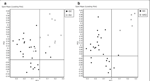
The PCA based on protein expression clearly
sepa-rated H. pylori isolated from GC from those isolated
from either DU or AAG, although there was a partial
overlap betweenH. pyloriisolated from patients affected
by DU and GC (Fig. 2).
Validation of selectedH. pyloriproteins
analysis of the in silico digested NapA protein. These 5 masses allowed to identify NapA among the proteins at 15 kDa (Mascot results with peptide tolerance at 0.5 Da: Score 93; Expect 0.0054; Sequence coverage 48%). In order to exclude the presence of the found 5 peptide se-quences in other proteins than NapA, the regions of simi-larity among other biological sequences were searched
with Basic Local Alignment Search Tool (BLAST) (http:// blast.ncbi.nlm.nih.gov/Blast.cgi). BLAST detected putative conserved domain of the ferritin-like superfamily and fer-ritin multi-domains, and confirmed the protein NapA at
Max Score 248 (Query cover 100%; E value 2e–82;
Iden-tity 100%; Accession AAG28154.1) (Supplementary re-sults, Additional file 1: Figure S1).
Fig. 2Principal component analysis of proteome maps of Helicobacter pylori isolates related to autoimmune atrophic gastritis (AAG), duodenal ulcer (DU) and gastric cancer (GC). The loading plots show an overview of the H. pylori spot maps of GCversusDU (a) and GCversusAAG (b). Each circle represents a spot map. AAG, DU and GC associated H. pylori spot maps are displayed ingrey,whiteandblack, respectively
Genetic interaction networks towards an understanding ofH. pyloriprotein profiles
We used the STRING software matching the H. pylori
strain 266995 to predict the protein-interactions based on the most differentially expressed proteins identified
by 2D-DIGE analysis among H. pylori isolated from
patients with either DU, AAG or GC. The obtained protein-protein interaction diagram (Fig. 4a, n. 33
pro-teins; p-value = 2.84e–10) revealed a widespread
con-nectivity among these differentially expressed proteins with relevance to proteins involved in: (i) organic sub-stance metabolic process (blue color); (ii) defense against extreme environment conditions (green color); (iii) oxidation reduction process (yellow color); (iv) chemical reactions involving various nitrogenous com-pounds (brown color), and (v) bacteria motility (red
color). Two proteins (the leucyl aminopeptidase,pepA,
and the ribosomal protein S12 methylthiotransferase
methylthiotransferase,rimO) were not interactive with
the other differentially expressed proteins. Both these proteins are presumably involved in the processing and regular turnover of intracellular proteins. The bifunctional
enzyme cysN/cysC (spot 111; gi|1706274), involved in bacterial sulfate assimilation pathway, as well as the 50S ribosomal protein L30 (spot 254; gi|226703094), did not
match with any H. pylori strains, the protein-interaction
for these proteins thus remaining uncertain.
To better understand the network ofH. pyloriproteins
associated with GC, we used the STRING software with the only up-regulated proteins found in GC-isolated
H. pylori. This allowed us to evidence that the molecular
pathway of these proteins was mainly related to nucleic
acid binding (Fig. 4b; 7 proteins,p-value = 0.0311).
Discussion
The scenario of molecular cross-talk between H. pylori
and host gastric mucosa is finely regulated allowing a microbial persistence in the host, but also confers a risk for important diseases. Genomics and proteomics
stud-ies showed an high variability among H. pylori strains
with strain-specific genes [18, 19] and proteins [20, 21] dispersed throughout the genome. In particular,
DU-promoting gene cluster (dupA cluster) and virB gene
forming a type IV secretory system (T4SS) have been
proposed as a risk marker for both DU disease and the production of the pro-inflammatory IL-8 cytokine [22],
while the intact H. pylori cag-PAI region has been
asso-ciated with both DU and GC [23, 24]. The complexity of
H. pyloriproteome is further increased whenH. pyloriis
analyzed in relation to gastric environment, in terms of both adaptation to hostile gastric conditions and host
re-sponse(s) to disease(s) [25–27]. Furthermore, factors and
molecular mechanisms linkingH. pylorito GC are yet to
be clearly elucidated.
The PCA analysis reported in our study showed a
good discrimination of H. pylori classification based on
patient’s disease, with the best result obtained analysing
H. pyloriisolated from patients with either GC or AAG,
rather than H. pylori isolated from patients with DU.
We compared by DIGE analysis paired groups of gastric
disease (i.e. DU versus GC and AAG versus GC). The
number of differentially expressed proteins of H. pylori
isolates was higher when comparing DU versus GC
(nr = 29) as compared with AAG versus GC (nr = 18),
this result showing a more pronounced molecular
di-versity between H. pylori strains from GC and DU.
This is in accord with the model of GC development, for which AAG, but not DU, is known to be a risk factor. Therefore, it is tempting to hypothesize that
similarly H. pylori strains isolated from GC are more
similar to H. pylori strains isolated from AAG than
those isolated from DU.
It is well accepted that different microorganisms may have different possibility to regulate cell metabolisms. In
our work, H. pyloriisolated from patients with DU was
found to regulate enzymes involved in metabolic path-ways that could neutralize particularly high acid secre-tion of the gastric microenvironment (i.e. log fold
increased expression; ispE: 3.25; groS: 3.13, metK: 2.92;
tuf: 2.92, amiE: 2.49; Table 2; Fig. 4a). Different
mecha-nisms allow the bacteria to proliferate in the highly acid gastric environment [28], and one of them involves am-monia generation from various substrates by enzymes such as urease (a nickel-containing enzyme composed of subunits UreA and UreB) [29] and amidases (i.g AmiE) [30]. Our work showed a higher content of AmiE and
UreB in the proteome ofH. pyloriisolated from DU and
AAG, respectively, as compared with GC. In particular, the AmiE enzyme is used as an alternative route for am-monia production necessary to maintain the pH homeo-stasis and to neutralize the gastric acidity [31], though ammonia may cause a direct tissue damage [32].
Diverse enzymes known to detoxify oxidants resulting from the high inflammatory status and to repair mole-cules [24, 33] have been found increased in content in
DU-H. pylori proteome: i.e. two ‘catalases’ (spots 267,
268), which protect cells against reactive oxygen species through degradation of hydrogen peroxide to water and
oxygen, and a ‘thioredoxin’ (spot 253), providing
elec-trons to peroxiredoxins to remove reactive oxygen and
nitrogen species [34]. Overall these H. pylori proteins
were up-regulated in DU-H. pylori and may play a role
in avoiding the higher acid and oxidative stress present in stomach microenvironment during DU with respect to that deriving from GC status. The reason for the oxi-dative stress behavior is that protein folding is severely affected by the gastric mucosa and inflammatory cells during DU.
Another protein strongly over-expressed in H. pylori
isolated from patients with DU was the putative heme
iron utilization protein, H. pyloriSJM 01705 (spot 149).
Iron must be acquired from the host, however, since an iron excess is toxic for bacteria, its acquisition is finely regulated by modulating the expression of this protein accordingly to the stomach conditions. In particular, this process may be particularly important in the case of
H. pylori isolated from patients with DU, where due
to stomach bleed, high levels of iron are present from hemoglobin degradation. Iron deficiency was shown to increase GC risk by increasing the virulence
phenotype of CagA-positive H. pylori [35].
Another pathway upregulated in H. pylori isolated
from patients with DU is involved in stress response with the up-regulated NapA and KatA proteins (spots 204 and 267, 268; Table 2; Fig. 4), which are both
pro-teins known to protect H. pylori DNA from oxidative
burst [36–38]. Moreover, NapA is also responsible for
the recruitment of neutrophils to the site of infection, resulting in an increased influx of oxyradicals leading to collateral tissue damage [36], and since phagocytes are
generally unable to kill the H. pylori, the production of
NapA is perpetuated with the concomitant increase in tissue damage and katA production. In agreement with this model, it is noted that peptic ulcer was less frequent in children and this had been related with a lower num-ber of neutrophils and CD3+ T-cells present in the gas-tric lamina propria of patients [39].
In the proteome of AAG-H. pylori compared with
GC-H. pylori, the metabolic pathway neutralizing the gastric acid microenvironment was the most decreased,
and indirectly it increased in GC-H. pylori (i.e. fold
change groL:−6.58; trxB:−5.35; tuf:−2.9; dnaG:−2.48;
atpA:−2.43; Table 2 and Fig. 4). Recently, Karlsson et al.
[40] found an increase in levels of the acid response
regulator ArsRS in H. pylori strain Nic25_A associated
(HCL) and acidity in the gastric lumen inhibits its secre-tion by negative feed-back. While in AAG condisecre-tions, the immune system attacks the parietal cells leading to hypochloridia (low HCL), which results in a loss of negative-feedback on gastrin secretion. In accord with this model, proteins involved in reduction of stomach
acidity were found less expressed in H. pylori isolated
from patients with AAG.
Urease B, a key enzyme for bacteria resistance to gastric acidity by catalyzing the hydrolysis of urea into
ammonia and CO2, is an immunogenic protein: its
epitope vaccination allowed a reduction in H. pylori
colonization and inflammation of the gastric mucosa [42]. We hypothesized that an increase in UreB
pro-duction in H. pylori from AAG patients compared with
H. pylori from GC patients could be beneficial since it
reduces gastric inflammation that is widely accepted to be related to GC pathogenesis. The importance of
ammonia in H. pylori metabolism and virulence is
underlined by the presence of several alternative routes for ammonia production, via enzymatic degradation of diverse amides and amino acids. Furthermore, network analyses with STRING showed that UreB protein are connected with the heat shock chaperone protein GroES (spot 227), which is known to induce a protective im-munity against mucosal infection [43]. Both AAG and GC are known to be associated with a severe inflamma-tory response, that is associated with increased levels of reactive oxygen and nitrogen radicals around the
colon-izing H. pylori. In a previous proteomics study it was
demonstrated that the infection with H. pylori strain
7.13 induces a severe inflammatory response in gerbils [11], that the authors associated with increased levels of reactive oxygen and nitrogen radicals at sites juxtaposed to colonizing organisms.
It is interesting to note that among the proteins highly
decreased in AAG-isolated H. pylori, there was a
fla-gellin A subunit (spot 42). This protein was known to polymerize together with flagellin B, and form the bacterial filaments, with an important role in both
bacterial motility and virulence [13, 44–46].
A putative elongation factor-Tu was detected in
H. pylori up-regulated proteome of both DU-H. pylori
and GC-H. pylori (spots 63 and 89). The major role of
this protein is to mediate the transfer of charged aminoacyl-tRNA to the A site of the ribosome during
peptide elongation. In our H. pylori samples, this
pro-tein showed two isoforms with a different accumulation in relation to patient gastric disease.
In regards to the biological processes, proteins
in-creasing in GC-H. pylori were mostly related to DNA
processes (replication, transcription and translation). In
particular, among the H. pylori proteins up-regulated in
GC isolates we identified an elongation factor (spots 63
and 89), a DNA primase involved in RNA modification
(spot 141), a DNA-directed RNA polymerase subunit α
(spot 95), a DNA-binding protein HU (spot 234), a tran-scriptional regulator (spot 262), a 50S ribosomal protein L21 (spot 233), a ribosomal protein S12 methyltioltrans-ferase (spot 232), and a 10 kDa chaperonin (spot 231) (Table 2; Fig. 4b). Interestingly, the DNA dependent RNA polymerase (RNAP) catalyzes the transcription of DNA into RNA, and it is composed of several subunits;
the subunit α of RNAP has been identified among
the proteins more specifically associated with gastric
H. pylori species rather than enterohepatic ones [47].
Moreover, the C-terminal domain of the α subunit of
RNAP, besides a primary role in the recruitment of RNA polymerase to various promoters, has a role in mediating interactions with several transcriptional regulators [48]. Concomitantly with these findings,
Lin et al. [49] identified the subunit α of RNAP like a
GC-related H. pylori antigen.
While the DNA primase encoded by the dnaG gene in an enzyme synthesizing short strands of RNA during DNA replication, and it is part of the replication
ma-chinery of the slowly growingH. pylori[50, 51]. Its
pres-ence may be related to a slow H. pylori growth related
to the extremes of the human gastric environment. In
addition, GC-H. pyloristrains increased the content of a
ribosomal protein. Xiao et al. [52] succeeded in
classify-ing differentH. pyloriorigins (P1 and P2) based on
ribo-somal proteins, which they estimated to represent the highest percentage (15%) of identified proteins. However,
the differential up-regulation in GC-H. pylori strains
may be only indicative of a higher demand of ribosomes, and, indirectly, a higher protein turnover as compared
with the DU-H. pyloristrains.
Conclusion
We have successfully performed a DIGE comparative
proteomics analysis ofH. pyloristrains isolated from
pa-tients affected by different gastric pathologies (AAG, DU or GC). Some of the identified proteins had not been
characterized in gastric disease-related H. pylori strains
before. The finding of differential protein profiles among
H. pylori related groups confirms the difference in H.
pyloristrains in relation to gastric disease. In particular,
in H. pyloriisolated from DU-patients a higher content
of proteins with antioxidant activity emerged (aroQ,
aspA, fldA, icd, oorA and scoB), as well as an
up-regulation of proteins belonging to metabolic pathways
counteracting the high acid environment (katA and
napA). While, in H. pylori isolated from AAG-patients
there was a significant decrease in proteins neutralizing hydrogen concentrations through organic substance
metabolic processes (dnaG, tuf, trxB and groL),
pathologies. In addition, a reduction of bacterial motility (flhA) was found to be associated with AAG-H. pylori
isolates. In GC-H. pylori strains it emerged an increase
in nucleic acid-binding proteins to be putatively involved in a higher demand of DNA- or protein-related pro-cesses. Some of the identified proteins may provide some new information in the understanding of the candidate
mechanism(s) associated with the differential H. pylori
behavior in human stomach disease(s), and indicate po-tential protein markers for the specific detection of DU
versusGC-related H. pylori. Some of our identified
pro-teins need to be further validated by functional analyses as well as at DNA transcriptional level, and it may be tempting to incorporate our protein expression data
with those of H. pylori genomic works in order to get
better insight into the differentialH. pyloripathogenesis.
Additional files
Additional file 1: Figure S1.List of peak masses enabling the identification of the neutrophil activating protein by mass spectrometry. The trypsin-digested peptides of the gel portion at ~ 15 kDa were also separated by MALDI-TOF to search for masses of the‘neutrophil activating protein, NapA’. The list of peak masses, which were generated by anin silicotripsin-digestion of the protein P43313 corresponding to the NapA, are listed together with both those found in the spot 204 digestion, and those detected in the digested 15 kDa bands. (PPT 167 kb)
Additional file 2: Table S1.Protein pairs of theHelicobacter pyloristrains isolated from patients affected by duodenal ulcer (DU) gastric cancer (GC) or autoimmune atrophic gastritis (AAG). Protein pairs fromH. pyloricases were labelled with either Cy3 or Cy5 dyes and mixed with a Cy2-labelled internal standard, containing equal amounts of the all protein extracts. (DOCX 13 kb)
Abbreviations
AAG:autoimmune atrophic gastritis; DU: duodenal ulcer; GC: gastric cancer; IARC: International Agency for Research on Cancer; IEF: isoelectric focusing; IPG: immobilized pH gradient; LC-MS/MS: liquid chromatography-tandem mass spectrometry; MALDI-TOF: matrix assisted laser desorption ionization-time of flight; PCA: principal component analysis; 1DE: one dimensional electrophoresis; 2DE: two dimensional electrophoresis; 2D-DIGE: two-dimensional difference in gel electrophoresis
Acknowledgements
We would like to thank Dr. Marica Garziera for her support to the study.
Funding
This work was supported by O. Repetto (5 × 1000 CRO grant) and L. Caggiari fellow (5 × 1000 Gastric cancer intramural grant).
Availability of data and materials
The datasets generated during the current study are available from the corresponding author on reasonable request.
Authors’contributions
Dr. VDR and Dr. OR designed the study, performed proteomics experiments and drafted the article; Dr. SZ and Dr. MC isolatedH. pyloristrains from biopsies; Dr. LC contributed to analyses; Dr. VC was involved in anatomical and cytological evaluation; Dr. RC selected and enrolled patients. All authors read and approved the final manuscript.
Consent for publication Not applicable.
Competing interests
The authors declare that they have no competing interests.
Ethics approval and consent to participate
All tested patients filled an informant consent reporting all the information about the research. Project was approved by CRO internal review board (IRB n°14).
Publisher’s Note
Springer Nature remains neutral with regard to jurisdictional claims in published maps and institutional affiliations.
Author details
1Facility of Bio-Proteomics, Immunopathology and Cancer Biomarkers, IRCCS CRO National Cancer Institute, Via F. Gallini 2, 33081 Aviano, Italy.
2
Microbiology-Immunology and Virology, IRCCS CRO National Cancer Institute, Aviano, Italy.3Pathology Gastroenterology, IRCCS CRO National Cancer Institute, Aviano, Italy.4Gastroenterology, IRCCS CRO National Cancer Institute, Aviano, Italy.
Received: 13 January 2017 Accepted: 13 April 2017
References
1. Naumann M, Sokolova O, Tegtmeyer N, Backert S. Helicobacter pylori: A paradigm pathogen for subverting host cell signal transmission. Trends Microbiol. 2017. pii: S0966-842X(16)30197-4. doi: 10.1016/j.tim.2016.12.004. [Epub ahead of print]
2. Mégraud F, Bessède E, Varon C. Helicobacter pylori infection and gastric carcinoma. Clin Microbiol Infect. 2015;21(11):984–90.
3. Alvarez-Arellano L, Maldonado-Bernal C. Helicobacter pylori and neurological diseases: Married by the laws of inflammation. World J Gastrointest Pathophysiol. 2014;5(4):400–4.
4. Magen E, Delgado JS. Helicobacter pylori and skin autoimmune diseases. World J Gastroenterol. 2014;20(6):1510–6.
5. Kim SS, Ruiz VE, Carroll JD, Moss SF. Helicobacter pylori in the pathogenesis of gastric cancer and gastric lymphoma. Cancer Lett. 2011;305(2):228–38. 6. Sasazuki S, Inoue M, Iwasaki M, Otani T, Yamamoto S, Ikeda S, Hanaoka T. Tsugane S; Japan Public Health Center Study Group. Effect of Helicobacter pylori infection combined with CagA and pepsinogen status on gastric cancer development among Japanese men and women: a nested case- control study. Cancer Epidemiol Biomarkers Prev. 2006;15(7):1341–7.
7. da Costa DM, Pereira Edos S, Rabenhorst SH. What exists beyond cagA and vacA? Helicobacter pylori genes in gastric diseases. World J Gastroenterol. 2015;21(37):10563–72.
8. Blanchard TG, Czinn SJ, Correa P, Nakazawa T, Keelan M, Morningstar L, Santana-Cruz I, Maroo A, McCracken C, Shefchek K, Daugherty S, Song Y, Fraser CM, Fricke WF. Genome sequences of 65 Helicobacter pylori strains isolated from asymptomatic individuals and patients with gastric cancer, peptic ulcer disease, or gastritis. Pathog Dis. 2013;68(2):39–43. 9. Romo-González C, Salama NR, Burgeño-Ferreira J, Ponce-Castañeda V, Lazcano-Ponce E, Camorlinga-Ponce M, Torres J. Differences in genome content among Helicobacter pylori isolates from patients with gastritis, duodenal ulcer, or gastric cancer reveal novel disease-associated genes. Infect Immun. 2009;77(5):2201–11.
10. Yamaoka Y. Roles of the plasticity regions of Helicobacter pylori in gastroduodenal pathogenesis. J Med Microbiol. 2008;57(Pt 5):545–53. 11. Franco AT, Israel DA, Washington MK, Krishna U, Fox JG, Rogers AB, Neish AS,
Collier-Hyams L, Perez-Perez GI, Hatakeyama M, Whitehead R, Gaus K, O’Brien DP, Romero-Gallo J, Peek Jr RM. Activation of beta-catenin by carcinogenic Helicobacter pylori. Proc Natl Acad Sci U S A. 2005;102(30):10646–51. 12. Pyndiah S, Lasserre JP, Ménard A, Claverol S, Prouzet-Mauléon V, Mégraud F,
Zerbib F, Bonneu M. Two-dimensional blue native/SDS gel electrophoresis of multiprotein complexes from Helicobacter pylori. Mol Cell Proteomics. 2007;6(2):193–206.
13. Jungblut PR, Bumann D, Haas G, Zimny-Arndt U, Holland P, Lamer S, Siejak F, Aebischer A, Meyer TF. Comparative proteome analysis of H. pylori. Mol Microbiol. 2000;36(3):710–25.
15. Fasciana T, Calà C, Bonura C, Di Carlo E, Matranga D, Scarpulla G, Manganaro M, Camilleri S, Giammanco A. Resistance to clarithromycin and genotypes in Helicobacter pylori strains isolated in Sicily. J Med Microbiol. 2015;64(11):1408–14.
16. Simula MP, De Re V. Hepatitis C virus-induced oxidative stress and mitochondrial dysfunction: a focus on recent advances in proteomics. Proteomics Clin Appl. 2010;4(10–11):782–93.
17. Franceschini A, Szklarczyk D, Frankild S, Kuhn M, Simonovic M, Roth A, Lin J, Minguez P, Bork P, von Mering C, Jensen LJ. STRING v9.1: protein-protein interaction networks, with increased coverage and integration. Nucleic Acids Res. 2013;41:D808–15. doi:10.1093/nar/gks1094.
18. Fernandez-Gonzalez E, Backert S. DNA transfer in the gastric pathogen Helicobacter pylori. J Gastroenterol. 2014;49(4):594–604.
19. Fischer W, Breithaupt U, Kern B, Smith SI, Spicher C, Haas R. A
comprehensive analysis of Helicobacter pylori plasticity zones reveals that they are integrating conjugative elements with intermediate integration specificity. BMC Genomics. 2014;15:310.
20. Austin CM, Maier RJ. Aconitase-mediated posttranscriptional regulation of Helicobacter pylori peptidoglycan deacetylase. J Bacteriol. 2013;195(23):5316–22. 21. Choi YW, Park SA, Lee HW, Kim DS, Lee NG. Analysis of growth phase-dependent proteome profiles reveals differential regulation of mRNA and protein in Helicobacter pylori. Proteomics. 2008;8(13):2665–75.
22. Jung SW, Sugimoto M, Shiota S, Graham DY, Yamaoka Y. The intact dupA cluster is a more reliable Helicobacter pylori virulence marker than dupA alone. Infect Immun. 2012;80(1):381–7.
23. Peek RM, Fiske C, Wilson KT. Role of innate immunity in Helicobacter pylori-induced gastric malignancy. Physiol Rev. 2010;90(3):831–58. 24. Kusters JG, van Vliet AHM, Kuipers EJ. Pathogenesis of Helicobacter pylori
infection. Clin Microbiol Rev. 2006;19(3):449–90.
25. Oluwasola AO. Genetic determinants and clinico-pathological outcomes of Helicobacter pylori infection. Ann Ib Postgrad Med. 2014;12(1):22–30. 26. Percival SL, Suleman L. Biofilms and Helicobacter pylori: dissemination and
persistence within the environment and host. World J Gastrointest Pathophysiol. 2014;5(3):122–32.
27. Roesler BM, Rabelo-Gonçalves EM, Zeitune JM. Virulence factors of Helicobacter pylori: a review. Clin Med Insights Gastroenterol. 2014;7:9–17.
28. Cover TL, Blaser MJ. Helicobacter pylori in health and disease. Gastroenterology. 2009;136(6):1863–73.
29. Scott DR, Marcus EA, Weeks DL, Sachs G. Mechanisms of acid resistance due to the urease system of Helicobacter pylori. Gastroenterology. 2002;123(1):187–95.
30. Skouloubris S, Labigne A, De Reuse H. The AmiE aliphatic amidase and AmiF formamidase of Helicobacter pylori: natural evolution of two enzyme paralogues. Mol Microbiol. 2001;40(3):596–609.
31. Basso D, Plebani M, Kusters JG. Pathogenesis of Helicobacter pylori infection. Helicobacter. 2010;15 Suppl 1:14–20.
32. el Nujumi AM, Rowe PA, Dahill S, Dorrian CA, Neithercut WD, McColl KE. Role of ammonia in the pathogenesis of the gastritis, hypergastrinaemia, and hyperpepsinogenaemia I caused by Helicobacter pylori infection. Gut. 1992;33(12):1612–6.
33. Wang G, Alamuri P, Maier RJ. The diverse antioxidant systems of Helicobacter pylori. Mol Microbiol. 2006;61(4):847–60.
34. Lu J, Holmgren A. The thioredoxin antioxidant system. Free Radic Biol Med. 2014;66:75–87.
35. Noto JM, Lee JY, Gaddy JA, Cover TL, Amieva MR, Peek Jr RM. Regulation of Helicobacter pylori virulence within the context of iron deficiency. J Infect Dis. 2015;211(11):1790–3.
36. Wang G, Hong Y, Olczak A, Maier SE, Maier RJ. Dual Roles of Helicobacter pylori NapA in inducing and combating oxidative stress. Infect Immun. 2006;74(12):6839–46.
37. Cooksley C, Jenks PJ, Green A, Cockayne A, Logan RP, Hardie KR. NapA protects Helicobacter pylori from oxidative stress damage, and its production is influenced by the ferric uptake regulator. J Med Microbiol. 2003;52(Pt 6):461–9. 38. Ramarao N, Gray-Owen SD, Meyer TF. Helicobacter pylori induces but
survives the extracellular release of oxygen radicals from professional phagocytes using its catalase activity. Mol Microbiol. 2000;38(1):103–13. 39. Bontems P, Aksoy E, Burette A, Segers V, Deprez C, Mascart F, Cadranel S.
NF-κB activation and severity of gastritis in Helicobacter pylori-infected children and adults. Helicobacter. 2014;19(3):157–67.
40. Karlsson R, Thorell K, Hosseini S, Kenny D, Sihlbom C, Sjöling Å, Karlsson A, Nookaew I. Comparative analysis of two Helicobacter pylori strains using
genomics and mass spectrometry-based proteomics. Front Microbiol. 2016;7:1757. eCollection 2016.
41. De Re V, Orzes E, Canzonieri V, Maiero S, Fornasarig M, Alessandrini L, Cervo S, Steffan A, Zanette G, Mazzon C, De Paoli P, Cannizzaro R. Pepsinogens to distinguish patients with gastric intestinal metaplasia and Helicobacter pylori infection among populations at risk for gastric cancer. Clin Transl Gastroenterol. 2016;7(7):e183.
42. Yang J, Dai LX, Pan X, Wang H, Li B, Zhu J, Li MY, Shi XL, Wang BN. Protection against Helicobacter pylori infection in BALB/c mice by oral administration of multi-epitope vaccine of CTB-UreI-UreB. Pathog Dis. 2015;73(5). doi: 10.1093/femspd/ftv026.
43. Ferrero RL, Thiberge JM, Kansau I, Wuscher N, Huerre M, Labigne A. The GroES homolog of Helicobacter pylori confers protective immunity against mucosal infection in mice. Proc Natl Acad Sci U S A. 1995;92(14):6499–503. 44. Hopf PS, Ford RS, Zebian N, Merkx-Jacques A, Vijayakumar S, Ratnayake D,
Hayworth J, Creuzenet C. Protein glycosylation in Helicobacter pylori: beyond the flagellins? PLoS One. 2011;6(9):e25722.
45. Franco AT, Friedman DB, Nagy TA, Romero-Gallo J, Krishna U, Kendall A, Israel DA, Tegtmeyer N, Washington MK, Peek Jr RM. Delineation of a carcinogenic Helicobacter pylori proteome. Mol Cell Proteomics. 2009;8(8):1947–58.
46. Lee HW, Choe YH, Kim DK, Jung SY, Lee NG. Proteomic analysis of a ferric uptake regulator mutant of Helicobacter pylori: regulation of Helicobacter pylori gene expression by ferric uptake regulator and iron. Proteomics. 2004;4(7):2014–27.
47. Fowsantear W, Argo E, Pattinson C, Cash P. Comparative proteomics of Helicobacter species: the discrimination of gastric and enterohepatic Helicobacter species. J Proteomics. 2014;97:245–55.
48. Borin BN, Tang W, Krezel AM. Helicobacter pylori RNA polymeraseα-subunit C-terminal domain shows features unique toɛ-proteobacteria and binds NikR/DNA complexes. Protein Sci. 2014;23(4):454–63.
49. Lin YF, Wu MS, Chang CC, Lin SW, Lin JT, Sun YJ, Chen DS, Chow LP. Comparative immunoproteomics of identification and characterization of virulence factors from Helicobacter pylori related to gastric cancer. Mol Cell Proteomics. 2006;5(8):1484–96.
50. Sharma A, Kamran M, Verma V, Dasgupta S, Dhar SK. Intracellular locations of replication proteins and the origin of replication during chromosome duplication in the slowly growing human pathogen Helicobacter pylori. J Bacteriol. 2014;196(5):999–1011.
51. Nitharwal RG, Verma V, Dasgupta S, Dhar SK. Helicobacter pylori chromosomal DNA replication: current status and future perspectives. FEBS Lett. 2011;585(1):7–17.
52. Xiao D, Zhang H, He L, Peng X, Wang Y, Xue G, Su P, Zhang J. High natural variability bacteria identification and typing: Helicobacter pylori analysis based on peptide mass fingerprinting. J Proteomics. 2014;98:112–22.
• We accept pre-submission inquiries
• Our selector tool helps you to find the most relevant journal
• We provide round the clock customer support
• Convenient online submission
• Thorough peer review
• Inclusion in PubMed and all major indexing services
• Maximum visibility for your research
Submit your manuscript at www.biomedcentral.com/submit