R E S E A R C H
Open Access
Pro-inflammatory adjuvant properties of
pigment-grade titanium dioxide particles
are augmented by a genotype that
potentiates interleukin 1
β
processing
Sebastian Riedle
1,2,3, Laetitia C. Pele
4, Don E. Otter
1,5, Rachel E. Hewitt
4,6, Harjinder Singh
2, Nicole C. Roy
1,2†and Jonathan J. Powell
4,6*†Abstract
Background:Pigment-grade titanium dioxide (TiO2) particles are an additive to some foods (E171 on ingredients lists), toothpastes, and pharma−/nutraceuticals and are absorbed, to some extent, in the human intestinal tract. TiO2 can act as a modest adjuvant in the secretion of the pro-inflammatory cytokine interleukin 1β(IL-1β) when triggered by common intestinal bacterial fragments, such as lipopolysaccharide (LPS) and/or peptidoglycan.
Given the variance in human genotypes, which includes variance in genes related to IL-1βsecretion, we investigated whether TiO2particles might, in fact, be more potent pro-inflammatory adjuvants in cells that are genetically susceptible to IL-1β-related inflammation.
Methods:We studied bone marrow-derived macrophages from mice with a mutation in the nucleotide-binding oligomerisation domain-containing 2 gene (Nod2m/m), which exhibit heightened secretion of IL-1βin response to the peptidoglycan fragment muramyl dipeptide (MDP). To ensure relevance to human exposure, TiO2was food-grade anatase (119 ± 45 nm mean diameter ± standard deviation). We used a short‘pulse and chase’format: pulsing with LPS and chasing with TiO2+/−MDP or peptidoglycan.
Results:IL-1βsecretion was not stimulated in LPS-pulsed bone marrow-derived macrophages, or by chasing with MDP, and only very modestly so by chasing with peptidoglycan. In all cases, however, IL-1βsecretion was augmented by chasing with TiO2in a dose-dependent fashion (5–100μg/mL). When co-administered with MDP or peptidoglycan, IL-1βsecretion was further enhanced for theNod2m/mgenotype. Tumour necrosis factorαwas triggered by LPS priming, and more so for theNod2m/mgenotype. This was enhanced by chasing with TiO2, MDP, or peptidoglycan, but there was no additive effect between the bacterial fragments and TiO2.
Conclusion:Here, the doses of TiO2that augmented bacterial fragment-induced IL-1βsecretion were relatively high. In vivo, however, selected intestinal cells appear to be loaded with TiO2, so such high concentrations may be ‘exposure-relevant’for localised regions of the intestine where both TiO2and bacterial fragment uptake occurs. Moreover, this effect is enhanced in cells fromNod2m/mmice indicating that genotype can dictate inflammatory signalling in response to (nano)particle exposure. In vivo studies are now merited.
Keywords:Nano, Particle, TiO2, E171, NOD2, IL-1β, TNF-α, Muramyl dipeptide, Peptidoglycan
* Correspondence:[email protected]
†Equal contributors 4
Biomineral Research Group, MRC Human Nutrition Research, Elsie Widdowson Laboratory, 120 Fulbourn Road, Cambridge CB1 9NL, UK
6Department of Veterinary Medicine, Biomineral Research Group, University
of Cambridge, Madingley Road, Cambridge CB3 0ES, UK Full list of author information is available at the end of the article
Background
Potential toxicological effects following exposure to ti-tanium dioxide (TiO2) are of current interest [1, 2].
TiO2 is a mineral pigment which, when used in a
particulate form, is valued for its properties as a whitening or brightening agent, and is included in some processed foods (E171 on ingredients lists), toothpastes, capsules and tablets. From these sources, the average daily intake of pigment-grade TiO2 for an adult in the UK is about
1012particles/day [3, 4], nominally ~0.04 mg/kg/day for a 70 kg adult. These findings are supported by a recent Dutch study with mean long term intakes of pigment-grade TiO2ranging from 0.06 mg/kg/day in elderly
sub-jects to 0.17 mg/kg/day for 7–69-year-olds [5]. In 2–6 year old children, however, it was higher at 0.67 mg/kg/day [5].
It is well established that particles of TiO2, likely
de-rived from sources of the Western lifestyle described above, accumulate in certain cells, such as macrophages in Peyer’s patches of the human small intestine [6–10]. Whether they have any deleterious impact in this envir-onment remains a matter of speculation, but, if they do, both cell accumulation and host factors are likely to be important [4]. Indeed, it has been often noted that the accumulation of these particles occurs where the earliest signs of Crohn’s disease have been reported [11]. With respect to cell accumulation and stimulation, the pristine particle is probably of limited relevance. The intestinal lumen is a ‘soup’ of proteins, bacterial fragments, ions, small organic molecules etc. and these will modify the surface of the particles through adsorptive interactions. Consistent with this, there are several reports of how TiO2 particles act as an adjuvant for cellular responses
to the bacterial-derived molecule lipopolysaccharide (LPS), either through formation of ‘conjugates’or by co-incubation [12–15].
More recently it has been shown that pigment-grade TiO2 is a modest trigger of the NLR family pyrin
domain-containing 3 (NLRP3) inflammasome and that this activity may contribute to intestinal inflammatory properties of the particle in murine models [16]. The inflammasome regulates the activation of caspase-1 which, in turn, determines cleavage of inactive pro-inter-leukin 1β(pro-IL-1β) to form mature pro-inflammatory interleukin 1β(IL-1β). If such a pro-inflammatory effect from oral TiO2 exposure translates significantly from
murine models to humans, it must be occurring in a small minority of the population because most children and adults do not have intestinal disease. In this respect some variants of human genotype could be important. Indeed, it is well recognised that inflammatory bowel diseases are complex polygenic disorders [17]. Certain mutations in the nucleotide-binding oligomerisation domain-containing 2 (NOD2) gene, for example, are as-sociated with an increased risk of the inflammatory
bowel disease, Crohn’s disease [18, 19]. Maeda et al. have shown that in mice at least one form ofNod2 mutation potentiates IL-1βprocessing and enhances risk of intes-tinal inflammation [20]. These mice carry a known Crohn’s disease-associated ‘knockin’ mutation in the
Nod2locus but also carry a duplication of the 3′end of the wild-type (WT)Nod2locus [21], and herein are des-ignated as Nod2m/mmice. Specifically, development of a modest pro-inflammatory phenotype in these animals is reportedly triggered by a bacterial peptidoglycan moiety, muramyl dipeptide (MDP), in an IL-1β-dependent fash-ion [20]. Since bacterial peptidoglycan is taken up by Peyer’s patch phagocytes [22, 23] it raises the possibility that TiO2 could act as an adjuvant for the
pro-inflammatory effects of peptidoglycan, and especially so where the genotype potentiates IL-1βprocessing. Hence, using bone marrow-derived macrophages (BMDMs) from WT andNod2m/mmice, we have tested these possi-bilities using an assay of short ‘pulse and chase’format, to determine if and how TiO2could amplify IL-1β
secre-tion at the cellular level.
Methods
Study design
The macrophage-stimulatory effects of dietary TiO2were
investigated, either alone or in combination with microbial-associated molecular patterns (MAMPs), using cells from WT andNod2m/mmice. MAMP concentrations were fixed whereas a range of TiO2concentrations was
in-vestigated. LPS pre-stimulation of cells was employed as this MAMP is abundant in the intestinal lumen and can prime cells for an inflammasome-driven response (IL-1β secretion), as described in the Introduction. Parameters assessed were overall cell viability, particle uptake, and secretion of the pro-inflammatory cytokines IL-1β and tumour necrosis factor alpha (TNF-α).
TiO2particles
Food- and pharmaceutical-grade TiO2particles with
an-atase crystal structure and a purity of not less than 99% were obtained from Sensient Colors (St. Louis, USA). According to the manufacturer, the TiO2 particles had
an average particle size of 300 nm and a maximum par-ticle size of 1.0μm, which had been determined using a sediograph instrument. We undertook further analysis of the powder, initially with transmission electron micros-copy. A 1 mg/mL suspension of TiO2powder in distilled
water (Life Technologies, Auckland, New Zealand) with 0.5% bovine serum albumin (BSA; Life Technologies) as a dispersant was prepared. A drop of the TiO2particle
analysis software iTEM (Olympus Soft Imaging Solutions, Münster, Germany) was used to record the images digitally and subsequently measure the diameter of the particles.
In addition, particle size under cell culture conditions was determined with nanoparticle tracking analysis, which is a method to analyse dispersed particles based on their Brownian motion, similar to analysis with dy-namic light scattering [24]. A 100 μg/mL TiO2 particle
suspension was prepared in tissue culture medium (TCM) consisting of RPMI 1640 medium (Sigma-Al-drich, Gillingham, UK) with 10% foetal bovine serum (FBS; PAA Laboratories, Yeovil, UK) and 1% penicillin-streptomycin antibiotics (Sigma-Aldrich). The suspen-sion was sonicated for 10 min to facilitate distribution of the TiO2 particles in the medium. The motion of the
particles in suspension was digitally recorded with a NanoSight NS500 instrument (NanoSight, Amesbury, UK). Three TiO2 suspensions were analysed
independ-ently. The particle sizes were calculated from the recorded videos with nanoparticle tracking analysis soft-ware (Nanosight).
Animals
For the cell culture experiments, bone marrow was ob-tained from 10 to 18 week old female C57BL/6 WT and
Nod2m/m mice. The original WT breeding pairs were purchased from the Jackson Laboratory (Bar Harbor, USA) and bred at the AgResearch Small Animal Colony (Hamilton, New Zealand). Breeding pairs for Nod2m/m
mice on a C57BL/6 background were kindly provided by Lars Eckmann [20], and backcrossed with WT mice for 10 generations at the AgResearch Small Animal Colony. The mice were kept under conventional conditions at all times [25].
Harvest of BMDMs and cell culture
For the bone marrow collection, the mice were eutha-nised with CO2 asphyxiation and cervical dislocation.
Femurs and tibias were collected, sterilised in 70% etha-nol for 10 s, and the bone marrow flushed out with cold RPMI 1640 medium (Life Technologies). Single cell sus-pensions were prepared by passing the cells repeatedly through a 19G needle (BD Biosciences, Singapore) and a 70μm cell strainer (BD Labware, Franklin Lakes, USA). Bone marrow cells were re-suspended in TCM consist-ing of RPMI 1640 medium (Life Technologies) with 10% FBS (Life Technologies), 1% penicillin-streptomycin anti-biotics (Life Technologies), and 10 μg/mL macrophage colony-stimulating factor (eBioscience, San Diego, USA). The cells were transferred to non-tissue culture treated 24-well plates (BD Labware) at a concentration of 1 × 10⁶cells/well in 1 mL TCM and cultured at 37 °C in 7% CO2/93% air. Half of the TCM was replaced every 3 days
with fresh TCM throughout the culture period. Bone mar-row cells were fully differentiated into BMDMs on day 7 and used for experiments between day 8 and day 10.
Stimulation of BMDMs with TiO2particles +/−
peptidoglycan or MDP
As previously noted, a short ‘pulse (LPS) and chase (TiO2 +/− peptidoglycan or MDP)’ format was used to
dissect out the point in the pathway that the particles might act as pro-inflammatory adjuvants of MAMPs. To that effect, harvested murine BMDMs from each geno-type, +/−LPS pre-stimulation, were exposed to a range of TiO2 particle concentrations +/− peptidoglycan or
MDP, as detailed below.
To activate the cells, especially for pro-IL-1β induc-tion, BMDMs were first primed in culture with 1 mL TCM containing 10 ng/mL LPS from Escherichia coli
O111:B4 (Sigma-Aldrich, Auckland, New Zealand) for 3 h at 37 °C in 7% CO2/93% air. Unprimed BMDMs
were cultured under identical conditions but without LPS. All cells were then washed in TCM before the TiO2
suspensions were added. A 1 mg/mL TiO2stock
suspen-sion was first prepared in distilled water and autoclaved. This stock suspension was used to prepare TiO2
suspen-sions in the TCM with final concentrations from 5μg/mL to 100 μg/mL. Similar concentrations have been used in previous studies that examined cytokine secretion by phagocytic cells after TiO2 exposure [13, 26–28]. The
TiO2 suspensions were sonicated in a water bath for
10 min before 1 mL of the respective TiO2 suspension
was added to the cells. When the BMDMs were co-stimu-lated with MAMPs, either synthetic MDP or peptidoglycan from Bacillus subtilis (both from Sigma-Aldrich) was added to the respective TiO2
sus-pensions in TCM, both at a final concentration of 10 μg/mL. The BMDMs were incubated with TiO2
particles in TCM with or without the co-stimulants for 3 h at 37 °C in 7% CO2/93% air.
Flow cytometry analysis of BMDMs
phycoerythrin-labelled F4/80 antibody (clone BM8; BioLegend), a specific marker for murine macrophages. In addition, 0.8 μg/mL propidium iodide (PI; Life Technologies) was added to each sample immediately before analysis for viability assessment. The cells were analysed with a FACS Calibur flow cytometer (BD Biosciences, San Jose, USA), and at least 12,000 events per sample were acquired with the CellQuest Pro soft-ware (BD Biosciences). Data analysis was performed with FlowJo (Tree Star, Ashland, USA). For details on the gat-ing strategy see Additional file 1. The percentage of vi-able cells in relation to the total number of detected events was assessed with PI staining. Cells that did not show PI staining (PI−) were considered to be viable cells. BMDMs were identified among the PI− cells based on the expression of F4/80, i.e. viable cells that expressed F4/80 (PI−F4/80+) were classified as viable BMDMs. The percentages of PI−F4/80+ cells in relation to the total number of viable cells are shown in Additional file 2. Uptake of TiO2 particles by BMDMs was assessed
with the median side scatter (SSC) intensity of the PI−F4/80+ cell populations. According to previous studies, an increase in SSC intensity indicated TiO2
particle uptake [12, 29, 30].
Validation of SSC analysis by flow cytometry as a measure of TiO2cellular uptake
To confirm that increases in SSC intensity did indeed in-dicate TiO2 particle uptake, we undertook correlative
studies with conventional flow cytometry and imaging cytometry which allows visualisation of TiO2 uptake by
individual cells [31]. This technique was not available in the laboratory that undertook the above work and is im-practical for a very large number of samples, so only the lower concentration range was investigated and corre-lated to ensure true discrimination from background.
To quantify TiO2 cellular uptake (i.e. association and
localisation) by peripheral myeloid cell populations, fresh leukocyte cones were purchased from the National Blood Service (Cambridge, UK). Peripheral blood mononuclear cells (PBMCs) were isolated by density centrifugation using the separating medium Lympho-prep (Axis Shield Diagnostics, Dundee, UK) and frozen until use. PBMCs from 3 leucocyte cones were thawed and rested for 2 h prior to incubation at 1 × 10⁶cells/mL with 0 μg/mL, 5 μg/mL, or 10 μg/mL TiO2 and
incubated for 24 h in RPMI 1640 medium (Sigma-Aldrich, Gillingham, UK) supplemented with 10% FBS (Sigma-Aldrich).
After incubation, cells were washed with ice cold PBS (Sigma-Aldrich) containing 1% BSA (Sigma-Aldrich) and stained for the human monocyte/myeloid cell markers CD14 Alexa Fluor 488 or CD11c fluorescein isothio-cyante (both from BD Biosciences), respectively. Single
stain compensation tubes and unstained PBMC tubes, with and without TiO2, were also prepared at this time
from PBMC samples for the generation of compensation matrices. After staining, PBMCs were washed with ice cold PBS containing 1% BSA, re-suspended in a small volume of PBS containing 2% paraformaldehyde (Sigma-Aldrich) solution, and placed on ice in the dark until acquisition.
Imaging cytometry analysis was undertaken using an ImageStreamX Mark I platform (Amnis-Merck-Milli-pore, Seattle, USA), equipped with 405 nm and 488 nm lasers for excitation, a 785 nm laser for a scatter signal with standard filter sets, multi magnification (20×/40×/ 60×) and extended depth of field. INSPIRE software (Amnis) was used for acquisition and IDEAS software (Amnis) for analysis. The machine passed all tests and was fully calibrated prior to acquisition of samples. Be-fore acquisition, cells were filtered through 35 μm cell strainers (BD Labware). A minimum of 10,000 events per sample were acquired. Compensation matrices were generated by running single stained cells (i.e. single cell surface marker) and analysed using IDEAS software. For analysis, TiO2 positive cells were identified and
quanti-fied using a spot count analysis of dark spots appearing within the cells based on bright-field images of CD14 positive (CD14+) cells. Briefly, cells were first plotted as area versus aspect ratio of the bright-field images and a single cell gate drawn, followed by a focused gate. CD14+ cells were then gated based on fluorescence intensity. A custom dark spot count mask was gener-ated to quantify CD14+ cells, with cells positive for 2 or more darks pots gated as dark spot positive.
Conventional flow cytometry analysis was performed using a CyAn ADP 9 colour analyser (Beckman Coulter, Brea, USA) equipped with 405 nm, 488 nm and 642 nm solid-state lasers and 11 detectors in standard configur-ation. Summit software was used for acquisition and analysis (Beckman Coulter, USA). At least 500,000 events were acquired on the flow cytometer using a lowered SSC setting on a logarithmic scale. Samples were filtered through 35μm cell strainers (BD Labware) directly prior to acquisition. For data analysis, events were first plotted as forward versus side scatter using SSC on a logarithmic scale, and a large gate was drawn excluding debris. Cells were then further gated for CD11c positivity based on fluorescence intensity for the mean fluorescence intensity (MFI) of the SSC signal of CD11c+myeloid cells.
Stimulation of PBMCs with monosodium urate crystals or silica nanoparticles
nanoparticles (SNPs) [32, 33], promote IL-1βprocessing in our short ‘pulse and chase’ format. Isolated PBMCs (n= 4) were thawed and rested overnight. Cells (1.106 cells/mL) were then subjected to LPS pre-stimulation (10 ng/mL, Escherichia coliO111:B4; Sigma-Aldrich) to induce the production of pro-IL-1β or with TCM as a negative control. Following 3 h, cells were washed and then challenged with 100 μg/mL MSU crystals (Caltag Medsystems, Buckingham, UK) or 100 μg/mL SNPs (InvivoGen, San Diego, USA) for a further 3 h. Fol-lowing this, cells were washed and replenished with fresh TCM for a further 21 h (3 + 21 h). Supernatants were collected at the 3 h and 3 + 21 h time points for IL-1β analyses.
Cytokine detection in cell supernatants
Cell supernatants were collected at the time points indi-cated and stored at −20 °C until required for cytokine analysis. IL-1β (TiO2 and exemplar
inflammasome-activating particles) and TNF-α(TiO2only) were
investi-gated with enzyme-linked immunosorbent assay (ELISA) using DuoSet ELISA kits (R&D Systems, Minneapolis, USA) according to the manufacturer’s instructions. The cytokine concentrations were generally determined with a FlexStation 3 microplate scanner (Molecular Devices, Sunnyvale, USA) and Soft Max Pro software (Molecular Devices).
Statistical analysis
All statistical comparisons were carried out using R (R Development Core Team, Vienna, Austria). For analysis of the flow cytometry results, the groups according to genotype (WT or Nod2m/m BMDMs) were compared with two-way analysis of variance (ANOVA) using co-st-imulation condition and TiO2 exposure as the two
fac-tors. For analysis of the cytokine secretion results without co-stimulation, the groups according to geno-type (WT or Nod2m/m BMDMs) were compared with one-way ANOVA using TiO2 exposure as the single
factor. For analysis of the cytokine secretion results with co-stimulation, the groups according to co-stimulation condition (MDP or peptidoglycan) were compared with two-way ANOVA using genotype and TiO2exposure as
the two factors. In instances where two-way ANOVA results showed a significant interaction effect or the one-way ANOVA results indicated a significant differ-ence between groups, pairwise group comparisons were performed with Tukey’s post-hoc test. Figures depict group means ± standard deviation (SD). Finally, paired T tests were used to compare supernatant levels of IL-1β for cells exposed to MSU crystals or SNPs versus non-particle-exposed control cells. Group means ± standard error of the mean (SEM) are depicted in the corresponding figure.
Results
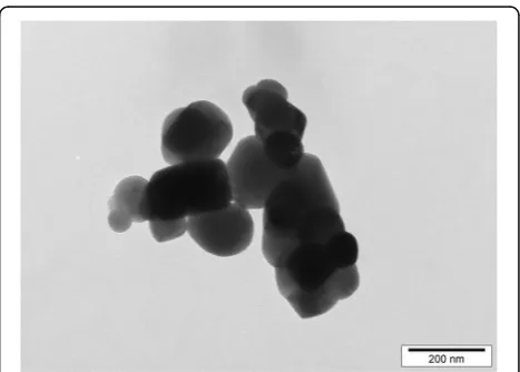
TiO2particle characterisation
Several images of TiO2 particles were obtained with
transmission electron microscopy and a representative image is shown in Fig. 1. The diameters of individual particles were measured with image analysis software. The average primary particle size was 119 nm with a SD of 45 nm, and the observed particle sizes ranged from 50 nm to 350 nm with a maximum frequency at 100 nm (Fig. 2a). Approximately 54% of the particles had a diam-eter between 125 nm and 200 nm, and about 40% had a diameter of 100 nm or less.
TiO2particles were suspended in TCM for the
sub-sequent cell culture experiments, so the particle sizes in TCM were also investigated, using nanoparticle tracking analysis. According to this method, the average particle size was 160 nm, and the sizes ranged between 20 nm and 450 nm (Fig. 2b). Ap-proximately 20% of the particles had a diameter of less than 100 nm. The slight increase in particle sizes versus electron microscopy measures probably results from the differing environments as, in solution, parti-cles have a hydration shell and are liable to adsorb TCM molecules. However, the possibility of a small degree of agglomeration in this environment cannot be precluded.
Cellular effects of TiO2particles
As intended with our short‘pulse and chase’style assay, BMDMs of both genotypes that were not primed with LPS (i.e. sham-pulsed) did not secrete meaningful amounts of IL-1βwhen chased for 3 h with TiO2from
Fig. 1Transmission electron microscopy image of TiO2particles.
Food- and pharmaceutical-grade anatase TiO2particles were suspended
0 μg/mL to 100μg/mL +/−MDP or peptidoglycan (IL-1β secretion always <5 pg/mL; data not shown). All subsequent data therefore refer to results with LPS-primed cells.
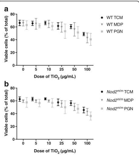
Cell viability
The viability of LPS-pulsed cells, from WT (Fig. 3a) and
Nod2m/m mice (Fig. 3b), was significantly reduced by chasing with TiO2particles, in a dose-responsive fashion
(p< 0.001 for trend, Fig. 3a and b). Addition of peptido-glycan or MDP during the chase phase marginally, but significantly, decreased cell viability further (p< 0.001 for trend), although there was no interaction effect with TiO2exposure (Fig. 3a and b).
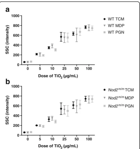
Particle uptake
Particle uptake was assessed by flow cytometric SSC in-tensities for LPS-pulsed viable (PI−) F4/80+WT (Fig. 4a) andNod2m/mBMDMs (Fig. 4b). During the chase phase, SSC intensities of WT and Nod2m/m BMDMs increased with increasing TiO2concentrations (p< 0.001 for trend)
but were unaffected by the presence of peptidoglycan or MDP (Fig. 4a and b). To confirm that such increases in SSC intensities did result from TiO2 uptake, as
antici-pated and as previously reported [12, 29, 30], we compared this form of analysis with imaging cytometry which allows visualisation of particle uptake [31]. Using PBMCs, and the lower end of the exposure range (where error would be greatest), increases in SSC intensity of myeloid-gated cells correlated positively and closely with observed TiO2uptake (r= 0.84,p< 0.01; Fig. 5).
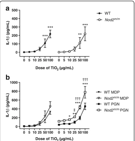
IL-1βsecretion
In LPS-pulsed BMDMs chased with TCM alone (i.e. zero dose TiO2in Fig. 6a), there was no secretion of mature
IL-1β, consistent with the role of LPS in stimulating pro-IL-1βbut not triggering the inflammasome [34, 35].
0 50 100 150 200 250 300 350 400
0.0 0.1 0.2 0.3
Size of TiO2 (nm)
Relative frequency
0 50 100 150 200 250 300 350 400
0 10 20 30 40 50
Size of TiO2 (nm)
Particle number (10
6)
a
b
Fig. 2Size determination of TiO2particles.aFood- and
pharmaceutical-grade anatase TiO2particles were suspended in distilled water with 0.5%
BSA at a concentration of 1 mg/mL. The particle suspension was analysed with transmission electron microscopy at 80 kV. TiO2particle
diameters were measured with image analysis software. The distribution of the particle diameters, grouped in sizes of 25 nm, is shown as a
relative frequency histogram,n= 133.bFood- and pharmaceutical-grade anatase TiO2particles were suspended in RPMI 1640 medium with
10% FBS and 1% penicillin-streptomycin at a concentration of 1 mg/mL. TiO2particle sizes were determined with nanoparticle tracking analysis,
and the size distribution of the particles is plotted as a line graph. Data represent mean ± SD from three independent experiments
0 5 10 25 50 100
0 20 40 60 80
Dose of TiO2 ( g/mL)
Viable cells (% of total)
WT TCM
WT MDP
WT PGN
0 5 10 25 50 100
0 20 40 60 80
Dose of TiO2 ( g/mL)
Viable cells (% of total)
Nod2m/m TCM
Nod2m/m MDP
Nod2m/m PGN
a
b
Fig. 3Viability of LPS-primed BMDMs after chasing with TiO2+/−
peptidoglycan or MDP. BMDMs from WT (a) andNod2m/m(b) mice were pre-stimulated for 3 h with LPS (10 ng/mL). Then BMDMs were incubated for 3 h with the indicated concentrations of TiO2particles
Again as anticipated, chasing LPS-primed BMDMs with TiO2 led to mature IL-1β secretion in a
dose-dependent fashion (p< 0.001; Fig. 6a) as these particles are a modest activator of the inflammasome [16, 28]. Pairwise group comparison with Tukey’s post-hoc test indicated significant IL-1β stimulation with TiO2 doses in TCM of ≥50 μg/mL (p between
<0.01 and <0.001; Fig. 6a).
Similarly, chasing LPS-primed BMDMs with TiO2+
peptidoglycan or MDP increased IL-1β secretion in a dose-dependent fashion, for cells of both genotypes (p< 0.001, Fig. 6b). However, genotype significantly influenced the extent of the IL-1β response (p< 0.01 for + MDP and p< 0.001 for + peptidoglycan). Fur-thermore, an interaction effect between genotype and TiO2exposure was observed for peptidoglycan (p< 0.001),
but not for MDP. Pairwise comparisons between groups with Tukey’s post-hoc test, when chasing with TiO2+
peptidoglycan, showed that the amount of IL-1βreleased by WT and Nod2m/mBMDMs differed significantly when the cells were similarly exposed to ≥10 μg/mL TiO2 (p
between <0.05 and <0.001; Fig. 6b).
TNF-αsecretion
LPS priming led to marked secretion of TNF-α even when chased with TCM alone (Fig. 7a) because, unlike IL-1β [36], there is no requirement for a second signal to enable protein formation and secretion of this cytokine. Chasing LPS-primed BMDMs with TiO2led to
further TNF-αsecretion in a dose-dependent fashion (p< 0.001; Fig. 7a) and, again, Tukey’s post-hoc test indicated significant TNF-αstimulation with TiO2doses in TCM of
≥50μg/mL (pbetween <0.05 and <0.001; Fig. 7a).
0 5 10 25 50 100
0 200 400 600 800 1000
Dose of TiO2 ( g/mL)
SSC (intensity)
WT TCM
WT MDP
WT PGN
0 5 10 25 50 100
0 200 400 600 800 1000
Dose of TiO2 ( g/mL)
SSC (intensity)
Nod2m/m TCM
Nod2m/m MDP
Nod2m/m PGN
a
b
Fig. 4Particle uptake by LPS-primed BMDMs after chasing with TiO2
+/−peptidoglycan or MDP. BMDMs from WT (a) andNod2m/m(b) mice were pre-stimulated for 3 h with LPS (10 ng/mL). Then BMDMs were incubated for 3 h with the indicated concentrations of TiO2
particles suspended in TCM alone (TCM), TCM + 10μg/mL MDP (MDP), or TCM + 10μg/mL peptidoglycan (PGN). Cells were stained with PI and F4/80 antibody for murine macrophages and analysed with flow cytometry, and median SSC intensities of PI−F4/80+cells were recorded. Data represent mean ± SD from two independent experiments with three replicates each,n= 6
Fig. 5Correlation of SSC intensity and dark spots in bright-field images by flow and imaging cytometry. PBMCs from human blood were incubated for 24 h with 0μg/mL, 5μg/mL, or 10μg/mL TiO2particles in TCM and stained with either CD11c or CD14 antibodies for human monocytes/myeloid
cells and analysed with conventional flow or imaging cytometry, respectively.aCorrelation between increases in SSC MFI of CD11c+myeloid cells
identified using conventional flow cytometry and the percentages of CD14+cells bearing dark spots in bright-field measured by imaging cytometry;
In contrast to IL-1β, the secretion of TNF-α by LPS-primed BMDMs that were chased with MDP or peptidoglycan was not affected by additional TiO2
ex-posure regardless of dose (i.e. the MAMPs rather than the particles dominated the scene for TNF-α se-cretion; Fig. 7b).
Although in all cases the genotype had a significant influence (p< 0.001) on TNF-α secretion, being greater for cells from Nod2m/m than WT mice, there was no interaction effect between genotype and TiO2
exposure (Fig. 7a and b).
Specificity of TiO2effect
Activation of the inflammasome is by no means specific to TiO2 particles although Pele et al. have shown that
correct design of in vitro experiments is critical. Notably, cell gorging of particles through extended particle
exposure (e.g. over 24 h) can lead to false positives [35]. SNPs and MSU crystals are considered exemplar particulate stimulants of the inflammasome, and we con-firmed that, with similar short exposures as for our TiO2
particles (3 h) and LPS priming, IL-1βsecretion was en-hanced compared to non-particle-exposed cells (Fig. 8).
Discussion
Relevance and context of our findings
The distal intestinal tract is bathed in high concentra-tions of MAMPs such as LPS and peptidoglycan (and their fragments) due to the continuous turnover of the microbiome. Since ingested particles, such as pigment-grade TiO2, are taken up by intestinal cells from this
distal environment it is important to consider interac-tions of these components (i.e. MAMPs + particles) when looking at potential cellular effects. In this work we have further considered the impact of genotype, namely one that imparts greater potential for an inflam-matory phenotype (Nod2m/m) than the WT version. We confirm that (a) primed cells fromNod2m/mmice secrete higher concentrations of pro-inflammatory cytokines,
a
b
***
** ***
***
0 5 10 25 50 100 0 5 10 25 50 100 0
100 200 300 400 500
Dose of TiO2 ( g/mL)
IL-1
(pg/mL)
WT
Nod2m/m
** *
*** ***
0 5 10 25 50 100 0 5 10 25 50 100 0
200 400 600 800 1000
Dose of TiO2 ( g/mL)
IL-1
(pg/mL)
WT MDP
Nod2m/m MDP
WT PGN
Nod2m/m PGN
Fig. 6IL-1βsecretion by LPS-primed BMDMs after chasing with TiO2
+/−peptidoglycan or MDP. BMDMs from WT andNod2m/mmice
were pre-stimulated for 3 h with LPS (10 ng/mL). Then BMDMs were incubated for 3 h with the indicated concentrations of TiO2particles
suspended in TCM alone (a) or suspended in TCM + 10μg/mL MDP (MDP) or TCM + 10μg/mL peptidoglycan (PGN) (b). Supernatant concentrations of IL-1βwere analysed by ELISA. Data represent mean ± SD from two independent experiments with three replicates each,n= 6.aResults were analysed with one-way ANOVA and Tukey’s post-hoc test; **p< 0.01, ***p< 0.001 compared to respective WT orNod2m/mcells incubated without TiO
2.bResults were
ana-lysed with two-way ANOVA and Tukey’s post-hoc test; *p< 0.05, **p < 0.01, ***p< 0.001 forNod2m/mcells compared to WT cells cultured
with the same TiO2concentration,†††p< 0.001 for WT andNod2m/m
cells compared to respective WT orNod2m/mcells incubated without
TiO2,‡p< 0.05 forNod2m/mcells compared toNod2m/mcells
incu-bated without TiO2
a
b
*** ***
*** *
0 5 10 25 50100 0 5 10 25 50100 0
100 200 300 400 500
Dose of TiO2 ( g/mL)
TN
(pg/m
L)
WT
Nod2m/m
0 5 10 25 50100 0 5 10 25 50100 0
500 1000 1500 2000
Dose of TiO2 ( g/mL)
TN
(pg/m
L)
WT MDP
Nod2m/m MDP WT PGN
Nod2m/m PGN
Fig. 7TNF-αsecretion by LPS-primed BMDMs after chasing with TiO2+/−peptidoglycan or MDP. BMDMs from WT andNod2
m/m
mice were pre-stimulated for 3 h with LPS (10 ng/mL). Then BMDMs were incubated for 3 h with the indicated concentrations of TiO2particles
namely IL-1β and TNF-α, in response to MDP-containing MAMPs than cells from WT mice [20] and (b) TiO2particles are mediators of inflammasome
acti-vation [12, 16, 28, 33]. Additionally, we show for the first time that, in primed cells exposed to peptidoglycan, the concentration of TiO2 that is required to trigger the
inflammasome and induce IL-1β secretion is lower for cells fromNod2m/m mice than it is from WT mice. This may have important implications as discussed below.
It is established that at least some ingested TiO2
parti-cles are taken up by intestinal cells, especially by macro-phages of large lymphoid follicles of the ileum termed Peyer’s patches [6–10]. Recent data suggest that cells of the large bowel can also scavenge particles of pigment-grade TiO2and that oral administration of this pigment
can lead to pre-cancerous lesions of the colon, termed aberrant crypt foci, in about a third of WT animals but not in controls without TiO2 exposure [37]. In that
work, intestinal mucosal levels of TNF-αand IL-1βwere modestly increased for animals fed TiO2versus controls
[37]. Whilst our data support these findings from a cell culture perspective they also show that particle dose is critical as a determinant of the cytokine response. The precise pathway of TiO2uptake by intestinal cells is still
not understood, but it is likely that particles in the lumen have their surfaces ‘decorated’ by soluble mole-cules of the intestinal lumen so that conjugates (with MAMPs for example), rather than pristine particles, are seen by intestinal cells. Moreover, it is not clear how basal macrophages of the human Peyer’s patches become loaded with particles such as TiO2 as, following M cell
uptake, particles should be scavenged by phagocytes that are more apical than the observed basal tissue-fixed macrophages [38]. However, despite the pathway not
being fully elucidated, the important point is that macro-phages of the Peyer’s patches do accumulate TiO2
particles in humans [9, 39]. If, as we show here, certain genotypes require a lesser cell dose of particles to re-spond in a pro-inflammatory fashion compared to other genotypes then, in vivo, the initiation of a cascade of inflammation may be host-dependent as well as dose-dependent.
Specificity of the IL-1βadjuvant effect to TiO2
nanoparticles
The ‘role’ of the TiO2 particles in the work presented
here involves boosting the pro-inflammatory effects of MAMPs via particle-activation of the inflammasome. Many materials activate the inflammasome, including other (nano)particles, and some of these will be more potent than pigment-grade TiO2given the modest
effi-cacy of the latter. For example, MSU crystals and silica particles are activators of the inflammasome (as exempli-fied here (Fig. 8) and [28, 32, 33]) and have direct relevance in terms of human exposure. MSU crystals may precipitate ectopically and are the cause of joint in-flammation in patients with gout, whilst silica exposure to the lungs is well established as an occupational hazard that leads to silicosis in miners. However, in terms of an adjuvant effect on MAMP-primed cells, TiO2 deserves
particular scrutiny because (a) humans are widely ex-posed to it orally [3, 5], (b) MAMPs are ubiquitous at high concentrations in the intestinal lumen which is un-like anywhere else in the body, and (c) pigment-grade TiO2is one of two major particle types that accumulates
in intestinal (Peyer’s patch) macrophages [7, 9, 39]. The second major particle type, namely aluminosilicate which is mostly in the kaolinite form [7], has not been obviously linked to inflammasome activation although this merits further careful assessment as prolonged macrophage exposure to kaolinite leads to modest IL-1β secretion even in the absence of MAMPS [40].
Interestingly, Winkler et al. have shown that food-grade silica induced production of pro-IL-1βand secre-tion of mature IL-1βwhen dendritic cells were exposed to these particles [41]. In other words, silica particles have the capacity to both prime IL-1βformation in the precursor (pro-) form and to induce cleavage to a ma-ture form via inflammasome activation. Although, unlike TiO2, this silica has not been demonstrated to
accumu-late in human intestinal immune cells [7], further studies are merited as there is significant oral exposure and perhaps intestinal cells other than those that have been so far characterised for particle accumulation in the in-testine are impacted.
In summary for this section, pigment-grade TiO2 is
especially relevant as a potential inflammasome adjuvant in intestinal tissue because of human exposure,
accumulation, and activity. However, other particles, such as aluminosilicates and silica, should not be ig-nored as there is certainly exposure and accumulation for the former and exposure and potential for activity for the latter.
IL-1βsecretion is not a simple consequence of TiO2-induced
cell death
Non-biological particles in a size range that enables phagocytosis, which includes pigment-grade TiO2, are
readily engulfed by macrophages and accumulate in lysosomes [7, 9, 42]. This in turn leads to lysosomal membrane disruption which is a trigger for two con-comitant events. The first is cathepsin-dependent IL-1β release which requires inflammasome activation, and the second is cell death which again is cathepsin-dependent but is independent of the inflammasome [42]. Hence, as expected, both events were observed in this study in a dose-dependent fashion when cells were exposed to TiO2. In vivo, cell death can lead to pro-IL-1β leakage
into the extracellular environment and its activation through ‘alternative’ pathways, such as cathepsin C-neutrophil proteases. However, this does not occur in ‘clean’cell culture media in vitro [42]. Moreover, a short ‘pulse and chase’ routine protects against such longer term complications. It is therefore anticipated that our observed IL-1β-inducing effect of TiO2 in LPS-primed
macrophages is independent of the concomitantly ob-served cell death. Regardless of mechanism, it does not alter the potential relevance of these findings to the in vivo situation where, as noted, pigment-grade TiO2
ac-cumulates in selective intestinal cells of humans.
In vivo relevance for health and disease
Notwithstanding the above, and as discussed earlier, TiO2is only a modest activator of the inflammasome, so
whether realistic oral exposure to TiO2leads to
interac-tions with MAMPs and whether intestinal cell loading of both materials is sufficient to trigger inflammation merits closer attention in a relevant genetically suscep-tible model. In particular, such work should focus on (a) the Peyer’s patches as sites of cellular TiO2accumulation
with the potential for early inflammatory processes [11] and (b) the colon, given the association of large bowel cancer with early inflammation and potential exacerba-tion of disease by TiO2[43].
In addition, our specific interest concerns inflamma-tory bowel disease, especially Crohn’s disease, and the potential for TiO2 as an adjuvant for pro-inflammatory
responses in recipient Peyer’s patch cells [7, 38, 39]. Although the murine model used here does not accurately mimic Crohn’s type mutations for NOD2because of the duplication of the 3′-end of the WT Nod2 locus [21], it does, nonetheless, have a heightened susceptibility to
inflammation in response to certain MAMPs, precisely as has been proposed for Crohn’s disease [44]. Further work with patient samples is therefore merited to scrutinise the potential for a TiO2adjuvant effect on MAMPs in terms
of IL-1βsecretion.
Conclusions
In summary, in this study we have shown that dietary TiO2particles have an impact on the production of the
pro-inflammatory cytokines IL-1β and TNF-α by LPS pre-stimulated murine macrophages in vitro, and that TiO2 particles can act as IL-1β-inducing adjuvants for
bacterial MAMPs that contain MDP moieties. We also demonstrated that the impact of this adjuvant effect is genotype-dependent. Primed macrophages fromNod2m/m
mice showed an elevated IL-1β response to incubation with TiO2particles and peptidoglycan compared to cells
from WT mice. Further work will need to consider if any human genotypes (sub-populations) are at greater inflam-matory risk than the background population from TiO2
exposure.
Additional files
Additional file 1:Flow cytometry gating strategy. (DOCX 321 kb)
Additional file 2:F4/80 expression of LPS-primed BMDMs after chasing with TiO2 +/−peptidoglycan or MDP. (PDF 129 kb)
Abbreviations
ANOVA:Analysis of variance; BMDM: Bone marrow-derived macrophage; BSA: Bovine serum albumin; FBS: Foetal bovine serum; IL-1β: Interleukin 1β; LPS: Lipopolysaccharide; MAMP: Microbial-associated molecular pattern; MDP: Muramyl dipeptide; MFI: Mean fluorescence intensity; MSU: Monosodium urate; NLRP3: NLR family pyrin domain-containing 3; NOD2: Nucleotide-binding oligomerisation domain-containing 2;Nod2m/m: Homozygous
Nod2gene mutation (as described); PBMC: Peripheral blood mononuclear cell; PBS: Phosphate-buffered saline; PGN: Peptidoglycan; PI: Propidium iodide; pro-IL-1β: pro-interleukin 1β; SD: Standard deviation; SEM: Standard error of the mean; SNP: Silica nanoparticle; SSC: Side scatter; TCM: Tissue culture medium; TiO2: Titanium dioxide; TNF: Tumour necrosis factor; WT: Wild-type
Acknowledgements
Funding
The research was mainly supported by the Riddet Institute through its Centre of Research Excellence funding which has been awarded to the Riddet Institute by the New Zealand government. Additional funding was provided by AgResearch, MRC Elsie Widdowson Laboratory (formerly MRC Human Nutrition Research, Grant number U105960399) and Nutrigenomics New Zealand, a collaboration between AgResearch, Plant & Food Research, and The University of Auckland (primarily supported by funding from the Ministry for Science & Innovation contract C11X1009). SR was supported by doctoral scholarships from Massey University and AgResearch. The funding bodies had no influence on the research or preparation of this manuscript.
Availability of data and materials
The datasets generated during and/or analysed during the current study are available from the corresponding author on reasonable request.
Authors’contributions
LCP and JJP developed the research hypothesis. LCP and SR designed the study with contributions from DEO, HS, NCR, and JJP. SR performed the experiments, analysed the results, and, together with LCP, designed the figures (except Figs. 5 and 8). REH and LCP provided the data and associated analyses for Figs. 5 and 8, respectively, and wrote the corresponding methods and results sections. All authors contributed to the interpretation of the results. SR and JJP wrote the manuscript with contributions from all authors. All authors read and approved the final manuscript.
Ethics approval
Collection of bone marrow from mice for this research was approved by the Grasslands Ethics Committee (Palmerston North, New Zealand), AgResearch Animal Ethics Committee, applications AE Tissue Collection 54 and 68 in compliance with the New Zealand Animal Welfare Act 1999.
Use of human blood for this research was approved by the ethics committee of the University of Cambridge (Cambridge, UK), Human Biology Research Ethics Committee, application HBREC.2015.10.
Consent for publication Not applicable.
Competing interests
The authors declare that they have no competing interests.
Publisher’s Note
Springer Nature remains neutral with regard to jurisdictional claims in published maps and institutional affiliations.
Author details
1
Food Nutrition & Health Team, Food & Bio-based Products Group, AgResearch, Grasslands Research Centre, Tennent Drive, Private Bag 11008, Palmerston North 4442, New Zealand.2Riddet Institute, Massey University, Private Bag 11222, Palmerston North 4442, New Zealand.3Present address:
Conreso GmbH, Neuhauser Str. 47, 80331, München, Germany.4Biomineral Research Group, MRC Human Nutrition Research, Elsie Widdowson Laboratory, 120 Fulbourn Road, Cambridge CB1 9NL, UK.5Present address: Center for Dairy Research, University of Wisconsin-Madison, 1605 Linden Drive, Madison, WI 53706-1565, USA.6Department of Veterinary Medicine, Biomineral Research Group, University of Cambridge, Madingley Road, Cambridge CB3 0ES, UK.
Received: 13 May 2017 Accepted: 23 November 2017
References
1. Shi H, Magaye R, Castranova V, Zhao J. Titanium dioxide nanoparticles: a review of current toxicological data. Part Fibre Toxicol. 2013;10:15. 2. Kreyling WG, Holzwarth U, Schleh C, Kozempel J, Wenk A, Haberl N, et al.
Quantitative biokinetics of titanium dioxide nanoparticles after oral application in rats: part 2. Nanotoxicology. 2017;11:443–53.
3. Lomer MCE, Hutchinson C, Volkert S, Greenfield SM, Catterall A, Thompson RPH, et al. Dietary sources of inorganic microparticles and their intake in healthy subjects and patients with Crohn's disease. Brit J Nutr. 2004;92:947–55.
4. Powell JJ, Faria N, Thomas-McKay E, Pele LC. Origin and fate of dietary nanoparticles and microparticles in the gastrointestinal tract. J Autoimmun. 2010;34:J226–J33.
5. Rompelberg C, Heringa MB, van Donkersgoed G, Drijvers J, Roos A, Westenbrink S, et al. Oral intake of added titanium dioxide and its nanofraction from food products, food supplements and toothpaste by the Dutch population. Nanotoxicology. 2016;10:1404–14.
6. Hummel TZ, Kindermann A, Stokkers PCF, Benninga MA, ten Kate FJW. Exogenous pigment in Peyer's patches of children suspected for inflammatory bowel disease. J Pediatr Gastr Nutr. 2014;58:477–80. 7. Powell JJ, Ainley CC, Harvey RSJ, Mason IM, Kendall MD, Sankey EA, et al.
Characterisation of inorganic microparticles in pigment cells of human gut associated lymphoid tissue. Gut. 1996;38:390–5.
8. Shepherd NA, Crocker PR, Smith AP, Levison DA. Exogenous pigment in Peyer's patches. Hum Pathol. 1987;18:50–4.
9. Thoree V, Skepper J, Deere H, Pele LC, Thompson RPH, Powell JJ. Phenotype of exogenous microparticle-containing pigment cells of the human Peyer's patch in inflamed and normal ileum. Inflamm Res. 2008;57:374–8. 10. Urbanski SJ, Arsenault AL, Green FH, Haber G. Pigment resembling
atmospheric dust in Peyer's patches. Mod Pathol. 1989;2:222–6. 11. Fujimura Y, Kamoi R, Iida M. Pathogenesis of aphthoid ulcers in Crohn's
disease: correlative findings by magnifying colonoscopy, electron microscopy, and immunohistochemistry. Gut. 1996;38:724–32. 12. Ashwood P, Thompson RPH, Powell JJ. Fine particles that adsorb
lipopolysaccharideviabridging calcium cations may mimic bacterial pathogenicity towards cells. Exp Biol Med. 2007;232:107–17.
13. Butler M, Boyle JJ, Powell JJ, Playford RJ, Ghosh S. Dietary microparticles implicated in Crohn's disease can impair macrophage phagocytic activity and act as adjuvants in the presence of bacterial stimuli. Inflamm Res. 2007;56:353–61.
14. Evans SM, Ashwood P, Warley A, Berisha F, Thompson RPH, Powell JJ. The role of dietary microparticles and calcium in apoptosis and interleukin-1β release of intestinal macrophages. Gastroenterology. 2002;123:1543–53. 15. Powell JJ, Harvey RSJ, Ashwood P, Wolstencroft R, Gershwin ME, Thompson
RPH. Immune potentiation of ultrafine dietary particles in normal subjects and patients with inflammatory bowel disease. J Autoimmun. 2000;14:99–105. 16. Ruiz PA, Morón B, Becker HM, Lang S, Atrott K, Spalinger MR, et al. Titanium
dioxide nanoparticles exacerbate DSS-induced colitis: role of the NLRP3 inflammasome. Gut. 2017;66:1216–24.
17. Khor B, Gardet A, Xavier RJ. Genetics and pathogenesis of inflammatory bowel disease. Nature. 2011;474:307–17.
18. Hugot JP, Chamaillard M, Zouali H, Lesage S, Cézard JP, Belaiche J, et al. Association of NOD2 leucine-rich repeat variants with susceptibility to Crohn's disease. Nature. 2001;411:599–603.
19. Ogura Y, Bonen DK, Inohara N, Nicolae DL, Chen FF, Ramos R, et al. A frameshift mutation inNOD2associated with susceptibility to Crohn's disease. Nature. 2001;411:603–6.
20. Maeda S, Hsu LC, Liu HJ, Bankston LA, Iimura M, Kagnoff MF, et al.Nod2 mutation in Crohn's disease potentiates NF-κB activity and IL-1βprocessing. Science. 2005;307:734–8.
21. Maeda S. Corrections and clarifications:Nod2mutation in Crohn's disease potentiates NF-κB activity and IL-1βprocessing [Maeda S et al. (2005) Science 307(5710): 734-738]. Science. 2011;333:288.
22. Powell JJ, Thomas-McKay E, Thoree V, Robertson J, Hewitt RE, Skepper JN, et al. An endogenous nanomineral chaperones luminal antigen and peptidoglycan to intestinal immune cells. Nat Nanotechnol. 2015;10:361–9. 23. Klasen IS, Melief MJ, van Halteren AG, Schouten WR, van Blankenstein M,
Hoke G, et al. The presence of peptidoglycan-polysaccharide complexes in the bowel wall and the cellular responses to these complexes in Crohn's disease. Clin Immunol Immunopathol. 1994;71:303–8.
24. Filipe V, Hawe A, Jiskoot W. Critical evaluation of nanoparticle tracking analysis (NTA) by NanoSight for the measurement of nanoparticles and protein aggregates. Pharm Res. 2010;27:796–810.
25. Roy N, Barnett M, Knoch B, Dommels Y, McNabb W. Nutrigenomics applied to an animal model of inflammatory bowel diseases: transcriptomic analysis of the effects of eicosapentaenoic acid- and arachidonic acid-enriched diets. Mutat Res. 2007;622:103–16.
27. Palomäki J, Karisola P, Pylkkänen L, Savolainen K, Alenius H. Engineered nanomaterials cause cytotoxicity and activation on mouse antigen presenting cells. Toxicology. 2010;267:125–31.
28. Winter M, Beer HD, Hornung V, Kärmer U, Schins RPF, Förster I. Activation of the inflammasome by amorphous silica and TiO2nanoparticles in murine
dendritic cells. Nanotoxicology. 2011;5:326–40.
29. Suzuki H, Toyooka T, Ibuki Y. Simple and easy method to evaluate uptake potential of nanoparticles in mammalian cells using a flow cytometric light scatter analysis. Environ Sci Technol. 2007;41:3018–24.
30. Zucker RM, Massaro EJ, Sanders KM, Degn LL, Boyes WK. Detection of TiO2
nanoparticles in cells by flow cytometry. Cytometry A. 2010;77:677–85. 31. Hewitt RE, Vis B, Pele LC, Faria N, Powell JJ. Imaging flow cytometry assays for
quantifying pigment grade titanium dioxide particle internalization and interactions with immune cells in whole blood. Cytometry A. 2017;91A:1009–20. 32. Martinon F, Pétrilli V, Mayor A, Tardivel A, Tschopp J. Gout-associated uric
acid crystals activate the NALP3 inflammasome. Nature. 2006;440:237–41. 33. Yazdi AS, Guarda G, Riteau N, Drexler SK, Tardivel A, Couillin I, et al.
Nanoparticles activate the NLR pyrin domain containing 3 (Nlrp3) inflammasome and cause pulmonary inflammation through release of IL-1α and IL-1β. Proc Natl Acad Sci U S A. 2010;107:19449–54.
34. Latz E, Xiao TS, Stutz A. Activation and regulation of the inflammasomes. Nat Rev Immunol. 2013;13:397–411.
35. Pele L, Haas CT, Hewitt R, Faria N, Brown A, Powell J. Artefactual nanoparticle activation of the inflammasome platform:in vitroevidence with a nano-formed calcium phosphate. Nanomedicine (Lond). 2015;10:1379–90. 36. Dinarello CA. Immunological and inflammatory functions of the
interleukin-1 family. Annu Rev Immunol. 2009;27:5interleukin-19–50.
37. Urrutia-Ortega IM, Garduno-Balderas LG, Delgado-Buenrostro NL, Freyre-Fonseca V, Flores-Flores JO, Gonzalez-Robles A, et al. Food-grade titanium dioxide exposure exacerbates tumor formation in colitis associated cancer model. Food Chem Toxicol. 2016;93:20–31.
38. Powell JJ, Thoree V, Pele LC. Dietary microparticles and their impact on tolerance and immune responsiveness of the gastrointestinal tract. Br J Nutr. 2007;98(Suppl 1):S59–63.
39. Lomer MCE, Thompson RPH, Powell JJ. Fine and ultrafine particles of the diet: influence on the mucosal immune response and association with Crohn's disease. Proc Nutr Soc. 2002;61:123–30.
40. Kato T, Toyooka T, Ibuki Y, Masuda S, Watanabe M, Totsuka Y. Effect of physicochemical character differences on the genotoxic potency of kaolin. Genes Environ. 2017;39:12.
41. Winkler HC, Kornprobst J, Wick P, von Moos LM, Trantakis I, Schraner EM, et al. MyD88-dependent pro-interleukin-1βinduction in dendritic cells exposed to food-grade synthetic amorphous silica. Part Fibre Toxicol. 2017;14:21. 42. Orlowski GM, Sharma S, Colbert JD, Bogyo M, Robertson SA, Kataoka H,
et al. Frontline science: multiple cathepsins promote inflammasome-independent, particle-induced cell death during NLRP3-dependent IL-1β activation. J Leukoc Biol. 2017;102:7–17.
43. Bettini S, Boutet-Robinet E, Cartier C, Comera C, Gaultier E, Dupuy J, et al. Food-grade TiO2impairs intestinal and systemic immune homeostasis,
initiates preneoplastic lesions and promotes aberrant crypt development in the rat colon. Sci Rep. 2017;7:40373.
44. de Souza HSP, Fiocchi C, Iliopoulos D. The IBD interactome: an integrated view of aetiology, pathogenesis and therapy. Nat Rev Gastroenterol Hepatol. 2017; doi:10.1038/nrgastro.2017.110.
• We accept pre-submission inquiries
• Our selector tool helps you to find the most relevant journal
• We provide round the clock customer support • Convenient online submission
• Thorough peer review
• Inclusion in PubMed and all major indexing services
• Maximum visibility for your research
Submit your manuscript at www.biomedcentral.com/submit