R E S E A R C H A R T I C L E
Open Access
The transcription factor Foxg1 regulates
telencephalic progenitor proliferation cell
autonomously, in part by controlling Pax6
expression levels
Martine N Manuel
1*, Ben Martynoga
1,2†, Mike D Molinek
1†, Jane C Quinn
1,3†, Corinne Kroemmer
1,
John O Mason
1, David J Price
1Abstract
Background:The transcription factor Foxg1 is an important regulator of telencephalic cell cycles. Its inactivation causes premature lengthening of telencephalic progenitor cell cycles and increased neurogenic divisions, leading to severe hypoplasia of the telencephalon. These proliferation defects could be a secondary consequence of the loss of Foxg1 caused by the abnormal expression of several morphogens (Fibroblast growth factor 8, bone morphogenetic proteins) in the telencephalon ofFoxg1 null mutants. Here we investigated whether Foxg1 has a cell autonomous role in the regulation of telencephalic progenitor proliferation. We analysedFoxg1+/+↔Foxg1 -/-chimeras, in which mutant telencephalic cells have the potential to interact with, and to have any cell non-autonomous defects rescued by, normal wild-type cells.
Results:Our analysis showed that theFoxg1-/-cells are under-represented in the chimeric telencephalon and the proportion of them in S-phase is significantly smaller than that of their wild-type neighbours, indicating that their under-representation is caused by a cell autonomous reduction in their proliferation. We then analysed the expression of the cell-cycle regulator Pax6 and found that it is cell-autonomously downregulated inFoxg1-/-dorsal telencephalic cells. We went on to show that the introduction intoFoxg1-/-embryos of a transgene designed to reverse Pax6 expression defects resulted in a partial rescue of the telencephalic progenitor proliferation defects. Conclusions:We conclude that Foxg1 exerts control over telencephalic progenitor proliferation by cell
autonomous mechanisms that include the regulation of Pax6, which itself is known to regulate proliferation cell autonomously in a regional manner.
Background
Each part of the central nervous system attains a charac-teristic size during embryogenesis. The vertebrate fore-brain (prosencephalon) grows larger than other parts of the neural tube, giving rise to rostral bilateral swellings known as telencephalic vesicles or, when considered together, the telencephalon. The telencephalon is parti-cularly large in mammals and generates the cerebral cor-tex and basal ganglia. The differential growth of each
part of the central nervous system depends critically on region-specific regulation of neural cell proliferation in the embryo. While there is extensive understanding of effector molecules such as cell cycle proteins that control the cell proliferation in general, we know much less about the mechanisms that specify the different rates at which cells divide in each part of the embryo.
The early neural plate and neural tube are patterned by region-specific expression of transcription factors, some of which exert control over cell proliferation as well as other aspects of regional development. For two of these transcription factors, Foxg1 and Pax6, there is strong evi-dence that one of their primary functions is to regulate telencephalic cell cycles [1-5]. Both are expressed in * Correspondence: Martine.Manuel@ed.ac.uk
†Contributed equally
1
Genes and Development Group, University of Edinburgh, Hugh Robson Building, George Square, Edinburgh EH8 9XD, UK
Full list of author information is available at the end of the article
progenitors in the developing telencephalon, with Foxg1 being activated slightly before Pax6 [4,6,7].
Foxg1 is one of the earliest transcription factors expressed specifically in the part of the neural plate that gives rise to the telencephalon and it remains expressed throughout the telencephalon during embryonic development. Its inactiva-tion leads to severe telencephalic hypoplasia [4]. In a pre-vious study we showed that the cell cycle lengthens prematurely and neurogenic divisions are increased in
Foxg1-/-telencephalon, thereby reducing the pool of prolif-erating progenitors [5]. These proliferation defects coincide with reduced expression of the pro-proliferative morphogen Fibroblast growth factor 8 (Fgf8) in the rostral telencephalon and expanded expression of several bone morphogenetic proteins (BMPs), which promote neural differentiation, from their normally dorsally restricted domain [5,8-10]. These observations suggest two possible explanations for the proliferation defects inFoxg1-/-telencephalon. First, it is possible that the proliferation defects ofFoxg1-/- telencepha-lic progenitors are secondary to abnormal expression of morphogens such as Fgf8 and BMPs. Second, telencephalic progenitors might require Foxg1 cell autonomously for their normal proliferation.
To test whether Foxg1 has a cell autonomous role in the regulation of telencephalic progenitor proliferation, we analysedFoxg1+/+↔Foxg1-/-chimeras, in which mutant telencephalic cells have the potential to interact with, and to have any cell non-autonomous defects rescued by, nor-mal wild-type cells. We found that theFoxg1-/-cells were under-represented in the chimeric telencephalon and that the proportion of them in S-phase was significantly smal-ler than that of their wild-type neighbours, indicating that their under-representation was caused by a cell-autonomous reduction in their proliferation.
We then examined the relationship between Foxg1 and Pax6, in view of their overlapping expression patterns and the fact that Pax6 is known to regulate cell autonomously the proliferation of cortical progenitors [2,3]. We found that Pax6 is cell-autonomously downregulated inFoxg1 -/-dorsal telencephalic cells. We tested the hypothesis that this contributes to the proliferation defects inFoxg1 -/-embryos by introducing a transgene designed to reverse Pax6 expression defects intoFoxg1-/-embryos and found that this partially rescued their telencephalic progenitor proliferation defects.
Materials and methods
Animals
Animal care followed institutional guidelines and UK Home Office regulations.
Derivation ofFoxg1-/-embryonic stem cells
Wild-type (Foxg1+/+) or null-mutant (Foxg1cre/lacZ) embryonic stem (ES) cells were derived using the
following protocol. Female mice (129Sv; Foxg1+/lacZ) were superovulated by intraperitoneal (i.p.) injection of 5U pregnant mare’s serum gonadotrophin (Intervet, Milton Keynes, UK) at the middle of the light cycle fol-lowed 47 hours later by i.p. injection of 5U human chor-ionic gonadotrophin (Intervet, UK). Females were mated with Foxg1+/crestud males homozygous for a reiterated
b-globin repeat transgene (Tg/Tg) [11]. Delayed implan-tation was induced 2.5 days post-coitum by i.p. injection of Tamoxifen (Sigma, Gillingham, UK; 10 μg/animal) and subcutaneous injection of Depo-Provera (Sigma; 1 to 3 mg/animal). At 7.5 days post-coitum, blastocysts were flushed from the uterus, transferred to a gelati-nised well containing N2B27 medium with 10 ng/ml leukaemia inhibitory factor and cultured at 37°C in 5% CO2. After approximately 5 days, inner cell mass
out-growths were detached from the bottom of each well using a fine pipette and disaggregated in trypsin (0.025% for 2 to 3 minutes at 37°C) to give individual clusters of 1 to 5 cells. Clusters were transferred to a fresh gelati-nised well containing N2B27 media containing leukae-mia inhibitory factor (10 ng/ml) with the addition of BMP4 (10 ng/ml). Primary ES cell colonies were visible after approximately 5 days in culture. ES cell lines were passaged in feeder-free conditions in BHK-21 Glasgow MEM (GMEM) with 15% fetal bovine serum and leu-kaemia inhibitory factor (1,000 U/ml). Cell lines were genotyped for presence ofcre,lacZandTg; their glucose phosphate isomerase 1 (GPI1) isotype was confirmed as described previously [2]. All ES cell lines used for chi-mera generation were karyotyped and found to have a normal chromosome complement.
Chimera production and tissue contribution analysis
described in Manuel et al., 2010. Foxg1cre/lacz and
Foxg1+/lacZ
embryos were aggregated with Foxg1+/+ embryos to give experimental and control chimeras, respectively. In these cases, the mutant cells were recog-nizable because the coding sequence of oneFoxg1allele is replaced by alacZreporter cassette [4].
Elevating Pax6 levels inFoxg1-/-embryos
The Pax77 transgene [15] was crossed into Foxg1 -/-embryos to increase their Pax6 levels. This transgene comprises five to seven copies of the humanPAX6locus including its upstream and downstream regulatory regions. Our previous studies showed that wild-type mice carrying thePax77transgene display elevated Pax6 levels at all its sites of expression [3,16].
Proliferation analyses
Pregnant females at E12.5 were sacrificed 30 minutes after injection with 200μl of 10 mg/ml bromodeoxyuri-dine (BrdU; Sigma; in 0.9% NaCl, i.p.). For analysis of proliferation in chimeras, wax coronal sections were immunostained with anti-BrdU following b-globin DNA-DNAin situhybridization. The percentage of Tg+ (that is, mutant) progenitors in S-phase was determined by counting the total number of BrdU+ Tg+ cells and BrdU-Tg+cells in the ventricular zone of the dorsal and ventral telencephalon of chimeras. To determine the percentage of Tg- (that is, wild-type) progenitors in S-phase, BrdU+Tg-and BrdU-Tg-cells were counted in 100-μm-wide sampling boxes in the ventricular zone of the dorsal and ventral telencephalon of chimeras.
For analysis of the effects of Pax6 overexpression on proliferation, BrdU+ and BrdU- cells were counted in 100-μm-wide sampling boxes along the ventricular zone of the dorsal telencephalon of wild-type embryos and the whole telencephalon ofFoxg1-/- andFoxg1-/-;Pax77 embryos at rostral, central and caudal levels. Each count was repeated on three to five non-adjacent sections from each embryo (n = 3 embryos of each genotype).
Quantitative reverse transcription-PCR
RNA was extracted from the telencephalon of Foxg1 -/-and Foxg1-/-;Pax77 littermates at E12.5 using Qiagen Rneasy kit (Qiagen, Crawley, UK). cDNA synthesis was performed as described in [17]. Analysis of totalPax6 levels by quantitative PCR was performed as described in [16].
Immunohistochemistry
Immunohistochemistry was carried out as described pre-viously [5]. The primary antibodies used were: anti-Pax6 (1:50 to 1:500; Developmental Studies Hybridoma Bank); anti-b-galactosidase (b-gal; 1:800; Invitrogen, Paisley, UK); and anti-BrdU (1:200; Becton Dickinson, Oxford, UK).
In situhybridisation
Antisense RNA probes forEmx1 andEmx2were digoxi-genin labelled. In situ hybridisations were on 10-μm paraffin sections [18].
Results
Foxg1 is required cell-autonomously for normal telencephalic progenitor proliferation
To test the hypothesis that Foxg1 is required cell autono-mously for cells to contribute normally to the telence-phalon, we made experimental and control chimeras by injectingFoxg1-/-orFoxg1+/+ES cells into wild-type blas-tocysts.Foxg1-/-andFoxg1+/+ES-derived cells carried a
b-globintransgene (Tg) identifiable by DNAin situ hybri-dization, which generated a labelled spot in their nuclei (Figure 1A-D). In each chimeric embryo, an estimate of the percentage of ES-derived cells in Foxg1 non-expressing regions was obtained by quantitative analysis of the GPI1 isozyme composition of the upper body [12]. ES cells produced the GPI1A isozyme, and so the percen-tage of GPI1 that was GPI1A in the upper body repre-sented the contribution of the ES-derived cells to regions of the embryo where the presence or absence of Foxg1 was predicted to have no effect.
We analysed three experimental and three control chi-meras at E12.5 and determined the percentage of Tg+ cells in the hippocampus, the cortex and the ventral tel-encephalon. The dorsal thalamus, in which Foxg1 is not normally expressed, was used as a control brain region. For each of these tissues in each chimera, the observed contribution of Tg+cells (obsTg+) was divided by the expected contribution ofTg+cells (expTg+) given by the percentage of GPI1A for that chimera. In control meras and in the dorsal thalamus of experimental chi-meras the obsTg+:expTg+ ratios were close to 1, which is the value anticipated in tissues where the contribution of ES-derived cells is no different to that throughout the body of the embryo. Foxg1-/- Tg+ cells were present throughout the telencephalon of experimental chimeras,
but they were significantly under-represented
(Figure 1E). The degree of under-representation was similar in all three tissues studied, that is, the hippocam-pus, the cortex and the ventral telencephalon.
We then tested whether the under-representation of
A
B
Foxg1
+/+l
Foxg1
+/+; Tg
+Foxg1
+/+l
Foxg1
-/-; Tg
+A
B
C
D
Contribution of Tg+ cells
Foxg1+/+<>Foxg1+/+E
g
0.6 0.8 1 1.2
b
served /expected
Foxg1 <>Foxg1
Foxg1+/+<>Foxg1
-/-0 0.2 0.4
Dorsal Thalamus Hippocampus Cortex Ventral
Telencephalon
ratio o
b
Figure 1Foxg1-/-mutant cells are underrepresented in the telencephalon ofFoxg1+/+;Tg-↔Foxg1-/-;Tg+chimeras.(A-D)Coronal sections through the thalamus (A,B) and the ventral telencephalon (C,D) of E12.5Foxg1+/+;Tg-↔Foxg1+/+;Tg+control (A,C) andFoxg1+/+;Tg-↔Foxg1-/-;Tg+ experimental chimeras (B,D) showingTg+cells (labelled with dark dots) derived from the ES cells. (D) Very few Tg+cells (arrowheads) are observed in the ventral telencephalon of experimental chimeras. Scale bar: 50μm.(E)Ratios of observed/expected contributions ofTg+cells in the dorsal
thalamus, the hippocampus, the cortex and the ventral telencephalon of control (Foxg1+/+;Tg
-↔Foxg1+/+;Tg+) and experimental (Foxg1+/+;Tg
-↔Foxg1-/-;
Tg+) chimeras.Tg+cells are significantly underrepresented in the telencephalon of mutant chimeras (mean ± s.e.m, n = 3 embryos of each genotype;
A
% BrdU labelled cells
0 10 20 30 40 50 60 70
Dorsal Thalamus Dorsal Telencephalon Ventral Telencephalon
B
Foxg1+/+, Tg
-Foxg1-/-, Tg+
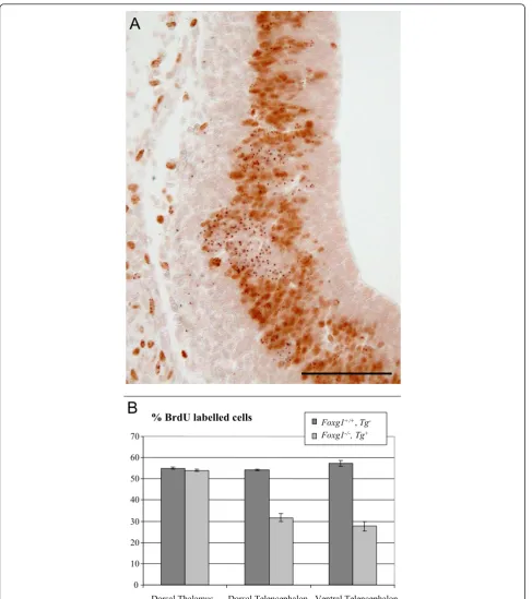
Figure 2Foxg1-/-cells display a cell autonomous proliferation defect.(A)Coronal section through the telencephalon of an E12.5Foxg1+/+;
Tg
-↔Foxg1-/-;Tg+
chimera showingTg+cells (labelled with dark dots) and BrdU+cells (with brown nuclei). Clusters of BrdU-Tg+cells are present among BrdU+Tg-andTg+progenitors. Scale bar: 50μm.(B)Percentages ofTg+cells in S-phase in the dorsal and ventral telencephalon, but not the dorsal thalamus, of experimental chimeras are significantly lower than the percentages ofTg-cells in S-phase (mean ± s.e.m, n = 3 embryos
Foxg1+/+
progenitors. These results indicate that Foxg1 is required cell autonomously for normal proliferation of tel-encephalic progenitors.
Foxg1 is required cell autonomously for normal telencephalic Pax6 expression
We examined the relationship between Foxg1 and the transcription factor Pax6, which is known to regulate tele-ncephalic progenitor proliferation and whose teletele-ncephalic expression begins shortly after that of Foxg1 [1-3,6,19,20]. Expression of Pax6 is normally restricted to dorsal telence-phalon. InFoxg1-/-embryos, progenitors throughout the entire telencephalon express Pax6 [4,5,21] and previous work has shown that this reflects the inability ofFoxg1 -/-telencephalic cells to develop ventral -/-telencephalic fates [21]. Here, we examined the levels of expression of Pax6 in the telencephalon ofFoxg1-/-embryos and in chimeras. Immunofluorescence for Pax6 showed that, whereas Pax6 is expressed in a rostro-lateralhighto caudo-mediallow gradi-ent in the dorsal telencephalon in wild-type embryos and control chimeras (Figure 3A,C), inFoxg1-/-embryos there is no obvious gradient and Pax6 appears to be expressed at a lower level throughout the telencephalon (Figure 3B). Comparison of Figure 3A and 3B shows a normal level of immunostaining in theFoxg1-/-prethalamus and eminentia thalami, a region that does not express Foxg1 and should not be affected in mutants, but comparatively lower immu-nostaining of the lateral telencephalon.
Regional reduction in immunostaining for Pax6 was very obvious inFoxg1-/-cells inFoxg1+/+↔Foxg1-/-chimeras. In rostro-lateral regions of the dorsal telencephalon of experimental chimeras, even very small groups ofFoxg1 -/-cells (recognized by their expression ofb-gal) expressed Pax6 at discernibly lower levels than their neighbours (Figure 3H-J). In more medial positions in the dorsal tele-ncephalon of experimental chimeras, where Pax6 levels are lower inFoxg1+/+ cells, Foxg1-/- cells appeared to express Pax6 at similar levels to their neighbouring
Foxg1+/+cells (Figure 3K-M). In control chimeras, levels of Pax6 immunostaining inb-gal-expressing cells and their neighbours were similar (Figure 3D-F). In summary,
Foxg1-/-cells express Pax6 at low levels, similar to those normally found in caudo-medial telencephalon, through all parts of the telencephalon with no increase in levels in rostro-lateral regions. Our findings from chimeras indicate that the generation of the normal graded increase of Pax6 in the rostro-lateral part of the dorsal telencephalon requires Foxg1 cell-autonomously.
Pax6 downregulation contributes to the proliferation defects ofFoxg1-/-telencephalic progenitors
As Pax6 is implicated in the control of cortical progeni-tor proliferation [1-3,19,20,22,23], we hypothesised that
Foxg1 might regulate telencephalic cell proliferation, at least in part, via its regulation of Pax6 levels. The cell autonomous inability of many Foxg1-/- dorsal telence-phalic cells to achieve normal Pax6 levels might contri-bute to their proliferation defects. To distinguish between this possibility and an alternative scenario in which loss of Foxg1 prevents normal proliferation inde-pendently of any change in Pax6 levels, we generated mice lacking Foxg1 but with elevated Pax6 levels. To do this, we used the Pax6 overexpressing line, Pax77, in which Pax6 levels are elevated within the physiological range in the normal domains of expression of Pax6 [3,15,16].
To confirm that this method successfully increased overall expression inFoxg1-/-mutants, we compared the levels of Pax6 mRNA in the telencephalon ofFoxg1 -/-and Foxg1-/-;Pax77+embryos at E12.5 by quantitative RT-PCR. We found that levels were increased about 2.25-fold in the telencephalon of Foxg1-/-;Pax77+ embryos compared toFoxg1-/-embryos (Figure 4A; Stu-dent’st-test,P < 0.05). This is a similar increase to that found when thePax77transgene is expressed on a wild-type background [16].
With immunohistochemistry, we observed more intense Pax6 labelling throughout the telencephalon in
Foxg1-/-;Pax77+embryos (Figure 4C) than in Foxg1 -/-embryos (Figure 4B). Whereas inFoxg1-/-embryos Pax6 immunostaining was much weaker throughout the tele-ncephalon than in the prethalamus and eminentia tha-lami, inFoxg1-/-;Pax77+embryos the intensity of staining in these regions was similar (Figure 4B,C). Pax6 immuno-labelling was increased across the telencephalon with no evidence for the restoration of a normal lateralhighto mediallow(Figure 4B,C) and rostralhighto caudallow(not shown) gradient of expression, indicating that thePax77
transgene and the endogenous Pax6 locus were
being regulated similarly to each other on aFoxg1 -/-background.
As shown in Figures 4B,C, the morphology of the
Pax6
200Pm
Foxg1
+/-
wt
CPax6
D E FMerge
D
Merge
50Pm
E
-gal
Pax6
Foxg1-LacZ
Pax6
Pax6
E
B C D
Merge
G F
K
H
Merge
Foxg1
-/-
wt
50Pm
Pax6
Pax6
E
-gal
E
-gal
Merge
Merge
G
H
I
K
J
L
M
B
Pax6
A
wt
Foxg1-/-PT ET
PT ET
Figure 3Pax6 is misregulated inFoxg1-/-cells.(A,B)Pax6 immunofluorescence on coronal sections through the telencephalon of wild-type
(wt) (A) andFoxg1-/-(B) embryos at E12.5. The characteristic lateralhighto mediallowgradient of Pax6 expression in the telencephalon is observed
in wild-type (A) but not in mutant embryos (C).(C,G)Pax6 immunofluorescence on coronal sections through the telencephalon of aFoxg1
+/+↔Foxg1+/-(C) control chimera and aFoxg1+/+↔Foxg1-/-experimental chimera (G).(D-F,H-J,K-M)Selected regions are shown at higher
magnification and co-labelled forb-gal expressed byFoxg1+/-(D-F) orFoxg1-/-cells (H-J,K-M). Throughout the whole telencephalon of control chimeras (D-F) and the dorso-medial telencephalon of experimental chimeras (K-M),b-gal+cells (arrows) express Pax6 at levels indistinguishable
from those in adjacentb-gal-cells. In the dorso-lateral telencephalon of experimental chimeras (H-J), however,Foxg1-/-(b-gal+) cells display
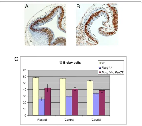
Pax77 embryos (Figure 5B) at E12.5 at three levels: (i) rostral (normally Pax6high): (ii) central; and (iii) caudal (normally Pax6low) levels.
Consistent with previous studies [4,5], we found that the average labelling index was significantly lower in the
Foxg1
-/-telencephalon than in the wild-type telencepha-lon at all three rostro-caudal levels (Figure 5C). At ros-tral and cenros-tral levels, average labelling indices were
significantly increased in Foxg1-/-;Pax77+telencephalon compared toFoxg1-/-telencephalon, although they were not restored completely to wild-type levels (Figure 5C). Caudally, the labelling index was not significantly differ-ent inFoxg1-/-;Pax77+ compared toFoxg1-/- telencepha-lon. At rostral, central and caudal levels, labelling indices were similar from dorsal to ventral in the wild-type,Foxg1-/-andFoxg1-/-;Pax77+telencephalon and in
Foxg1
-/-; Pax77
Foxg1
-/-Pax6 expression
0 0.5 1 1.5 2 2.5 3
ra
ti
o
Pax6 / G
APD
H
Foxg1
-/-Foxg1-/-; Pax77
A
B
C
Figure 4Pax6expression levels are increased inFoxg1-/-;Pax77telencephalon compared toFoxg1-/-telencephalon.(A)Levels of total
Pax6mRNA (endogenousPax6and humanPAX6) in the telencephalon of E12.5Foxg1-/-;Pax77embryos andFoxg1-/-embryos determined by real-time quantitative RT-PCR and normalised againstGAPDHmRNA levels (mean ± s.e.m, n = 3 in each case). TotalPax6mRNA levels are significantly increased inFoxg1-/-;Pax77telencephalon compared toFoxg1-/-telencephalon (Student’st-test,P< 0.05).(B,C)Coronal sections through one hemisphere of the forebrain ofFoxg1-/-(B) andFoxg1-/-;Pax77(C) embryos at E12.5, showing the expression of total Pax6
the wild-type dorsal telencephalon and so data from all dorsal to ventral sampling areas were combined for each genotype to generate the histograms in Figure 5C.
We conclude that raising Pax6 levels throughout the telencephalon ofFoxg1-/-embryos raises proliferation rates in the direction of normal in rostral and central parts of the telencephalon. It does not, however, have a detectable effect in caudal telencephalon.
It is possible that Foxg1 and/or Pax6 directly regulate the expression of genes that regulate proliferation, such as cell cycle genes. Alternatively, they might affect
proliferation indirectly by controlling the expression of other transcription factors that themselves regulate prolif-eration. Previous work has shown that Emx1 and Emx2, two transcription factors implicated in the control of corti-cal progenitor proliferation [24], are misregulated in the
Foxg1-/-telencephalon [25]. WhereasEmx1andEmx2are expressed in a rostro-laterallowto caudo-medialhigh gradi-ent in the dorsal telencephalon in wild-type embryos [25] (Figure 6A,D), inFoxg1-/-embryos there is no obvious gra-dient and they appear to be expressed at a high level throughout the telencephalon [25] (Figure 6B,E). Since an
% Brdu+ cells
0 10 20 30 40 50 60 70
Rostral Central Caudal wt
Foxg1-/-Foxg1-/- ; Pax77
A
B
C
earlier study implicated Pax6 as a regulator ofEmx1and
Emx2expression [26], we wondered whether the observed rise in proliferation rate in the rostral and central telence-phalon of Foxg1-/-;Pax77embryos might result from a restoration of normalEmx1andEmx2expression.In situ hybridisation forEmx1andEmx2expression in the tele-ncephalon ofFoxg1-/-;Pax77embryos (Figure 6C,F) did not show any obvious change compared to Foxg1 -/-mutants (Figure 6B,E). It is unlikely, therefore, that elevat-ing Pax6 levels affects proliferation via a change inEmx1 orEmx2expression.
Discussion
Foxg1 and Pax6 are transcription factors essential for early brain development and are implicated particularly strongly in the regulation of telencephalic progenitor proliferation [1-5]. Here we provide evidence linking the activities of the two factors in the regulation of progeni-tor proliferation. We show that Foxg1 regulates cell autonomously both proliferation and levels of Pax6 expression in telencephalic progenitors. Pax6 is itself already known to regulate telencephalic cell proliferation by cell autonomous mechanisms [2,3]. We show that
raising Pax6 levels in Foxg1-/-embryos partially reverses their telencephalic proliferation defects. This suggests that reduced proliferation in Foxg1-/-telencephalic pro-genitors can be explained, at least in part, by their reduced Pax6 levels.
Our evidence that Foxg1 regulates cell proliferation cell autonomously is based on data from chimeras in which the proportions of mutant cells are relatively low even in areas that do not normally express Foxg1. The advantage of the mutant cells being greatly outnumbered by the wild-type cells is that it increases the probability of rescu-ing any cell non-autonomous defects that they might have inFoxg1-/-telencephalon, arising, for example, from altered production of intercellular signals such as Fgf8 or BMPs by surrounding cells [5,8-10]. In chimeras, the labelling indices of mutant telencephalic cells (that is, the percentages of mutant cells in S-phase of the cell cycle) were around 30% (Figure 2B), which is the same as the labelling indices of mutant cells in fullFoxg1-/-mutants (Figure 5C). This means that the proliferative defects of
Foxg1-/- cells might be accounted for entirely by cell autonomous defects, but it does not exclude the possibi-lity that cell non-autonomous proliferation-enhancing
A
B
C
D
E
F
wt
Foxg1
-/-Foxg1
-/-; Pax77
Emx
1
Emx
2
Figure 6Increased Pax6 levels do not restore normalEmx1andEmx2expression in the telencephalon ofFoxg1mutant embryos.(A-F)
In situhybridisation forEmx1(A-C) andEmx2(D-F) on coronal sections through the telencephalon of wild-type (wt) (A,D),Foxg1-/-(B,E) and
processes such as intercellular signalling are defective in fullFoxg1-/-mutants.
When examined in detail, the relationship between Foxg1 and Pax6 is not straightforward. Interestingly, while loss of Foxg1 lowers overall Pax6 expression in the telencephalon, the magnitude of the effect is regio-nal: the greatest reduction is in those areas where Pax6 expression is normally highest, that is, rostro-laterally. The consequence is to abolish the normal gradient of expression of Pax6 across the telencephalon. Since in normal telencephalon Foxg1 expression levels are not linearly related to Pax6 expression levels - for example, Foxg1 is normally expressed in some ventral regions where Pax6 is not [4,7] - it seems most likely that Foxg1 is an essential requirement for activation of normal tele-ncephalic Pax6 expression in combination with addi-tional factors. Together these factors might activate Pax6 expression and raise its levels rostro-laterally; Foxg1 is a required component in this process and its loss causes Pax6 expression to fall to basal levels nor-mally found caudo-medially.
The ideal rescue experiment would have involved reactivation of the graded expression of Pax6 across the telencephalon in a Foxg1-/-embryo. This is, however, not feasible with existing tools. Our approach increased Pax6 levels in Foxg1-/- telencephalon in a controlled manner within a physiological range but did not restore the gradient of expression. Immunohistochemistry suggested that levels were raised throughout the telence-phalon to those normally seen in the lateral telencepha-lon, prethalamus and eminentia thalami. Interestingly, while this raised overall proliferation rates in the
Foxg1
-/-telencephalon, effects were again regional with the greatest rescue seen rostrally, coinciding with the region where Pax6 is normally highest [7].
Previous studies have shown that normal levels of Pax6 are particularly important for regulating prolifera-tion in the rostral part of the telencephalon where Pax6 levels are normally highest [3,19]. The simplest explana-tion for the failure of caudal telencephalic progenitors to increase their proliferation in response to elevation of Pax6 levels is that they are not competent to respond to this increase and their proliferation is regulated mainly by Foxg1-dependent factors other than Pax6.
Even rostrally, elevation of Pax6 levels inFoxg1-/- tele-ncephalon did not restore normal proliferation. There are several possible explanations for this. Probably the best is that Foxg1 regulates telencephalic progenitor proliferation through pathways that do not involve Pax6 as well as through pathways that do involve Pax6. The Pax6-independent pathways might be cell autonomous or cell non-autonomous. While our chimera experi-ments provide clear evidence that Foxg1 regulates cell proliferation cell autonomously, they do not exclude the
possibility of cell non-autonomous defects with the potential to influence telencephalic progenitor prolifera-tion in Foxg1-/-embryos. It is known, for example, that
Foxg1
-/-embryos have reduced expression of the pro-proliferative intercellular signalling molecule Fgf8 [5]. Cell autonomous actions of Foxg1 might include direct regulation of the transcription of cell cycle genes in tele-ncephalic progenitors but there is currently little evi-dence on which to base strong hypotheses. For example, while previous studies have shown that Foxg1 can inhi-bit TGF-beta-mediated anti-proliferative responses through suppressing p21 transcription and P21 is expressed in an expanded domain in Foxg1mutants, we have shown previously that P21 is not upregulated in
Foxg1
-/-telencephalic cells in chimeras [21].
Conclusions
In their original description of the functions of Foxg1, Xuanet al. [4] described a major proliferation defect as the most prominent feature of the Foxg1-/-phenotype. Subsequent work has reinforced this conclusion and has added important information on the importance of Foxg1 for normal development of telencephalic dorso-ventral structures [5,21,27]. Here we focussed on the gene’s pro-proliferative function. We conclude that Foxg1 exerts control over telencephalic progenitor pro-liferation by cell autonomous mechanisms that include the regulation of Pax6, which itself regulates prolifera-tion cell autonomously in a regional manner.
Abbreviations
β-gal:β-galactosidase; BMP: bone morphogenetic protein; BrdU: bromodeoxyuridine; ES: embryonic stem; Fgf: Fibroblast growth factor; GPI: glucose phosphate isomerase; i.p.: intraperitoneal.
Acknowledgements
We thank J Hebert, S McConnell, E Lai, L Shen, V van Heynigen and J West for provision of transgenic mouse lines; R Smith and animal house staff for expert technical assistance; and members of the Price and Mason labs for advice and encouragement. This work was funded by the Wellcome Trust, MRC and BBSRC.
Author details
1Genes and Development Group, University of Edinburgh, Hugh Robson
Building, George Square, Edinburgh EH8 9XD, UK.2National Institute for
Medical Research, The Ridgeway, Mill Hill, London NW7 1AA, UK.3School of
Animal and Veterinary Science, Charles Sturt University, Boorooma Street, Locked Bag 588, Wagga Wagga, NSW 2678, Australia.
Authors’contributions
MNM participated in design and supervision, carried out some of the experiments and co-wrote the paper. BM, JCQ and MDM designed and carried out some of the experiments. CK carried out some of the experiments. JOM participated in design, supervision and analysis. DJP participated in design and supervision and co-wrote the paper.
Competing interests
The authors declare that they have no competing interests.
References
1. Estivill-Torrus G, Pearson H, van Heyningen V, Price DJ, Rashbass P:Pax6 is required to regulate the cell cycle and the rate of progression from symmetrical to asymmetrical division in mammalian cortical progenitors.
Development2002,129:455-466.
2. Quinn JC, Molinek M, Martynoga BS, Zaki PA, Faedo A, Bulfone A, Hevner RF, West JD, Price DJ:Pax6 controls cerebral cortical cell number by regulating exit from the cell cycle and specifies cortical cell identity by a cell autonomous mechanism.Dev Biol2007,302:50-65.
3. Manuel M, Georgala PA, Carr CB, Chanas S, Kleinjan DA, Martynoga B, Mason JO, Molinek M, Pinson J, Pratt T, Quinn JC, Simpson TI, Tyas DA, van Heyningen V, West JD, Price DJ:Controlled overexpression of Pax6 in vivo negatively autoregulates the Pax6 locus, causing cell-autonomous defects of late cortical progenitor proliferation with little effect on cortical arealization.Development2007,134:545-555.
4. Xuan S, Baptista CA, Balas G, Tao W, Soares VC, Lai E:Winged helix transcription factor BF-1 is essential for the development of the cerebral hemispheres.Neuron1995,14:1141-1152.
5. Martynoga B, Morrison H, Price DJ, Mason JO:Foxg1 is required for specification of ventral telencephalon and region-specific regulation of dorsal telencephalic precursor proliferation and apoptosis.Dev Biol2005,
283:113-127.
6. Shimamura K, Rubenstein JL:Inductive interactions direct early regionalization of the mouse forebrain.Development1997,124:2709-2718. 7. Manuel M, Price DJ:Role of Pax6 in forebrain regionalization.Brain Res
Bull2005,66:387-393.
8. Dou CL, Li S, Lai E:Dual role of brain factor-1 in regulating growth and patterning of the cerebral hemispheres.Cereb Cortex1999,9:543-550. 9. Hanashima C, Shen L, Li SC, Lai E:Brain factor-1 controls the proliferation
and differentiation of neocortical progenitor cells through independent mechanisms.J Neurosci2002,22:6526-6536.
10. Seoane J, Le HV, Shen L, Anderson SA, Massague J:Integration of Smad and forkhead pathways in the control of neuroepithelial and glioblastoma cell proliferation.Cell2004,117:211-223.
11. Lo CW, Coulling M, Kirby C:Tracking of mouse cell lineage using microinjected DNA sequences: analyses using genomic Southern blotting and tissue-sectionin situhybridizations.Differentiation1987,
35:37-44.
12. West JD, Flockhart JH:Genotypically unbalanced diploid< = = >diploid foetal mouse chimaeras: possible relevance to human confined mosaicism.Genet Res1994,63:87-99.
13. Keighren M, West JD:Analysis of cell ploidy in histological sections of mouse tissues by DNA-DNAin situhybridization with digoxigenin-labelled probes.Histochem J1993,25:30-44.
14. Quinn JC, West JD, Hill RE:Multiple functions for Pax6 in mouse eye and nasal development.Genes Dev1996,10:435-446.
15. Schedl A, Ross A, Lee M, Engelkamp D, Rashbass P, van Heyningen V, Hastie ND:Influence of PAX6 gene dosage on development: overexpression causes severe eye abnormalities.Cell1996,86:71-82. 16. Manuel M, Pratt T, Liu M, Jeffery G, Price DJ:Overexpression of Pax6
results in microphthalmia, retinal dysplasia and defective retinal ganglion cell axon guidance.BMC Dev Biol2008,8:59.
17. Barnett MW, Old RW, Jones EA:Neural induction and patterning by fibroblast growth factor, notochord and somite tissue inXenopus.Dev
Growth Differ1998,40:47-57.
18. Christoffels VM, Keijser AG, Houweling AC, Clout DE, Moorman AF:
Patterning the embryonic heart: identification of five mouse Iroquois homeobox genes in the developing heart.Dev Biol2000,224:263-274. 19. Georgala PA, Manuel M, Price DJ:The generation of superficial cortical
layers is regulated by levels of the transcription factor Pax6.Cereb Cortex
2010,21:81-94.
20. Warren N, Caric D, Pratt T, Clausen JA, Asavaritikrai P, Mason JO, Hill RE, Price DJ:The transcription factor, Pax6, is required for cell proliferation and differentiation in the developing cerebral cortex.Cereb Cortex1999,
9:627-635.
21. Manuel M, Martynoga B, Yu T, West JD, Mason JO, Price DJ:The transcription factor Foxg1 regulates the competence of telencephalic cells to adopt subpallial fates in mice.Development2010,137:487-497. 22. Gotz M, Stoykova A, Gruss P:Pax6 controls radial glia differentiation in
the cerebral cortex.Neuron1998,21:1031-1044.
23. Sansom SN, Griffiths DS, Faedo A, Kleinjan DJ, Ruan Y, Smith J, van Heyningen V, Rubenstein JL, Livesey FJ:The level of the transcription factor Pax6 is essential for controlling the balance between neural stem cell self-renewal and neurogenesis.PLoS Genet2009,5:e1000511. 24. Bishop KM, Garel S, Nakagawa Y, Rubenstein JL, O’Leary DD:Emx1 and
Emx2 cooperate to regulate cortical size, lamination, neuronal differentiation, development of cortical efferents, and thalamocortical pathfinding.J Comp Neurol2003,457:345-360.
25. Muzio L, Mallamaci A:Foxg1 confines Cajal-Retzius neuronogenesis and hippocampal morphogenesis to the dorsomedial pallium.J Neurosci
2005,25:4435-4441.
26. Muzio L, DiBenedetto B, Stoykova A, Boncinelli E, Gruss P, Mallamaci A:
Emx2 and Pax6 control regionalization of the pre-neuronogenic cortical primordium.Cereb Cortex2002,12:129-139.
27. Danesin C, Peres JN, Johansson M, Snowden V, Cording A, Papalopulu N, Houart C:Integration of telencephalic Wnt and hedgehog signaling center activities by Foxg1.Dev Cell2009,16:576-587.
doi:10.1186/1749-8104-6-9
Cite this article as:Manuelet al.:The transcription factor Foxg1 regulates telencephalic progenitor proliferation cell autonomously, in part by controlling Pax6 expression levels.Neural Development20116:9.
Submit your next manuscript to BioMed Central and take full advantage of:
• Convenient online submission
• Thorough peer review
• No space constraints or color figure charges
• Immediate publication on acceptance
• Inclusion in PubMed, CAS, Scopus and Google Scholar
• Research which is freely available for redistribution