R E V I E W
Open Access
Generation of functional human
hepatocytes in vitro: current status
and future prospects
Tomoko Yamaguchi
1,2, Juntaro Matsuzaki
2,3, Takeshi Katsuda
2, Yoshimasa Saito
1, Hidetsugu Saito
1and
Takahiro Ochiya
2,4*Abstract
Liver and hepatocyte transplantation are the only effective therapies for late-stage liver diseases, in which the liver loses its regenerative capacity. However, there is a shortage of donors. As a potential alternative approach, functional hepatocytes were recently generated from various cell sources. Analysis of drug metabolism in the human liver is important for drug development. Consequently, cells that metabolize drugs similar to human primary hepatocytes are required. This review discusses the current challenges and future perspectives concerning hepatocytes and hepatic progenitor cells that have been reprogrammed from various cell types, focusing on their functions in transplantation models and their ability to metabolize drugs.
Keywords:Hepatocyte, Regeneration, Progenitor cells, Drug metabolism
Background
The prognosis of patients with end-stage liver cirrhosis and fulminant hepatitis is poor unless they receive a liver transplant [1]. Unfortunately, there is a shortage of transplantable organs, and consequently, alternatives have been explored. Although the resected human liver has an enormous regenerative capacity [2], the functions of primary human hepatocytes decrease upon conven-tional two-dimensional culture on an extracellular matrix-coated surface. Functional human hepatocytes can be generated in vitro due to recent technological ad-vances in the stem cell research field [3]. This approach could be an abundant source of cells for therapeutic ap-plications. In addition, in vitro culture of human hepato-cytes and/or their progenitors may help to increase understanding of liver development and regeneration following injury, to estimate the risk of drug-induced liver injury, to analyze the interactions between hepato-cytes and hepatitis virus, to elucidate the mechanisms
underlying liver carcinogenesis, and to assist the devel-opment of personalized therapies for patients with hepa-tocellular carcinoma. This review discusses the current challenges associated with therapeutically relevant ap-proaches for regenerating hepatocytes in vitro and future perspectives for hepatocytes and hepatic progenitor cells reprogrammed from various cell types. Particular focus is paid to the functions of these cells in transplantation models and their ability to metabolize drugs.
Main text
Animal models for hepatocyte transplantation experiments
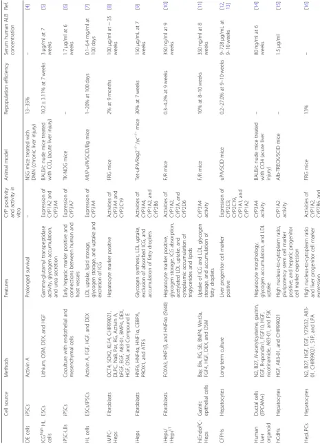
Evaluation of the repopulation rate and hepatic function of transplanted human primary hepatocytes has in-creased over the past two decades with the development of various mouse models (Table1). There are three main mouse models: albumin (ALB) uroplasminogen activator (uPA) transgenic mice, mice with knockout of the fumarylacetoacetate hydrolase (Fah) gene, and ALB thy-midine kinase transgenic-NOD-SCID-interleukin com-mon mice gamma chain knockout (TK-NOG) mice [19].
In uPA/SCID mice, constitutive expression of uPA in hepatocytes causes liver injury and permits selective ex-pansion of transplanted human hepatocytes. However,
© The Author(s). 2019Open AccessThis article is distributed under the terms of the Creative Commons Attribution 4.0 International License (http://creativecommons.org/licenses/by/4.0/), which permits unrestricted use, distribution, and reproduction in any medium, provided you give appropriate credit to the original author(s) and the source, provide a link to the Creative Commons license, and indicate if changes were made. The Creative Commons Public Domain Dedication waiver (http://creativecommons.org/publicdomain/zero/1.0/) applies to the data made available in this article, unless otherwise stated.
* Correspondence:[email protected]
2
Division of Molecular and Cellular Medicine, National Cancer Center Research Institute, 5-1-1 Tsukiji, Chuo-ku, Tokyo 104-0045, Japan
4Institute of Medical Science, Tokyo Medical University, 6-1-1 Shinjuku,
Shinjuku-ku, Tokyo 160-8402, Japan
Table 1 Comparison of potential cell sources for cell-based treatment of liver failure Cel l sour ce Method s Features CYP posi tivity an d activ ity in vit ro An imal mod el Repop ula tion efficie ncy Serum hum an ALB conc entration Ref. DE cell s iPSC s Activin A Prolon ged survival – NS G mice treated with D MN (chro nic liver inju ry) 13 – 35% – [ 4 ] ICG high HL cells ESCs Lithium, O SM, DEX , and H GF Gamma-glutam yl transpe ptidase activity, glyco gen accu mulation , and ure a se cretion Exp ression of CYP 1A2 an d CYP 3A4 BAL B/c nude mi ce treated with CCl 4 (acute liv er injury ) 10.2 ± 3.11 % at 7 weeks 3 μ g/ml at 7 we eks [ 5 ] iPSC -LBs iPSC s Coculture with end othe lial an d mesen chymal cells Early hep atic mar ker posi tive and connections betw een hum an and host vesse ls Exp ression of CYP 3A7 TK-N OG mice – 1.7 μ g/ml at 6 we eks [ 6 ] HL cells ESCs /iPS Cs Activin A, FGF , HGF, an d DEX LDL uptak e, lipid storage , glycoge n storage, and uptak e and excret ion of ICG Exp ression of CYP 3A4 MUP -uP A /SCID/Bg mi ce 1 – 20% at 100 days 0.1 – 6. 4 mg /ml at 100 days [ 7 ] iMPC -Heps Fi broblasts OCT4, SOX 2, K LF4, CHIR9 9021, DLPC, NaB, Par, RG, Ac tivin A, bFGF, EGF , A83-0 1, BMP4, D EX, HGF, OSM, and Comp ound E Hepat ocyte mar ker positive Ac tivities of CYP 3A4 an d CYP 2C19 FR G mice 2% at 9 month s 100 μ g/ml at ~ 35 we eks [ 8 ] iHeps Fi broblasts HNF6, HNF4 α , HNF1 α , CE BPA, PROX1 , and ATF5 Glycogen syn thesi s, LD L uptak e, exclusio n of absorbed ICG, and accumu lation of fatty droplets Ac tivities of CYP 3A4, CYP 1A2, and CYP 2B6 Tet-uPA/R ag2 − / − /γ c − / − mice 30% at 7 we eks 150 μ g/mL at 7 we eks [ 9 ] iHeps / iHeps LT Fi broblasts FOXA3 , HNF1 β , and HNF4 α (SV4 0) Hepat ocyte mar ker positive, glycoge n storage, ICG abs orpt ion, acetylated LDL uptake, and cytoplasmic accu mulation of triglycerides and lipid s Ac tivities of CYP 1A2, CYP 2A, an d CYP 2D6 F/ R mice 0.3 – 4. 2% at 9 we eks 350 ng/ml at 9 we eks [ 10 ] hiEn doPC-Heps Gas tric ep ithel ial cells Bay, Bix, RG , SB, BM P4, Wnt 3a, FGF4, HGF, D EX, and OSM Uptake of ICG and LD L, glycoge n storage, and accu mulat ion of fatty dropl ets CYP 3A4 activ ity F/ R mice 10% at 8 – 10 we eks 350 ng/ml at 8 we eks [ 11 ] CFPHs Hep atocy tes Long-term cu lture Liver proge nitor cell mar ker positive Exp ression of CYP 2C9, CYP 2C19, CYP 1A1, and CYP 1A2 uP A/SCID mi ce 0.2 – 27 .0% at 9 – 10 weeks 9 – 72 8 μ g/mL at 9 – 10 weeks [ 12 , 13 ]
Human liver organoid
Table
1
Comparison
of
potential
cell
sources
for
cell-based
treatment
of
liver
failure
(Continued)
Cel
l
sour
ce
Method
s
Features
CYP
posi
tivity
an
d
activ
ity
in
vit
ro
An
imal
mod
el
Repop
ula
tion
efficie
ncy
Serum
hum
an
ALB
conc
entration
Ref.
CYP
3A4
Prol
iHHs
Hep
atocy
tes
Wnt3a
,
N2,
B2
7,
N
-ac
etylc
ysteine,
gastrin
,
EGF,
FGF10,
HGF,
nicoti
namide
,
A83-0
1,
FSK
,
and
Y2763
2
Progeni
tor-a
ssociated
gen
e
expressi
on
and
bip
henot
ypic
cells
CYP
2B6
activ
ity
FR
G
mice
64
±
21.8%
(at
P4)
at
4
mont
hs
5.8
±
4.5
mg/ml
(at
P4)
at
4
mon
ths
[
17
]
Hep-O
rgs
Fe
tal
an
d
cryo
preserved
prim
ary
hep
atocy
tes
RSPO1
cond
itioned
me
dium,
B27,
EGF,
N
-ac
etylc
ysteine,
gastrin
,
CHIR9
9021,
HGF,
FGF7,
FGF
10,
A83-01,
nic
otinami
de,
Rho
inhib
i-tor
g-27,
632,
and
TGF
α
Network
s
of
bil
e
cana
liculi,
PAS
staining,
and
LD
L
uptak
e
CYP
3A4
activ
ity
an
d
CYP
2E1
exp
ression
FN
RG
mi
ce
–
200
μ
g/ml
after
90
day
s
[
18
uPA/SCID mice have some disadvantages. Repopulation of human hepatocytes in the liver of these mice is de-creased due to deletion of the uPA transgene by hom-ologous recombination. In addition, hemizygotes cannot be used as hosts because homologous recombination oc-curs more frequently in hemizygotes than in homozy-gotes. To overcome these disadvantages, Tateno et al. established a novel host strain that expresses a transgene comprising the ALB promoter/enhancer and uPA cDNA and is of a SCID background (cDNA-uPA/SCID mice) [20]. Tesfaye et al. also generated a novel mouse strain that expresses the uPA gene under the control of the major urinary protein promoter and is of a SCID/beige background (MUP-uPA/SCID/Bg mice) [21]. cDNA-uPA/SCID mice have the following advantages: their body is larger than that of uPA/SCID mice, it is easier to perform animal experiments, and the frequency of renal damage is decreased. MUP-uPA/SCID/Bg mice provide a long time window (up to 12 months) for hepatocyte engraftment and are efficiently infected with hepatitis B virus or hepatitis C virus [22]. Tet-uPA/Rag2−/−/γc−/− mice are easily bred, remain healthy prior to induction of liver injury, and have no time-window limit for liver cell transplantation.
In Fah-knockout mice, deletion of Fah, which func-tions in the tyrosine catabolic pathway, causes accumu-lation of toxic fumarylacetoacetate, resulting in liver injury. Liver disease can be controlled by administering 2-(2-nitro-4-trifluoromethylbenzoyl)-1,3-cyclohexane-dione in these mice. Azuma et al. generated Fah−/−/ Rag2−/−/Il2rg−/− (FRG) mice by crossing Fah-knockout mice and Rag2−/−/Il2rg−/− mice, which are immunodefi-cient and lack B, T, and NK cells [23]. The capacity for liver xeno-repopulation is reduced in Fah−/−Rag2−/− (F/ R) mice due to the presence of NK cells [24]. However, F/R mice are easy to bred and tolerate hepatocyte trans-plantation. Fah−/− NOD Rag1−/−Il2rg−/− (FNRG) mice are more immunodeficient than FRG mice [25].
A herpes simplex virus type 1 thymidine kinase (HSVtk) transgene was expressed in the liver of highly immunodeficient NOG mice. Ganciclovir can control the hepatotoxic transgene in TK-NOG mice. In addition, TK-NOG mice mimic liver zonation and drug metabol-ism in the repopulated liver [26].
Azuma et al. intrasplenically transplanted human he-patocytes into FRG mice [23]. Human hepatocytes repo-pulated the livers of these mice with a repopulation rate of > 80%. Hasegawa et al. intrasplenically transplanted human liver cells into TK-NOG mice [26]. The repopu-lation rate was 43% in the livers of these mice. Tateno et al. intrasplenically transplanted human hepatocytes into cDNA-uPA/SCID mice [20]. The repopulation rate was > 70% in the livers of these mice. Thus, transplanted ma-ture human hepatocytes demonstrate a high capacity to
regenerate the injured liver in mice, which indicates the feasibility of mouse models for checking the function of in vitro-derived cells.
Potential alternative cell sources for hepatocyte transplantation therapy
To overcome the shortage of donor hepatocytes, many attempts have been made to generate functional hepato-cytes from multiple types of cells (Table 1). However, there is controversy regarding the usefulness of these cells for transplantation therapy. Liu et al. generated hu-man induced pluripotent stem cell (iPSC) lines from dif-ferent sources and intravenously transplanted definitive endoderm (DE) cells differentiated from these iPSC lines
into NOD/Lt-SCID/IL-2Rγ−/−(NSG) mice that had been
treated with dimethylnitrosamine (DMN) for 4 weeks (liver cirrhosis model) [4, 27–30]. The engraftment per-centage, calculated as the percentage of human hepatic cells expressing ALB, was 13% in the livers of mice transplanted with 2 × 10⁶DE cells and 35% in the livers of mice transplanted with 7 × 10⁶DE cells. Woo et al. re-ported that embryonic stem cells (ESCs) treated with lithium and cultured in the presence of hepatocyte growth factor (HGF), oncostatin M (OSM), and dexa-methasone (DEX) differentiated into cells with a hepatocyte-like (HL) morphology that expressed ALB and keratin 18, and that HL cells with high liver function were enriched using indocyanine green (ICG) [5,31–34]. When HL ICGhigh cells were transplanted into CCl4 -in-toxicated BALB/c mice (acute liver injury model), the percentage of human ALB-positive cells was lower at day 35 (10.2 ± 3.11%) than at day 3 (20.2 ± 4.45%) after transplantation. Takebe et al. revealed that hepatic endo-derm cells derived from human iPSCs formed a three-dimensional spherical tissue mass termed iPSC-derived liver buds (iPSC-LBs), which expressed early hepatic marker genes, upon culture with human umbilical vein endothelial cells and human mesenchymal stem cells [6]. In vitro-derived human iPSC-LBs integrated with the host vasculature within 48 h after transplantation. Hu-man iPSC-LBs began producing ALB at approximately day 10 post-transplantation in TK-NOG mice and in-creased the concentration of ALB to 1.983μg/ml by day 45. Carpentier et al. demonstrated that HL cells differen-tiated from iPSCs via a multistep protocol were positive for α-1-antitrypsin (AAT) and Forkhead box a2 (FOXA2), which are endoderm cell markers, as well as hepatocyte nuclear factor 4 alpha (HNF4α), which is a master regulator of hepatic differentiation. Upon trans-plantation of HL cells into the spleen of MUP-uPA/ SCID/Bg mice, the human ALB concentration at day 10 post-engraftment was 50–3900μg/ml [7,35,36].
intermediary pluripotent stage, could be an alternative to iPSCs for generation of functional hepatocytes. Zhu et al. transduced human fibroblasts with retroviruses ex-pressing OCT4, SOX2, and KLF4 and then replated these cells into a medium containing established growth factors and CHIR99021 (a GSK-3β inhibitor) for repro-gramming into endoderm cells [8]. Upon addition of A83-01 (a transforming growth factor-β inhibitor) and Compound E (a Notch signaling inhibitor) to inhibit bil-iary differentiation, these cells differentiated into in-duced multipotent progenitor cell hepatocytes (iMPC-Heps) that expressed hepatocyte markers. Following transplantation of iMPC-Heps into FRG mice, human ALB was detected in mouse serum at 2 months post-transplantation and reached a concentration of 104μg/ ml after 6 months, with a liver repopulation efficiency of 2%. Du et al. demonstrated that overexpression of HNF6, HNF4α, and HNF1αinduced differentiation of fi-broblasts into cells that were morphologically similar to hepatocytes (3H cells). They also overexpressed CEBPA, PROX1, and ATF5 in 3H cells and observed a dramatic morphological change of fibroblasts into epithelial cells within 1 week (iHeps) [9]. iHeps were intrasplenically transplanted into Tet-uPA/Rag2−/−/γc−/− mice [37]. The concentration of human ALB in mouse serum gradually increased and peaked at 313 ng/ml at 7 weeks post-transplantation, with a repopulation efficiency of ap-proximately 30%. Huang et al. reported that overexpres-sion of FOXA3, HNF1β, and HNF4αinduced high levels of hepatic gene expression in fibroblasts at 12 days after induction (iHeps) [10]. When iHeps transfected with the SV40 large T antigen were transplanted into F/R mice, staining of human Fah and AAT showed that these cells repopulated 0.3–4.2% of the liver parenchyma in surviv-ing mice [23]. Transdifferentiation of fibroblasts was in-duced via gene transfer in these three reports. On the other hand, Wang et al. demonstrated that treatment with four small molecules (Bay K 8644, Bix01294, RG108, and SB431542) converted gastric epithelial cells into induced endodermal progenitor cells (hiEndoPCs) with a multilineage differentiation capacity [11]. Trans-planted hiEndoPC-derived hepatic cells (hiEndoPC-Heps) with hepatocyte-specific functions rescued liver failure in F/R mice. Moreover, human ALB levels were comparable to those from either hESC-Heps, with a maximum repopulation efficiency of 10%.
Several recent studies proposed that hepatocytes are a source of expandable hepatic cells. In 2008, Utoh et al. identified a small population of replicative hepatocytes, termed colony-forming parenchymal hepatocytes (CFPHs), in long-term cultures of human adult hepato-cytes. The frequency of these cells was 0.01–0.09% de-pending on donor age [12, 13]. When CFPHs were transplanted into uPA/SCID mice, they engrafted into
ml after 4 months. The repopulated ProliHHs expressed phase I and II enzymes and transporters at levels com-parable with those in primary human hepatocytes after transplantation.
Hu et al. established human fetal hepatocyte organoids with a typical grape-like structure [18]. They also estab-lished organoids from cryopreserved primary human he-patocytes, which had small lumina and contained large cells with a hepatocyte morphology. Notably, ALB secre-tion by the latter organoids was comparable with that by primary human hepatocytes. Organoids were trans-planted like hepatocyte transplantations into FNRG mice via splenic injection [44,45]. At 90 days after transplant-ation, the serum human ALB in mice transplanted with human fetal hepatocyte organoids had increased by 200-fold to more than 200μg/ml on average. Fu et al. re-vealed that three-dimensional spheroid formation en-hanced hepatic differentiation in vitro [16]. Zhang et al. reported that ProliHHs matured in three-dimensional organoid culture [17]. Thus, three-dimensional culture may contribute to the maturation of hepatocytes.
Potential application of in vitro-generated hepatic cells for drug development studies
Primary human hepatocytes are the gold standard for drug development studies. Olson et al. compared drug toxicities between humans and various animals, includ-ing dogs, primates, rats, mice, and guinea pigs [46]. Their analysis indicated that the overall concordance be-tween human and animal toxicity was 71%. Many in vitro models of the liver have been used, including liver slices, hepatic cell lines, and primary hepatocytes. Liver tissue slices exhibit zone-specific cytochrome p450 (CYP) activity and phase II enzyme expression; however, these are unstable [47]. Although hepatic cell lines pro-vide an unlimited number of cells, their expression levels of phase I and II enzymes decrease upon repeated pas-sage [48]. Consequently, human hepatocytes that can metabolize drugs and toxicity screening platforms are re-quired. However, the use of primary human hepatocytes is hampered by the limited number of donors and the small number of cells that are obtained. In addition, it is difficult to maintain the proliferative capacity and func-tion of hepatocytes in vitro [49].
Stem cell-derived hepatocytes reportedly exhibit sub-stantial CYP enzyme activity; however, their applicability for drug testing remains controversial. Liu et al. demon-strated that human iPSC-derived hepatocytes exhibited activities of major CYP enzymes, such as CYP1A2, CYP2C9, CYP2C19, and CYP2D6, similar to primary he-patocytes [4]. Woo et al. reported that ICGhigh HL cells were positive for ALB, keratin 18, HNF4α, and CYP1A2 and that expression of enzymes related to phase I and II drug metabolism, namely, CYP3A4 and glutathione
S-transferase 1/2, was enhanced in these cells according to quantitative PCR [30]. Carpentier et al. demonstrated that HL cells exhibited various hepatocyte-specific functions, including uptake of low-density lipoprotein (LDL), storage of lipids based on Oil Red O staining, storage of glycogen based on periodic acid-Schiff staining, and uptake and ex-cretion of ICG; however, HL cells were mainly negative for CYP2D6 and only a few cells were weakly positive for CYP3A4 [32]. These studies collectively suggest that stem cell-derived hepatic cells are useful for pharmaceutical studies. However, they did not demonstrate the inducibil-ity of CYP enzyme activities, which is a major criterion for application of cultured hepatic cells in drug development studies. A few groups described CYP inducibility in terms of enzymatic activity [50–52]. However, the number of such studies is very small, and consequently, the useful-ness of stem cell-derived hepatocytes for pharmaceutical studies remains controversial.
Hepatocyte-derived expandable hepatic cells could be used instead of primary human hepatocytes in pharma-ceutical studies. Kim et al. reported that omeprazole treat-ment significantly increased CYP1A2 activity in hCdH-derived hepatocytes relative to that in hCdHs to a similar level as that in primary human hepatocytes [15]. Fu et al. demonstrated that omeprazole treatment increased CYP1A2 expression by 80 ± 11-fold to 193 ± 27-fold, CITCO treatment increased CYP2B6 expression by 10 ± 2-fold to 26 ± 4-fold, and rifampicin treatment increased CYP3A4 expression by 47 ± 2-fold to 96 ± 5-fold (in com-parison with the DMSO-treated control) in HepLPCs-Heps [16]. Furthermore, HepLPCs-Heps metabolized acetaminophen, bupropion, diclofenac, OH-testosterone, and OH-coumarin Glu to a similar extent as primary hepatocytes. Zhang et al. reported that CYP2B6 metabolic activity in ProliHHs increased after maturation, in accordance with increased mRNA expression of genes involved in CYP2B6 metabolism [17]. These reports strongly suggest that hepatocyte-derived expandable cells have an advantage over stem cell-derived hepatic cells in terms of CYP inducibility.
Future perspectives
the quality of cells. Serum human ALB levels and re-population efficiencies in several animal models of liver disease provide reliable data to evaluate cell functions. Secretion of ALB by transplanted cells is higher in re-cent studies than in older studies (Table1). The safety of cell replacement therapy must also be considered. In particular, the risk of tumor formation following trans-plantation of cells reprogrammed via gene transfer must be thoroughly investigated. Generation of mature hepatocyte-derived progenitors via treatment with small molecules is currently the best strategy in terms of cell function and safety. Further studies are required to de-termine whether mature hepatocytes obtained from pa-tients with severe liver disease such as cirrhosis can be converted into progenitors with sufficient functions.
In vitro culture of functional hepatocytes may facilitate the evaluation of drug metabolism, which would acceler-ate the safety assessment of new drugs. Personalized as-sessment of the hepatic side effects of drugs may also be possible using in vitro models generated using a person’s own hepatocytes. Therefore, in vitro drug metabolism should be considered when selecting a strategy to gener-ate hepatocytes.
The rapid development of genome editing technologies means that genetic changes can be introduced into hep-atocyte progenitors in a site-specific manner, including
correction of disease-causing gene mutations in patient-derived hepatocytes. This approach may enable us to cure congenital/inherited metabolic diseases. On the other hand, the introduction of specific mutations into non-diseased hepatocyte progenitors could be used to generate ideal disease models. This approach could be used to in-vestigate the mechanisms underlying liver carcinogenesis.
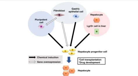
Conclusion
In vitro-expandable hepatocytes are required as thera-peutic alternatives to liver transplantation and for drug development. Three strategies have been proposed to generate functional hepatocytes: (i) generation of hepa-tocytes from ESCs or iPSCs, (ii) transdifferentiation of fi-broblasts and other differentiated cells into hepatocytes, and (iii) chemical induction of hepatocyte progenitors from mature hepatocytes (Fig. 1). Standardized methods to evaluate cell functions are required to compare these methods. The coming decade will reveal which strategy holds the most promise for translation into clinical applications.
Abbreviations
Il2rg−/−; hCdH: Human chemically derived hepatic progenitor;
HepLPC: Hepatocyte-derived liver progenitor-like cell; Hep: HepLPC-derived hepatocyte; HGF: Hepatocyte growth factor; hiEndoPC: Human induced endodermal progenitor cell; hiEndoPC-Hep: hiEndoPC-derived hepatic cell; HL: Hepatocyte-like; HNF4α: Hepatocyte nuclear factor 4 alpha; ICG: Indocyanine green; iMPC-Hep: Induced multipotent cell progenitor hepatocyte; iPSC: Induced pluripotent stem cell; iPSC-LB: iPSC-derived liver bud; LDL: Low-density lipoprotein; NSG:NOD/Lt-SCID/IL-2Rγ−/−;
OSM: Oncostatin M; ProliHH: Proliferating human hepatocyte; YAC: Y27632, A83-01, and CHIR99021
Authors’contributions
TY and JM wrote the manuscript. TK, YS, HS, and TO revised the manuscript. All authors read and approved the final manuscript.
Funding
This research was funded by a Grant-in-Aid for Scientific Research (C) (17 K09471, to JM) from the Japan Society for the Promotion of Science (JSPS).
Availability of data and materials
Not applicable
Ethics approval and consent to participate
Not applicable
Consent for publication
Not applicable
Competing interests
The authors declare that they have no competing interests.
Author details
1Division of Pharmacotherapeutics, Keio University Faculty of Pharmacy,
1-5-30 Shibakoen, Minato-ku, Tokyo 105-8512, Japan.2Division of Molecular
and Cellular Medicine, National Cancer Center Research Institute, 5-1-1 Tsukiji, Chuo-ku, Tokyo 104-0045, Japan.3Division of Gastroenterology and
Hepatology, Department of Internal Medicine, Keio University School of Medicine, 35 Shinanomachi, Shinjuku-ku, Tokyo 160-8582, Japan.4Institute of
Medical Science, Tokyo Medical University, 6-1-1 Shinjuku, Shinjuku-ku, Tokyo 160-8402, Japan.
Received: 10 March 2019 Accepted: 22 May 2019
References
1. Dhawan A, Puppi J, Hughes RD, Mitry RR. Human hepatocyte transplantation: current experience and future challenges. Nat Rev Gastroenterol Hepatol. 2010;7(5):288–98.
2. Michalopoulos GK. The liver is a peculiar organ when it comes to stem cells. Am J Pathol. 2014;184(5):1263–7.
3. Tanaka M, Miyajima A. Liver regeneration and fibrosis after inflammation. Inflamm Regen. 2016;36:19.
4. Liu H, Kim Y, Sharkis S, Marchionni L, Jang YY. In vivo liver regeneration potential of human induced pluripotent stem cells from diverse origins. Sci Transl Med. 2011;3(82):82ra39.
5. Woo DH, Kim SK, Lim HJ, Heo J, Park HS, Kang GY, Kim SE, You HJ, Hoeppner DJ, Kim Y, et al. Direct and indirect contribution of human embryonic stem cell-derived hepatocyte-like cells to liver repair in mice. Gastroenterology. 2012;142(3):602–11.
6. Takebe T, Sekine K, Enomura M, Koike H, Kimura M, Ogaeri T, Zhang RR, Ueno Y, Zheng YW, Koike N, et al. Vascularized and functional human liver from an iPSC-derived organ bud transplant. Nature. 2013;499(7459):481–4. 7. Carpentier A, Tesfaye A, Chu V, Nimgaonkar I, Zhang F, Lee SB,
Thorgeirsson SS, Feinstone SM, Liang TJ. Engrafted human stem cell-derived hepatocytes establish an infectious HCV murine model. J Clin Invest. 2014;124(11):4953–64.
8. Zhu S, Rezvani M, Harbell J, Mattis AN, Wolfe AR, Benet LZ, Willenbring H, Ding S. Mouse liver repopulation with hepatocytes generated from human fibroblasts. Nature. 2014;508(7494):93–7.
9. Du Y, Wang J, Jia J, Song N, Xiang C, Xu J, Hou Z, Su X, Liu B, Jiang T, et al. Human hepatocytes with drug metabolic function induced from fibroblasts by lineage reprogramming. Cell Stem Cell. 2014;14(3):394–403.
10. Huang P, Zhang L, Gao Y, He Z, Yao D, Wu Z, Cen J, Chen X, Liu C, Hu Y, et al. Direct reprogramming of human fibroblasts to functional and expandable hepatocytes. Cell Stem Cell. 2014;14(3):370–84.
11. Wang Y, Qin J, Wang S, Zhang W, Duan J, Zhang J, Wang X, Yan F, Chang M, Liu X, et al. Conversion of human gastric epithelial cells to multipotent endodermal progenitors using defined small molecules. Cell Stem Cell. 2016;19(4):449–61.
12. Utoh R, Tateno C, Yamasaki C, Hiraga N, Kataoka M, Shimada T, Chayama K, Yoshizato K. Susceptibility of chimeric mice with livers repopulated by serially subcultured human hepatocytes to hepatitis B virus. Hepatology. 2008;47(2):435–46.
13. Yamasaki C, Tateno C, Aratani A, Ohnishi C, Katayama S, Kohashi T, Hino H, Marusawa H, Asahara T, Yoshizato K. Growth and differentiation of colony-forming human hepatocytes in vitro. J Hepatol. 2006;44(4):749–57. 14. Huch M, Gehart H, van Boxtel R, Hamer K, Blokzijl F, Verstegen MM, Ellis
E, van Wenum M, Fuchs SA, de Ligt J, et al. Long-term culture of genome-stable bipotent stem cells from adult human liver. Cell. 2015; 160(1–2):299–312.
15. Kim Y, Kang K, Lee SB, Seo D, Yoon S, Kim SJ, Jang K, Jung YK, Lee KG, Factor VM, et al. Small molecule-mediated reprogramming of human hepatocytes into bipotent progenitor cells. J Hepatol. 2019;70(1):97–107. 16. Fu GB, Huang WJ, Zeng M, Zhou X, Wu HP, Liu CC, Wu H, Weng J, Zhang
HD, Cai YC, et al. Expansion and differentiation of human hepatocyte-derived liver progenitor-like cells and their use for the study of hepatotropic pathogens. Cell Res. 2019;29(1):8–22.
17. Zhang K, Zhang L, Liu W, Ma X, Cen J, Sun Z, Wang C, Feng S, Zhang Z, Yue L, et al. In vitro expansion of primary human hepatocytes with efficient liver repopulation capacity. Cell Stem Cell. 2018;23(6):806–819 e804.
18. Hu H, Gehart H, Artegiani B, LO-I C, Dekkers F, Basak O, van Es J, Chuva de Sousa Lopes SM, Begthel H, Korving J, et al. Long-term expansion of functional mouse and human hepatocytes as 3D organoids. Cell. 2018; 175(6):1591–1606 e1519.
19. Grompe M, Strom S. Mice with human livers. Gastroenterology. 2013;145(6): 1209–14.
20. Tateno C, Kawase Y, Tobita Y, Hamamura S, Ohshita H, Yokomichi H, Sanada H, Kakuni M, Shiota A, Kojima Y, et al. Generation of novel chimeric mice with humanized livers by using hemizygous cDNA-uPA/SCID mice. PLoS One. 2015;10(11):e0142145.
21. Tesfaye A, Stift J, Maric D, Cui Q, Dienes HP, Feinstone SM. Chimeric mouse model for the infection of hepatitis B and C viruses. PLoS One. 2013;8(10): e77298.
22. Wang Z, Wu N, Tesfaye A, Feinstone S, Kumar A. HCV infection-associated hepatocellular carcinoma in humanized mice. Infect Agent Cancer. 2015;10:24. 23. Azuma H, Paulk N, Ranade A, Dorrell C, Al-Dhalimy M, Ellis E, Strom S, Kay
MA, Finegold M, Grompe M. Robust expansion of human hepatocytes in Fah-/-/Rag2-/-/Il2rg-/- mice. Nat Biotechnol. 2007;25(8):903–10.
24. He Z, Zhang H, Zhang X, Xie D, Chen Y, Wangensteen KJ, Ekker SC, Firpo M, Liu C, Xiang D, et al. Liver xeno-repopulation with human hepatocytes in Fah-/-Rag2-/- mice after pharmacological immunosuppression. Am J Pathol. 2010;177(3):1311–9.
25. Stevens KR, Scull MA, Ramanan V, Fortin CL, Chaturvedi RR, Knouse KA, Xiao JW, Fung C, Mirabella T, Chen AX, et al. In situ expansion of engineered human liver tissue in a mouse model of chronic liver disease. Sci Transl Med. 2017;9(399):eaah5505.
26. Hasegawa M, Kawai K, Mitsui T, Taniguchi K, Monnai M, Wakui M, Ito M, Suematsu M, Peltz G, Nakamura M, et al. The reconstituted‘humanized liver’ in TK-NOG mice is mature and functional. Biochem Biophys Res Commun. 2011;405(3):405–10.
27. Locke JE, Sun Z, Warren DS, Sheets TP, Holzer H, Shamblott MJ, Montgomery RA, Cameron AM. Generation of humanized animal livers using embryoid body-derived stem cell transplant. Ann Surg. 2008;248(3): 487–93.
28. Nishibe Y, Kaneko H, Suzuki H, Abe T, Matsuura Y, Takaku H. Baculovirus-mediated interferon alleviates dimethylnitrosamine-induced liver cirrhosis symptoms in a murine model. Gene Ther. 2008;15(13):990–7.
30. Ogiso T, Nagaki M, Takai S, Tsukada Y, Mukai T, Kimura K, Moriwaki H. Granulocyte colony-stimulating factor impairs liver regeneration in mice through the up-regulation of interleukin-1beta. J Hepatol. 2007;47(6):816–25. 31. Lickert H, Domon C, Huls G, Wehrle C, Duluc I, Clevers H, Meyer BI, Freund
JN, Kemler R. Wnt/(beta)-catenin signaling regulates the expression of the homeobox gene Cdx1 in embryonic intestine. Development. 2000;127(17): 3805–13.
32. Gadue P, Huber TL, Paddison PJ, Keller GM. Wnt and TGF-beta signaling are required for the induction of an in vitro model of primitive streak formation using embryonic stem cells. Proc Natl Acad Sci U S A. 2006; 103(45):16806–11.
33. Hay DC, Fletcher J, Payne C, Terrace JD, Gallagher RC, Snoeys J, Black JR, Wojtacha D, Samuel K, Hannoun Z, et al. Highly efficient differentiation of hESCs to functional hepatic endoderm requires ActivinA and Wnt3a signaling. Proc Natl Acad Sci U S A. 2008;105(34):12301–6.
34. Klein PS, Melton DA. A molecular mechanism for the effect of lithium on development. Proc Natl Acad Sci U S A. 1996;93(16):8455–9.
35. Tesfaye M, Silverstein KA, Nallu S, Wang L, Botanga CJ, Gomez SK, Costa LM, Harrison MJ, Samac DA, Glazebrook J, et al. Spatio-temporal expression patterns of Arabidopsis thaliana and Medicago truncatula defensin-like genes. PLoS One. 2013;8(3):e58992.
36. Weglarz TC, Degen JL, Sandgren EP. Hepatocyte transplantation into diseased mouse liver. Kinetics of parenchymal repopulation and identification of the proliferative capacity of tetraploid and octaploid hepatocytes. Am J Pathol. 2000;157(6):1963–74.
37. Song X, Guo Y, Duo S, Che J, Wu C, Ochiya T, Ding M, Deng H. A mouse model of inducible liver injury caused by tet-on regulated urokinase for studies of hepatocyte transplantation. Am J Pathol. 2009;175(5):1975–83. 38. Katsuda T, Kawamata M, Hagiwara K, Takahashi RU, Yamamoto Y, Camargo
FD, Ochiya T. Conversion of terminally committed hepatocytes to culturable bipotent progenitor cells with regenerative capacity. Cell Stem Cell. 2017; 20(1):41–55.
39. Suarez-Causado A, Caballero-Diaz D, Bertran E, Roncero C, Addante A, Garcia-Alvaro M, Fernandez M, Herrera B, Porras A, Fabregat I, et al. HGF/c-Met signaling promotes liver progenitor cell migration and invasion by an epithelial-mesenchymal transition-independent, phosphatidyl inositol-3 kinase-dependent pathway in an in vitro model. Biochim Biophys Acta. 2015;1853(10 Pt A):2453–63.
40. Kitade M, Factor VM, Andersen JB, Tomokuni A, Kaji K, Akita H, Holczbauer A, Seo D, Marquardt JU, Conner EA, et al. Specific fate decisions in adult hepatic progenitor cells driven by MET and EGFR signaling. Genes Dev. 2013;27(15):1706–17.
41. Kwon YJ, Lee KG, Choi D. Clinical implications of advances in liver regeneration. Clin Mol Hepatol. 2015;21(1):7–13.
42. Wu H, Zhou X, Fu GB, He ZY, Wu HP, You P, Ashton C, Wang X, Wang HY, Yan HX. Reversible transition between hepatocytes and liver progenitors for in vitro hepatocyte expansion. Cell Res. 2017;27(5):709–12.
43. Huch M, Dorrell C, Boj SF, van Es JH, Li VS, van de Wetering M, Sato T, Hamer K, Sasaki N, Finegold MJ, et al. In vitro expansion of single Lgr5+ liver stem cells induced by Wnt-driven regeneration. Nature. 2013;494(7436): 247–50.
44. Billerbeck E, Mommersteeg MC, Shlomai A, Xiao JW, Andrus L, Bhatta A, Vercauteren K, Michailidis E, Dorner M, Krishnan A, et al. Humanized mice efficiently engrafted with fetal hepatoblasts and syngeneic immune cells develop human monocytes and NK cells. J Hepatol. 2016;65(2):334–43. 45. Grompe M. Fah knockout animals as models for therapeutic liver
repopulation. Adv Exp Med Biol. 2017;959:215–30.
46. Olson H, Betton G, Robinson D, Thomas K, Monro A, Kolaja G, Lilly P, Sanders J, Sipes G, Bracken W, et al. Concordance of the toxicity of pharmaceuticals in humans and in animals. Regul Toxicol Pharmacol. 2000; 32(1):56–67.
47. Soldatow VY, Lecluyse EL, Griffith LG, Rusyn I. In vitro models for liver toxicity testing. Toxicol Res (Camb). 2013;2(1):23–39.
48. Guguen-Guillouzo C, Guillouzo A. General review on in vitro hepatocyte models and their applications. Methods Mol Biol. 2010;640:1–40. 49. Bale SS, Moore L, Yarmush M, Jindal R. Emerging in vitro liver technologies
for drug metabolism and inter-organ interactions. Tissue Eng Part B Rev. 2016;22(5):383–94.
50. Inamura M, Kawabata K, Takayama K, Tashiro K, Sakurai F, Katayama K, Toyoda M, Akutsu H, Miyagawa Y, Okita H, et al. Efficient generation of
hepatoblasts from human ES cells and iPS cells by transient overexpression of homeobox gene HEX. Mol Ther. 2011;19(2):400–7.
51. Pettinato G, Ramanathan R, Fisher RA, Mangino MJ, Zhang N, Wen X. Scalable differentiation of human iPSCs in a multicellular spheroid-based 3D culture into hepatocyte-like cells through direct Wnt/beta-catenin pathway inhibition. Sci Rep. 2016;6:32888.
52. Takayama K, Inamura M, Kawabata K, Katayama K, Higuchi M, Tashiro K, Nonaka A, Sakurai F, Hayakawa T, Furue MK, et al. Efficient generation of functional hepatocytes from human embryonic stem cells and induced pluripotent stem cells by HNF4alpha transduction. Mol Ther. 2012;20(1):127– 37.
Publisher’s Note