that TN without NVC is more prevalent in younger individuals (6). These findings suggest contributions of genetic and epi-genetic factors to the pathophysiology of classical TN, especially in patients with-out severe NVC. Rare cases of familial idiopathic trigeminal pain with an autoso-mal dominant inheritance pattern (7–10) support a genetic component for TN.
The molecular basis for TN has thus far remained elusive. Its pathophysiol-ogy has been modeled by the “ignition theory” (11,12), which posits that injured trigeminal neurons fire spontaneously and have a lower threshold for evoked afterdischarges leading to paroxysmal pain. Intraoperative microneurographic recordings in trigeminal nerve fibers after stimulation of the known trigger zone in a TN patient demonstrated after-discharges that were absent when unaf-fected facial areas were stimulated (13), loss, demyelination and dysmyelination
consistent with NVC of the trigeminal nerve (2). However, recent studies have suggested that only severe neurovascular contact, causing displacement or atrophy of the nerve, is associated with pain in classical TN (3,4), and some studies have questioned whether NVC is necessary and sufficient for TN, since pain can occur and recur in the absence of nerve com-pression (5,6). A recent study reported InTRODUCTIOn
Trigeminal neuralgia (TN) is character-ized by recurring episodes of unilateral and lancinating facial pain along one or more branches of the trigeminal nerve (1). Neurovascular compression (NVC) of the trigeminal nerve is prevalent in TN with no other known comorbidities, and has been classically regarded as causing TN (1). Ultrastructural studies on bi-opsied trigeminal roots showed axonal
of Trigeminal neuralgia
Brian S Tanaka,
1,2,3Peng Zhao,
1,2,3Fadia B Dib-Hajj,
1,2,3Valerie Morisset,
4Simon Tate,
4Stephen G Waxman,
1,2,3and Sulayman D Dib-Hajj
1,2,31Department of Neurology; 2Center for Neuroscience and Regeneration Research, Yale University School of Medicine, New Haven,
Connecticut, United States of America; 3Rehabilitation Research Center, Veterans Affairs Connecticut Healthcare System, West
Haven; and 4Convergence Pharmaceuticals Ltd, Cambridge, United Kingdom
Idiopathic trigeminal neuralgia (TN) is a debilitating pain disorder characterized by episodic unilateral facial pain along the territory of branches of the trigeminal nerve. Human pain disorders, but not TN, have been linked to gain-of-function mutations in peripheral voltage-gated sodium channels (NaV1.7, NaV1.8 and NaV1.9). Gain-of-function mutations in NaV1.6, which is
ex-pressed in myelinated and unmyelinated central nervous system (CNS) and peripheral nervous system neurons and supports neuronal high-frequency firing, have been linked to epilepsy but not to pain. Here, we describe an individual who presented with evoked and spontaneous paroxysmal unilateral facial pain and carried a diagnosis of TN. Magnetic resonance imaging showed unilateral neurovascular compression, consistent with pain in areas innervated by the second branch of the trigeminal nerve. Genetic analysis as part of a phase 2 clinical study in patients with TN conducted by Convergence Pharmaceuticals Ltd revealed a previously undescribed de novo missense mutation in NaV1.6 (c.A406G; p.Met136Val). Whole-cell voltage-clamp
re-cordings show that the Met136Val mutation significantly increases peak current density (1.5-fold) and resurgent current (1.6-fold) without altering gating properties. Current-clamp studies in trigeminal ganglia (TRG) neurons showed that Met136Val increased the fraction of high-firing neurons, lowered the current threshold and increased the frequency of evoked action potentials in response to graded stimuli. Our results demonstrate a novel NaV1.6 mutation in TN, and show that this mutation potentiates
transient and resurgent sodium currents and leads to increased excitability in TRG neurons. We suggest that this gain-of-function NaV1.6 mutation may exacerbate the pathophysiology of vascular compression and contribute to TN.
Online address: http://www.molmed.org doi: 10.2119/molmed.2016.00131
Address correspondence to Sulayman D Dib-Hajj, PhD, Neuroscience and Regeneration Research Center, VA Connecticut Healthcare System, 950 Campbell Avenue, Bldg. 34, West Haven, CT 06516 USA. Phone: 203-937-3802; Fax: 203-937-3801; E-mail: sulayman. [email protected].
reagent (1.5 μL) and DNA (0.6 μg total) were each mixed with 50 μL Opti-MEM (serum- and antibiotic-free) and incu-bated at room temperature (RT, 20° ± 1°C) for 5 min. The two solutions were com-bined and incubated at RT for 20 min. The combined mixture (100 μL/well) was added to the ND7/23 cell culture medium (0.5 mL Dulbecco’s modified Eagle medium [DMEM] plus 10% fetal bovine serum [FBS] per well) and incu-bated at 37°C for 3 h. Culture medium was replaced and cells were incubated at 37°C for 48 h before electrophysiological recordings.
Voltage-clamp in ND7/23 cells. ND7/23 cells with robust green fluorescence and WT or Met136Val were recorded on the same day under similar conditions (21–23). Whole-cell voltage -clamp recordings were per-formed using an EPC10-double ampli-fier and Patchmaster software (HEKA Electronik) at RT (20° ± 1°C). The ex-tracellular solution contained (in mM): 140 NaCl, 3 KCl, 1 MgCl2, 1 CaCl2, 10 N-2- hydroxyethylpiperazine-N′ -2-ethanesulfonic acid (HEPES), 5 CsCl, 20 TEA-Cl, 0.1 CdCl2, pH 7.3 with NaOH (osmolarity adjusted to 320 mOsm with sucrose). Tetrodotoxin (TTX) (500 nM) was added to the bath to block endogenous TTX-sensitive sodium currents in ND7/23 cells (21–23). Patch pipettes were fabri-cated from borosilicate glass (1.65/1.1, OD/ID; World Precision Instruments) using a Sutter Instruments P-97 puller and had a resistance of 0.8-1.5 MΩ. Transfected ND7/23 cells were held at –120 mV for all parameters examined. To minimize voltage errors, 70–90% series resistance compensation was applied. Currents were acquired 5 min after establishing whole-cell configuration. Recordings were acquired at 10 kHz with low-pass Bessel filter and digitized at 50 kHz. Linear leak and residual capacitance artifacts were removed using P/6 subtraction.
To measure current-voltage (I-V) relationships, a range of potentials from –80 mV to +40 mV in 5 mV increments were applied from the holding potential (–120 mV) for 100 ms at 5 s intervals. checks were performed prior to PCR to
ensure high-quality DNA. Bioinformat-ics analysis of the sequencing data and detection of mutations in the target chan-nels were performed using GATK (Broad Institute of Massachusetts Institute of Technology, https://www.broadinstitute. org/gatk/). The incidence of mutations was evaluated in a matching population using the 1000 Genome database.
Plasmid and animals
A plasmid carrying human NaV1.6 (hNaV1.6), purchased from Genionics, carries optimized codons for expression in mammalian cells. The channel was ren-dered resistant to tetrodotoxin (TTX-R) by a Tyr371Ser substitution so that it could be studied in voltage-clamp in iso-lation in ND7/23 cells (a rat DRG/mouse neuroblastoma hybrid cell line) and tri-geminal ganglion (TRG) neurons from NaV1.8-null mice (20). The Tyr371Ser and Met136Val substitutions were introduced into the channel using Quick Change XL site-directed mutagenesis kit (Stratagene). The identity of the insert was confirmed by Sanger sequencing at the Keck se-quencing facility at Yale University.
Animal care followed a protocol approved by the Institutional Animal Care and Use Committee at the Veterans Administration Connecticut Healthcare System, West Haven. TRG from adult NaV1.8-null mice were isolated and used for recording the resurgent current, while TRGs were isolated from male Sprague-Dawley albino rats (4–6 wks old, Harlan) and used for current-clamp studies.
Transient transfection of ND7/23 cells. hNaV1.6R (WT) and hNaV 1.6-M136V (called Met136Val hereafter) were cotransfected with enhanced green fluorescent protein (EGFP) constructs into ND7/23 cells as previously de-scribed (21–23). Briefly, cells were plated at low density on 10 mm coverslips in 24-well plates and incubated at 37°C for 24 h before transfection. Cells were cotransfected with WT or Met136Val (0.5 ug/well) and EGFP (0.1 ug/well) (pEGFP, Clontech). Lipofectamine 2000 and consistent with the predictions of
ignition theory, the period of intense afterdischarge was followed by a pro-longed period refractory to additional stimulation. Voltage-gated sodium channel blockers carbamazepine and oxcarbazepine are the first-line pharma-cotherapy for classical TN (1); thus, it is reasonable to suggest an involvement of voltage-gated sodium channels in its pathophysiology.
Rare and common painful disorders, but not TN, have been genetically and functionally linked to gain-of-function mutations in peripheral sodium channels NaV1.7, NaV1.8 and NaV1.9 (14–16). Although gain-of-function mutations of NaV1.6 have been shown to cause infan-tile epileptic encephalopathy (17–19), there has been no genetic evidence thus far for a role for this channel in human pain disorders. We report here a novel NaV1.6 mutation in an individual with TN. Functional testing shows that the substitution confers gain-of-function attributes on the channel and increases the excitability of trigeminal ganglion neurons that express the mutant channel. This data support a role of NaV1.6 in the pathophysiology of TN, adding to the spectrum of excitability disorders linked to this channel.
MaTERIaL anD METHODS
Molecular Genetics
The study was approved by an insti-tutional review board. Genomic analy-sis was performed as part of a phase 2 clinical study in patients with trigeminal neuralgia conducted by Convergence Pharmaceuticals Ltd (NCT01540630; Tate
described for transfected ND7/23 cells above. TRG neurons were held at –100 mV and linear leak currents were subtracted using the P/6 method. To minimize volt-age errors, we applied 80–90% series re-sistance compensation. Recordings were acquired 8 min after establishing whole-cell configuration to allow steady-state dialysis between the cytoplasm and pi-pette solution. Recordings were acquired at 10 kHz with low-pass Bessel filter and digitized at 50 kHz.
Resurgent currents were recorded with a two-step protocol where the membrane was depolarized to +30 mV for 20 ms from a holding potential of –100 mV, followed by repolarization pulses to voltages from –10 to –80 mV in 5 mV increments for 100 ms, before returning to the holding potential. Only cells that showed slow rise and slow decay upon repolarization, typical of resurgent current (25), were in-cluded in our analysis. Resurgent current density was calculated by normalizing maximal peak resurgent current with cell capacitance.
Isolation and transfection of TRG neurons from adult rat. TRG from adult male Sprague-Dawley albino rats (4–6 wks old, Harlan) were isolated and cultured as described previously (24). WT and Met136Val NaV1.6R channels were transiently transfected into TRG neurons, along with EGFP, by electroporation with AmaxaTM Basic Neuron SCN Nucleofec-tor Kit (Lonza) using program SCN-BNP6 on Nucleofector IIS (Lonza), as described previously (26). The ratio of sodium channel to GFP constructs was 10:1 (2 μg of WT or Met136Val plus 0.2 μg of GFP). Transfected neurons were allowed to re-cover for 5 min at 37°C in 0.1 mL of calci-um-free DMEM (Invitrogen), then diluted in TRG media containing 1.5 mg/mL bovine serum albumin and 1.5 mg/mL trypsin inhibitor. The cell suspension (100 μl) was plated on each of 12 mm cir-cular poly-D-lysine/laminin-coated cov-erslips (BD Biosciences) and incubated at 37°C in 5% CO2 for 45 min for neurons to attach to the coverslips. TRG media (final volume of 1 mL/well), supplemented with 50 ng/mL (final concentration) each Isolation and transfection of NaV
1.8-null TRG neurons. TRG neurons from 4-to-8-wk-old NaV1.8-null mice were harvested and dissociated as previously described (24). Briefly, dissected trigem-inal ganglia were placed in ice-cold ox-ygenated complete saline solution (CSS) (in mM: 137 NaCl, 5.3 KCl, 1 MgCl2, 25 sorbitol, 3 CaCl2, 10 HEPES; pH 7.2). After removal of roots and desheathing, ganglia were minced into small pieces and incubated with gentle agitation at 37°C for 20 min in oxygenated CSS con-taining 1.5 mg/mL Collagenase A (Roche Applied Science) and 0.6 mM ethylenedi-aminetetraacetic acid (EDTA), followed by a 17-min incubation with gentle agitation at 37°C in oxygenated CSS con-taining 1.5 mg/mL Collagenase D (Roche Applied Science), 0.6 mM EDTA and 30 U/mL papain (Worthington Biochem-ical). The solution was then aspirated and the ganglia carefully triturated in TRG media (DMEM/Fl2 [1:1, Invitrogen] with 100 U/mL penicillin, 0.1 mg/mL strep-tomycin [Invitrogen], 2 mM L-glutamine [Invitrogen] and 10% FBS [Hyclone]) containing 1.5 mg/mL bovine serum al-bumin (Sigma-Aldrich) and 1.5 mg/mL trypsin inhibitor (Sigma-Aldrich). After filtration through a 70-μm nylon mesh (BD Biosciences), the cell suspension was centrifuged at 900 × rpm for 3 min and cell pellet was ready for transfection. TRG neurons were cotransfected with EGFP and WT or Met136Val NaV1.6R constructs using Nucleofector IIS (Lonza) and Basic Neuron SCN Nucleofector Kit (VSPI-1003; Amaxa). Recordings were conducted 40–48 h after transfection.
Resurgent current recordings in TRG neurons. Transfected small TRG neurons (20–25 μm) with robust green fluorescence and no apparent neurites were selected for whole-cell patch-clamp recordings of the resurgent current within 40–48 h after transfection, using an EPC10-double amplifier and Patch-master software (HEKA Electronik) at room temperature (20° ± 1°C). The com-position of the extracellular and pipette solutions and physical characteristics of the patch pipettes were the same as Current density was calculated by
normalizing maximal peak currents with cell capacitance. Peak inward currents obtained from activation protocols were converted to conductance values using the equation G = I/(Vm – ENa), where G is the conductance, I is the peak inward current, Vm is the membrane potential used to elicit the response and ENa is the reversal potential for sodium. Con-ductance data were normalized by the maximum conductance value and fit with a Boltzmann equation, G = Gmin + (Gmax-Gmin)/(1 + exp[Vm-V1/2]/k), where V1/2 is the midpoint of activation and k is the slope factor. Ramp currents were elicited with ramp depolarizations from –120 to +20 mV at a rate of 0.2 mV/ms. The ramp current was normalized to the maximal peak inward current recorded during the activation protocol for each cell.
Steady-state fast inactivation was assessed with a series of 100 ms pre-pulses (–140 mV to 0 mV in 10 mV increments) followed by a 40 ms step depolarization to –10 mV to activate the remaining noninactivated channels. Peak inward currents from steady-state fast inactivation were normalized by the maximum current amplitude and fit with a Boltzmann equation, I = Imin + 1/ (1 + exp[Vm-V1/2]/k), where I is the cur-rent amplitude measured during the test depolarization, V1/2 is the midpoint of fast-inactivation and k is the slope factor.
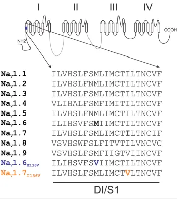
methionine 136 by valine (Met136Val) in transmembrane segment 1 of domain 1 (DI/S1). This residue is highly conserved in all human NaVs except for NaV1.8, where it is leucine (Figure 1). No other mutations in neuronal sodium channels NaV1.1-1.3 and NaV1.7 or CaV2.1 and 2.2 were found in this individual.
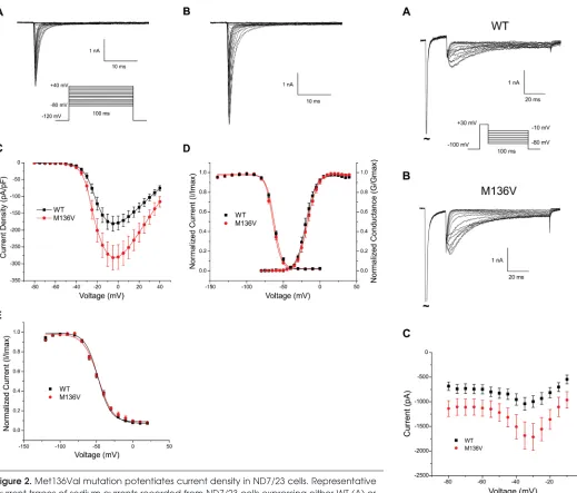
Voltage-Clamp Characterization of the Met136Val Channel
The location of the mutation and its conservation among sodium channels suggested that it can have an effect at the channel level. Sodium currents were recorded in the voltage-clamp mode from ND7/23 cells transiently transfected with either hNaV1.6R WT or Met136Val mutant channels. Figures 2A and B show represen-tative traces of sodium current from WT and Met136Val mutant channels, respec-tively. The peak current density at –5 mV (Figure 2C) was significantly increased for Met136Val (294 ± 26.5 pA/pF, n = 14) by 1.5-fold compared with WT channels (189.4 ± 11.3 pA/pF, n = 15, p = 0.03). Met136Val did not alter the voltage- dependence of activation (V1/2: WT, –19.1 ± 0.2 mV, k = 6.5 ± 0.2, n = 9; Met136Val, –16.8 ± 0.1 mV, k = 6.9 ± 0.1, n = 10), fast inactivation (V1/2: WT, –62.5 ± 0.5 mV, k = 5.3 ± 0.3, n = 12; Met136Val, –64.2 ± 0.8 mV, k = 5.3 ± 0.4, n = 12) (Figure 2D), or slow inactivation (V1/2: WT, –47.3 ± 1.0, k = 10.1 ± 0.7, n = 10; Met136Val, –48.0 ± 1.5, k = 8.8 ± 1.1, n = 9) (Figure 2E).
We examined the response of WT and Met136Val channels to slow depolariza-tions using a ramp stimulus from a hold-ing potential of –120 mV to +20 mV over 600 ms for a ramp rate of 0.2 mV/ms. The ramp current was normalized to the maxi-mal peak inward current recorded during the activation protocol for each cell. Peak ramp current of Met136Val measured 1.6 ± 1.3% (n = 6) of the peak current, and was not significantly different from WT (3.0 ± 2.2%, n = 6). Persistent current, measured 70 ms after the onset of a step depolarization to –10 mV and normalized to the peak inward current, also showed no significant difference (WT: 1.6 ± 0.2%, n = 15; Met136Val: 1.5 ± 0.3%, n = 14). the first action potential elicited by a
se-ries of depolarizing current injections (200 ms) that increased in 5 pA increments. Action potential frequency was deter-mined by depolarizing current injections (500 ms). Voltage threshold was deter-mined by assessing the first derivative of the membrane potential (dV/dt) and when the slope reached 20 mV/ms.
Data analysis
Data were analyzed offline with FitMaster (HEKA Electronik), Excel (Microsoft) and Origin 8.5 (Microcal Soft-ware) software and presented as mean ± standard error (SE). Statistical significance was determined by unpaired Student t test for voltage-clamp and current-clamp. Mann-Whitney test was used for compari-son of firing frequencies between WT and Met136Val in response to stimuli ranging from 25 to 500 pA. Two-proportion z-test was used for comparison of spontaneous activity. Fisher’s exact test was used for comparing high/low firing groups of cells expressing Met136Val and WT channels.
RESULTS
Clinical Phenotype and Identification of Met136Val Mutation
The subject was a 64-year-old Caucasian female of European heritage. She pre-sented with classical TN in the second branch of the nerve for more than one year, with both spontaneous and evoked pain paroxysms, and no family history of similar symptoms. A diagnosis mon-itoring committee consisting of three experts on TN (27) confirmed the pres-ence of NVC on the affected side by MRI and the diagnosis of TN. Whole-exome sequencing with good coverage (40X) of a set of genes encoding voltage-gated sodium channels (NaV1.1, 1.2, 1.3, 1.6 and 1.7) and calcium channels (CaV2.1 and 2.2) identified a single nucleotide substitution c.A406G (reference sequence NM_0141913) in the second coding exon of SCN8A, the gene that encodes NaV1.6. This mutation has not been previously reported and is absent from the 1000 Ge-nome database. This mutation substitutes of mNGF (Alomone Labs) and GDNF
(Peprotec), was then added and the cells maintained at 37°C in a humidified 5% CO2 incubator.
Current-clamp recordings in rat TRG neurons. Whole-cell current-clamp recordings were performed in transfected TRG neurons. The electrophysiologist was blinded to the sodium channel ex-pressed in the TRG neurons until data were pooled into WT and Met136Val groups. Recordings were acquired at 10 kHz with a low-pass Bessel filter and digitized at 50 kHz using a HEKA EPC10-double amplifier. The extra-cellular solution contained (in mM): 140 NaCl, 3 KCl, 2 MgCl2, 2 CaCl2, 10 HEPES, 10 dextrose; pH 7.3 with NaOH ( osmolarity adjusted to 320 mOsm w/sucrose). Patch pipettes (0.9–1.5 MΩ) were fabricated from borosilicate glass (1.65/1.1, OD/ID; World Precision In-struments) using a Sutter Instruments P-97 puller and fire polished. Pipette solution contained (in mM): 140 KCl, 0.5 EGTA, 5 HEPES, 3 Mg-ATP, 10 dex-trose; pH 7.30 with KOH (adjusted to 310 mOsm with sucrose). TTX was not used in the bath for current-clamp re-cordings. Pipette potential was zeroed before seal formation, and the voltages were not corrected for liquid junction potential.
n = 12). The average peak amplitude of the resurgent current was significantly larger (1.6X) in neurons transfected with Met136Val compared with WT channels (Met136Val: 1.8 ± 0.3 nA, n = 15; WT: 1.1 ± 0.1 nA, n = 12; p < 0.05) (Figure 3C). Adjusting for cell capacitance, the peak resurgent current density was signifi-cantly increased in Met136Val mutant channels (89.8 ± 11.5 pA/pF, n = 15;
p < 0.05) compared with NaV1.8-null TRG neurons transfected with WT channels (59.5 ± 6.1 pA/pF, n = 12).
Current-Clamp analysis of TRG neurons
To assess the effects of Met136Val on the excitability of adult TRG neurons, which express a full complement of sodium channels, current-clamp record-ings were performed on TRG neurons transfected with Met136Val or WT chan-nels. Input resistance was not signifi-cantly different among neurons express-ing Met136Val (649 ± 75 MΩ, n = 29) and WT channels (601 ± 93 MΩ, n = 29). The resting potential of TRG neurons expressing Met136Val channels (–56.7 ± 1.2 mV, n = 29) was not significantly dif-ferent from that of TRG neurons express-ing WT channels (–57.1 ± 0.8 mV, n = 29).
To assess the effects of the variant channels on the current threshold of TRG neurons, we injected a series of depolarizing current pulses to evoke the first all-or-none action potential. TRG neurons that spontaneously fired were excluded from the analysis of evoked data. The current threshold of TRG neurons expressing Met136Val channels was significantly reduced compared with neurons expressing WT channels (Met136Val: 88.4 ± 10.2 pA, n = 29; WT: 128.3 ± 15.6 mV, n = 29; p < 0.05).
Voltage thresholds were determined by measuring the voltage at which the slope of the voltage trace (dV/dt) increases sharply at 20 mV/ms. Voltage threshold in TRG neurons expressing Met136Val channels (–20.3 ± 1.5 mV, n = 29) was sim-ilar to neurons expressing WT channels (–19.2 ± 1.5 mV, n = 29). Action potential amplitude was measured as the difference from a holding potential of –100 mV,
followed by a range of repolarization pulses from –10 to –80 mV. Cells that showed slow rise and slow decay, typical of resurgent current and distinct from tail currents (25,28), were used for analysis.
Resurgent current was observed in 71% of NaV1.8-null TRG neurons trans-fected with Met136Val (15 of 21 cells), whereas 50% of neurons transfected with the WT channels (12 of 24 cells) showed resurgent currents. The range in peak amplitude of the resurgent current was larger in neurons transfected with Met136Val (0.7 to 4.8 nA, n = 15) com-pared with WT channels (0.3 to 2.0 nA, Resurgent Current of Met136Val
Channel in TRG neurons
NaV1.6 channels produce resurgent currents that facilitate repetitive firing of neurons, including DRG neurons (25, 28–30). To examine whether enhanced peak current density of NaV1.6 channels is paralleled by increased resurgent current in the native neuronal background, we expressed WT and Met136Val channels in NaV1.8-null TRG neurons. Representa-tive traces of resurgent sodium currents are shown from WT ( Figure 3A) and Met136Val mutant channels ( Figure 3B). Resurgent currents were elicited by briefly depolarizing to +30 mV for 20 ms
Figure 1. Sequence alignment of domain I transmembrane segment 1. Alignment of the sequence of DI/S1 from human NaV channels shows that Met136 is highly conserved in all
different from that in neurons expressing WT channels (112.4 ± 2.2 mV, n = 29). The half-width of action potentials evoked in TRG neurons expressing Met136Val channels (2.8 ± 0.2 ms, n = 29) was not between the resting potential and the
peak of the action potential. The action potential amplitude of TRG neurons expressing Met136Val channels (112.0 ± 2.5 mV, n = 29) was not significantly
Figure 2. Met136Val mutation potentiates current density in ND7/23 cells. Representative current traces of sodium currents recorded from ND7/23 cells expressing either WT (A) or Met136Val (B) mutant channels. Cells were held at –120 mV and inward sodium currents were elicited by step depolarizations from –80 mV to +20 mV in 5 mV increments every 5 s. (C) Peak current-voltage relationship for WT (n = 15) and Met136Val (n = 14) chan-nels showing significant increase in peak current density of M136V mutant chanchan-nels. (D) Comparison of voltage-dependent activation and steady-state fast inactivation of WT and Met136Val mutant channels. (E) Comparison of steady-state slow inactivation of WT and Met136Val mutant channels. Cells were held at –120 mV and a series of depolariz-ing pulses (30 s) from –120 mV to +20 mV in 10 mV increments was applied, followed by 20 ms return to –120 mV and then a 20 ms pulse to –10 mV to elicit a test response. Data represent mean ± SE.
Figure 3. Resurgent sodium current in-creased in NaV1.8-null TRG neurons
expressing Met136Val channels. Rep-resentative traces of resurgent currents recorded from NaV1.8-null TRG neurons
expressing either WT (A) or Met136Val (B) mutant channels. TRG neurons were held at –100 mV and depolarized to +30 mV for 20 ms, followed by repolarization to a range of pulses (–10 mV to –80 mV) to elicit resurgent current. (C) Average peak amplitudes of resurgent current recorded from WT (n = 12) and Met136Val (n = 15) show an average 60% increase in NaV
potential from each spontaneously firing cell was used to determine the voltage threshold. Although the voltage thresh-old of spontaneously firing cells was neurons expressing Met136Val or WT
channels fired spontaneously at com-parable frequencies of 4.2 ± 0.9 Hz and 4.2 ± 0.8 Hz, respectively. A single action significantly different from neurons
ex-pressing WT channels (WT: 3.0 ± 0.3 ms, n = 29).
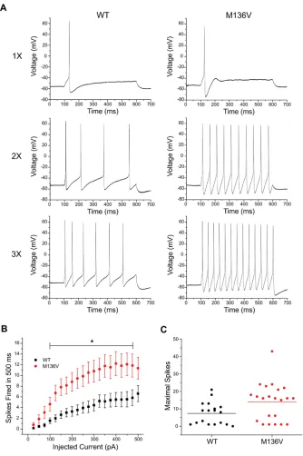
The effect of the mutation on the excitability of TRG neurons was also assessed by analyzing the number of action potentials evoked by a series of depolarizing current injections (25–500 pA) for 500 ms. TRG neurons expressing Met136Val channels fired a greater number of action potentials compared with WT channels in response to similar stimuli. Figure 4 shows rep-resentative responses of TRG neurons expressing WT or Met136Val channels evoked at one, two and three times the current threshold. Repetitive spike firing significantly increased in neurons ex-pressing Met136Val between 100 pA and 475 pA current injections compared with WT (Figure 4B, p < 0.05). Interestingly, a scatter plot of the number of maximal action potentials fired during the series of depolarizing current injections shows low- and high-firing groups for TRG neurons that express WT or Met136Val channels. Low-firing neurons (8 cells in each group) are comparable between the two groups, firing 1–6 APs. The majority of the high-firing neurons (10 cells) expressing WT channels fired 8–21 APs and only 2 neurons fired > 15 APs. In contrast, all high- firing neurons that expressed the Met136Val (14 cells) fired ≥ 15 APs. Using a threshold of 15 APs, there were significantly more high-firing cells in TRG neurons expressing Met-136Val channels (14/22) compared with WT channels (2/18, p = 0.001; Table 1).
Neurons that are transfected with NaV1.6 channels demonstrate a propen-sity for spontaneous firing. Spontaneous action potentials recorded for 30 s in a representative TRG neuron express-ing Met136Val channels is shown in Figure 5A, as well as an expansion of the time scale of the same trace illustrating a single action potential (Figure 5B). Spontaneous firing was observed in 41% and 48% of TRG neurons transfected with Met136Val and WT channels, re-spectively (Met136Val, 20/49 cells; WT, 27/56 cells, p = 0.40; Figure 5C). TRG
myelinated fibers of the central and peripheral nervous system (34–37). NaV1.6 is also expressed in small DRG neurons that produce unmyelinated C-fibers, and total loss of this channel in DRG neurons slows C-fiber conduc-tion in the sciatic nerve (38), suggesting that it contributes to the transmission of noxious signals in these fibers. We have previously shown that NaV1.6 is widely expressed in TRG neurons, giving rise to myelinated and unmyelinated fibers, and is localized in the nerve endings of unmyelinated fibers in the cornea (38), which are thought to be almost entirely nociceptive (39). Animal studies support a role for NaV1.6 in nerve injury models (40), toxin-induced pain (41), chemo-therapy-induced pain (42) and local DRG inflammatory pain (43,44). Thus, NaV1.6 is poised to play an important role in the pathophysiology of chronic pain, including in TN.
NaV1.6 channels manifest distinctive gating properties consistent with a critical role in regulating firing in the different neurons in which they are ex-pressed. NaV1.6 produces a rapidly rep-riming fast-inactivating TTX-S current (45–47) and a persistent current (47,48), which allow this channel to contribute to the overshoot of action potential in sensory neurons (49). Voltage-clamp re-cordings show that the Met136Val mu-tation significantly increases the peak current density. The increase in current density may have contributed to the reduction in current threshold for firing action potentials (~ 30%).
NaV1.6 produces a resurgent current in both CNS neurons and DRG neurons (28,29) as a result of channel reopening upon repolarization of the membrane (25). The resurgent current shortens refractory periods, thus supporting repetitive and burst firing of neurons (50), and has been implicated in human pain conditions caused by mutations in NaV1.7 (30,51,52) and in an animal model of radicular pain induced by local DRG inflammation (53). Our data show that Met136Val channels produce, on average, a 1.6-fold increase in the resurgent current in TRG neurons; determined to be similar between TRG
neurons expressing Met136Val or WT channels (Met136Val: –28.3 ± 1.2 mV, n = 20; WT: –28.7 ± 0.9 mV, n = 27), this was much lower than the voltage thresh-old measured for evoked action poten-tials by approximately –10 mV.
DISCUSSIOn
Genetic and functional evidence support a role for the SCN8A c.406A > G (p.Met136Val) mutation in TN. The mutation substitutes a highly conserved residue in DI/S1 of the channel, and has not been reported in the 1000 Genome database. The Met136Val substitution pro-duces a significant increase in peak tran-sient and resurgent currents of NaV1.6, reduces the threshold for action potential in TRG neurons, and enhances the neu-ronal evoked response and the fraction of neurons that fire at a higher rate than those expressing WT channels. Compared to other DI-S1 sodium channel mutations that have been linked to familial febrile seizures (31), erythromelalgia (32) and myotonia (33), the Met136Val mutation does not alter gating properties, which points to isoform-specific effects of muta-tions in this transmembrane segment. The increased peak transient and resurgent currents that confer hyperexcitability at the neuronal level combined with the rare occurrence of this mutation increase our confidence in concluding that Met136Val is a clinically significant variant that con-tributes to the pathophysiology of TN.
The paroxysmal nature of TN, the oc-currence of intense afterdischarges and favorable response to carbamazepine are consistent with a role for voltage-gated sodium channels. NaV1.6 channels are predominant at axon initial segments (AISs) and mature nodes of Ranvier in
Table 1. Proportion of low- and high-firing TRG neurons expressing Met136Val or WT channels.
hNaV1.6R hNaV1.6R-M136V
Low-firing 16 8
High-firing 2 14*
P value 0.001
collaboration of the Paralyzed Veterans of America with Yale University.
DISCLOSURE
VM and ST are employees of Convergence Pharmaceuticals, a Biogen company.
REFEREnCES
1. Cruccu G, Bonamico LH, Zakrzewska JM. (2010) Cranial neuralgias. Handb. Clin. Neurol. 97:663–78. 2. Devor M, Govrin-Lippmann R, Rappaport ZH.
(2002) Mechanism of trigeminal neuralgia: an ultrastructural analysis of trigeminal root speci-mens obtained during microvascular decompres-sion surgery. J. Neurosurg. 96:532–43.
3. Maarbjerg S, Wolfram F, Gozalov A, Olesen J, Bendtsen L. (2015) Significance of neurovascular contact in classical trigeminal neuralgia. Brain. 138:311–19.
4. Antonini G, et al. (2014) Magnetic resonance imaging contribution for diagnosing symptom-atic neurovascular contact in classical trigeminal neuralgia: a blinded case-control study and meta-analysis. Pain. 155:1464–71.
5. Lee A, McCartney S, Burbidge C, Raslan AM, Burchiel KJ. (2014) Trigeminal neuralgia occurs and recurs in the absence of neurovascular com-pression. J. Neurosurg. 120:1048–54.
6. Ko AL, et al. (2015) Trigeminal neuralgia without neurovascular compression presents earlier than trigeminal neuralgia with neurovascular compression. J. Neurosurg. 123:1519–27. 7. Bozkurt M, et al. (2012) Management of bilateral
trigeminal neuralgia with trigeminal radiofre-quency rhizotomy: a treatment strategy for the life-long disease. Acta. Neurochir. (Wien.) 154: 785–91; discussion 791–2.
8. Wang Y, Yu CY, Huang L, Riederer F, Ettlin D. (2011) Familial neuralgia of occipital and inter-medius nerves in a Chinese family. J. Headache
Pain. 12:497–500.
9. Riederer F, Sandor PS, Linnebank M, Ettlin DA. (2010) Familial occipital and nervus intermedius neu-ralgia in a Swiss family. J Headache Pain 11:335–8. 10. El Otmani H, Moutaouakil F, Fadel H, Slassi I.
(2008) [Familial trigeminal neuralgia]. Rev.
Neurol. (Paris) 164:384–7.
11. Rappaport ZH, Devor M. (1994) Trigeminal neuralgia: the role of self-sustaining discharge in the trigeminal ganglion. Pain. 56:127–38. 12. Devor M, Amir R, Rappaport ZH. (2002)
Patho-physiology of trigeminal neuralgia: the ignition hypothesis. Clin. J. Pain. 18:4–13.
13. Burchiel KJ, Baumann TK. (2004) Pathophysiology of trigeminal neuralgia: new evidence from a tri-geminal ganglion intraoperative microneurographic recording. Case report. J. Neurosurg. 101:872–3. 14. Dib-Hajj SD, Yang Y, Black JA, Waxman SG.
(2013) The NaV1.7 sodium channel: from
molecule to man. Nat. Rev. Neurosci. 14:49–62.
ignition theory of TN. According to igni-tion theory, TN is triggered by ectopic afterdischarges that are either spontaneous or low-threshold evoked phenomena leading to paroxysmal pain (11,12). An increase in peak transient and resurgent current could be linked to the evoked re-petitive firing, which would be consistent with the evoked pain phenomenon in TN. The high prevalence of spontaneous firing of TRG neurons overexpressing WT channels may have masked the real effect of mutant channels in rendering TRG neurons spontaneously firing when both WT and mutant channels were present at physiological levels. Addition-ally, we cannot formally discount the possible role of inflammatory mediator sensitization of primary afferents in TN due to NVC, which could lead to sponta-neous excitability of TRG neurons.
COnCLUSIOn
The genetic and functional data pre-sented in this study support a link of a gain-of-function mutation in NaV1.6 to human pain disorders, expand-ing the clinical-genetic spectrum of excitability disorders caused by this channel. Early-onset seizures and in-tellectual disabilities associated with human NaV1.6 mutations in infantile epileptic encephalopathy (64) may mask any pain phenotype in these in-dividuals. Whole-exome screening of a number of relevant ion channel genes and functional testing in relevant cell background increase confidence in the utility of this approach for discovering pathogenic mutations in pain disorders of unknown etiology.
aCknOWLEDGMEnTS
We thank the subject for participating in this study. We thank Lawrence Macala and Palak Shah for technical support. This work was supported by the Medical Research Service and Rehabilitation Re-search Service, Department of Veterans Affairs (SGW and SDH), and by funding to Yale University from Convergence Pharmaceuticals. The Center for Neuro-science and Regeneration Research is a the range of resurgent current amplitude
in cells expressing WT channels is 0.3– 2.4 nA, whereas resurgent currents in neu-rons that express the Met136Val channels range from 0.7–4.8 nA. We previously showed that roughly half of the small DRG neurons that are transfected with WT NaV1.6 channels produce resurgent currents (29). This is consistent with data showing that production of resurgent currents is cell-type-dependent (50), sug-gesting a cell-type-specific expression of an open channel blocker necessary for generating the resurgent current. The low-firing group of TRG neurons de-tected by the repetitive firing protocol possibly do not express an endogenous open channel blocker and do not pro-duce a resurgent current, but such a blocker is likely present in the high-firing group of TRG neurons that produce a resurgent current. The increase in re-surgent current of Met136Val channels provides the physiological basis for the enhanced evoked firing of TRG neurons that express these channels.
One possible mechanism that could link the mutation described here to TN involves ephaptic transmission, or electrotonic cross-talk between nearby axons, which has been proposed (54,55) as a mechanism contributing to hemi-facial spasm. There have been multiple demonstrations of ephaptic interactions that can lead to ectopic impulse activity, not only between closely apposed unin-jured axons (56) but also between inunin-jured axons (57,58) and between neighboring dysmyelinated axons (59). It has been suggested that demyelination due to compression is present in some cases of trigeminal neuralgia (2,60,61). It is well established that myelinated axons can produce large external electrical fields in regions where myelin is focally absent (62). Moreover, it has been demonstrated that increases in sodium channel density increase the likelihood of action potential generation in demyelinated axons (63).
and Nav1.7 sodium channels in mouse spinal sensory neurons. J .Physiol. (Lond.) 551:741–50. 46. Sangameswaran L, et al. (1997) A novel
tetrodo-toxin-sensitive, voltage-gated sodium channel expressed in rat and human dorsal root ganglia.
J. Biol. Chem. 272:14805–9.
47. Smith MR, Smith RD, Plummer NW, Meisler MH, Goldin AL. (1998) Functional analysis of the mouse Scn8a sodium channel. J. Neurosci. 18:6093–102.
48. Rush AM, Dib-Hajj SD, Waxman SG. (2005) Elec-trophysiological properties of two axonal sodium channels, Nav1.2 and Nav1.6, expressed in mouse spinal sensory neurons. J. Physiol. (Lond.) 564:803–15. 49. Rush AM, Cummins TR, Waxman SG. (2007)
Multiple sodium channels and their roles in electrogenesis within dorsal root ganglion neurons. J. Physiol. (Lond.) 579(Pt 1):1–14. 50. Lewis AH, Raman IM. (2014) Resurgent
cur-rent of voltage-gated Na(+) channels. J. Physiol. 592:4825–38.
51. Faber CG, et al. (2012a) Gain of function NaV1.7
mutations in idiopathic small fiber neuropathy.
Ann. Neurol. 71:26–39.
52. Han C, et al. (2012b) Functional profiles of SCN9A variants in dorsal root ganglion neurons and superior cervical ganglion neurons correlate with autonomic symptoms in small fibre neuropathy. Brain. 135:2613–28.
53. Xie W, et al. (2016) Upregulation of the sodium channel NaVbeta4 subunit and its contributions to mechanical hypersensitivity and neuronal hyperexcitability in a rat model of radicular pain induced by local DRG inflammation. Pain. 157:879–91.
54. Nielsen VK. (1984) Pathophysiology of hemifa-cial spasm: I. Ephaptic transmission and ectopic excitation. Neurology. 34:418–26.
55. Sanders DB. (1989) Ephaptic transmission in hemifacial spasm: a single-fiber EMG study.
Muscle Nerve. 12:690–4.
56. Ramon F, Moore JW. (1978) Ephaptic
transmission in squid giant axons. Am. J. Physiol. 234: C162–69.
57. Seltzer Z, Devor M. (1979) Ephaptic transmission in chronically damaged peripheral nerves.
Neurology. 29:1061–4.
58. Lisney SJ, Pover CM. (1983) Coupling between fibres involved in sensory nerve neuromata in cats. J. Neurol. Sci. 59:255–64.
59. Rasminsky M. (1980) Ephaptic transmission between single nerve fibres in the spinal nerve roots of dystrophic mice. J. Physiol. 305:151–69. 60. Love S, Hilton DA, Coakham HB. (1998) Central
demyelination of the Vth nerve root in trigeminal neuralgia associated with vascular compression.
Brain Pathol. 8:1–11; discussion 11–12. 61. Lagares A, Rivas JJ, Jimenez L, Cicuendez M,
Avendano C. (2010) Central demyelination in the pathogenesis of trigeminal neuralgia associated with cerebellopontine angle tumors: case report with ultrastructural trigeminal root analysis.
Neurosurgery. 66: E841–42. 31. Mantegazza M, et al. (2005) Identification of an
Nav1.1 sodium channel (SCN1A) loss-of-function mutation associated with familial simple febrile seizures. Proc. Natl. Acad. Sci. U.S.A. 102: 18177–182.
32. Cheng X, Dib-Hajj SD, Tyrrell L, Waxman SG. (2008) Mutation I136V alters electrophysiological properties of the NaV1.7 channel in a family with onset of erythromelalgia in the second decade.
Mol. Pain. 4:1.
33. Petitprez S, et al. (2008) A novel dominant mutation of the Nav1.4 alpha-subunit domain I leading to sodium channel myotonia. Neurology. 71:1669–75.
34. Caldwell JH, Schaller KL, Lasher RS, Peles E, Levinson SR. (2000) Sodium channel Nav1.6 is
localized at nodes of ranvier, dendrites, and syn-apses. Proc. Natl. Acad. Sci. U.S.A. 97:5616–20. 35. Schafer DP, Custer AW, Shrager P, Rasband MN.
(2006) Early events in node of Ranvier formation during myelination and remyelination in the PNS. Neuron Glia Biol. 2:69–79.
36. Black JA, et al. (1996) Spinal sensory neurons express multiple sodium channel alpha-subunit mRNAs. Mol. Brain Res. 43:117–31.
37. Dib-Hajj SD, Cummins TR, Black JA, Waxman SG. (2010) Sodium channels in normal and pathological pain. Annu. Rev. Neurosci. 33:325–47. 38. Black JA, Renganathan M, Waxman SG. (2002)
Sodium channel Nav1.6 is expressed along
nonmyelinated axons and it contributes to conduction. Mol. Brain Res. 105:19–28. 39. Chen X, Gallar J, Pozo MA, Baeza M,
Belmonte C. (1995) CO2 stimulation of the cor-nea: a comparison between human sensation and nerve activity in polymodal nociceptive afferents of the cat. Eur. J. Neurosci. 7:1154–63.
40. Henry MA, Freking AR, Johnson LR, Levinson SR. (2007) Sodium channel Nav1.6 accumulates at the site of infraorbital nerve injury. BMC
Neurosci. 8:56.
41. Deuis JR, et al. (2013) An animal model of oxaliplatin-induced cold allodynia reveals a cru-cial role for Na1.6 in peripheral pain pathways.
Pain. 154:1749–57.
42. Sittl R, et al. (2012) Anticancer drug oxaliplatin induces acute cooling-aggravated neuropathy via sodium channel subtype NaV1.6-resurgent and
persistent current. Proc. Natl. Acad. Sci. U.S.A. 109:6704–9.
43. Xie W, Strong JA, Ye L, Mao JX, Zhang JM. (2013) Knockdown of sodium channel Na1.6 blocks mechanical pain and abnormal bursting activity of afferent neurons in inflamed sensory ganglia. Pain. 154:1170–80.
44. Xie W, Strong JA, Zhang JM. (2015) Local knockdown of the Na1.6 sodium channel reduces pain behaviors, sensory neuron excitability, and sympathetic sprouting in rat models of neuro-pathic pain. Neuroscience. 291:317–30. 45. Herzog RI, Cummins TR, Ghassemi F, Dib-Hajj
SD, Waxman SG. (2003) Distinct repriming and closed-state inactivation kinetics of Nav1.6 15. Dib-Hajj SD, Waxman SG. (2014) Translational
pain research: Lessons from genetics and genomics. Sci. Transl. Med. 6:249sr244. 16. Dib-Hajj SD, Black JA, Waxman SG. (2015)
NaV1.9: a sodium channel linked to human pain.
Nat. Rev. Neurosci. 16:511–19.
17. Blanchard MG, et al. (2015) De novo gain-of- function and loss-of-function mutations of SCN8A in patients with intellectual disabilities and epilepsy. J. Med. Genet. 52:330–7
18. Estacion M, et al. (2014) A novel de novo mutation of SCN8A (Nav1.6) with enhanced channel activation in a child with epileptic encephalopathy. Neurobiol. Dis. 69:117–23. 19. Veeramah KR, et al. (2012) De Novo Pathogenic
SCN8A Mutation Identified by Whole-Genome Sequencing of a Family Quartet Affected by Infantile Epileptic Encephalopathy and SUDEP.
Am. J. Hum. Genet. 90:502–10.
20. Stirling LC, et al. (2005) Nociceptor-specific gene deletion using heterozygous NaV1.8-Cre
recombinase mice. Pain. 113:27–36.
21. Wittmack EK, et al. (2004) Fibroblast growth factor homologous factor 2B: association with Nav1.6 and selective colocalization at nodes of Ranvier of dorsal root axons. J. Neurosci. 24:6765–75. 22. Wittmack EK, Rush AM, Hudmon A,
Waxman SG, Dib-Hajj SD. (2005) Voltage-gated sodium channel Nav1.6 is modulated by p38 mitogen-activated protein kinase. J. Neurosci. 25:6621–30.
23. Gasser A, et al. (2010) Two Nedd4-binding motifs underlie modulation of sodium channel Nav1.6 by p38 MAPK. J. Biol. Chem. 285:26149–61. 24. Estacion M, et al. (2011) Intra- and interfamily
phenotypic diversity in pain syndromes associ-ated with a gain-of-function variant of NaV1.7.
Mol. Pain. 7:92.
25. Raman IM, Bean BP. (1997) Resurgent sodium current and action potential formation in disso-ciated cerebellar Purkinje neurons. J. Neurosci. 17:4517–26.
26. Dib-Hajj SD, et al. (2009) Transfection of rat or mouse neurons by biolistics or electroporation.
Nat. Protoc. 4:1118–26.
27. Zakrzewska JM, et al. (2013) Novel design for a phase IIa placebo-controlled, double-blind ran-domized withdrawal study to evaluate the safety and efficacy of CNV1014802 in patients with trigeminal neuralgia. Trials. 14:402.
28. Raman IM, Sprunger LK, Meisler MH, Bean BP. (1997) Altered subthreshold sodium currents and disrupted firing patterns in Purkinje neurons of Scn8a mutant mice. Neuron. 19:881–91. 29. Cummins TR, Dib-Hajj SD, Herzog RI,
Waxman SG. (2005) Nav1.6 channels generate resurgent sodium currents in spinal sensory neu-rons. FEBS Letters. 579:2166–70.
62. Waxman SG, Pappas GD, Bennett MV. (1972) Morphological correlates of functional differen-tiation of nodes of Ranvier along single fibers in the neurogenic electric organ of the knife fish Stern archus. J. Cell Biol. 53:210–24.
63. Waxman SG, Wood SL. (1984) Impulse conduc-tion in inhomogeneous axons: effects of variaconduc-tion in voltage-sensitive ionic conductances on inva-sion of demyelinated axon segments and preter-minal fibers. Brain Res. 294:111–22.
64. Wagnon JL, Meisler MH. (2015) Recurrent and Non-Recurrent Mutations of SCN8A in Epileptic Encephalopathy. Front Neurol. 6:104.