The enzymatic activity of IMPDH is composed of two separate isoforms, type 1 and 2 (2). The IMPDH2 isoform is associated with aggressive cancerous disease in experimental cancer (3–6), and related to poor survival in osteosarcoma patients (7). Mycophenolic acid (MPA) acts as a nonnucleoside, noncompetitive, reversible inhibitor of IMPDH with five-fold higher potency of inhibiting IMPDH2 than IMPDH1. It has been reported to be able to inhibit cancer cell proliferation and induce apoptosis in several experimen-tal models of human solid tumors and hematological malignancies by depleting guanine nucleotide pools (5,8–10).
In the last decade, interest into the importance of cell cycle velocity het-erogeneity of cancers has increased. Although it was initially thought that mass (1). This impressive proliferative
capacity is, however, dependent on adequate supply of nucleotides. Cellular nucleotide synthesis is biochemically complex, but requires various enzymes that can be targeted clinically, including inosine monophosphate dehydroge-nase (IMPDH), which is a rate-limiting enzyme in de novo synthesis of guanine. InTRODUCTIOn
Uncontrolled cell proliferation resulting from cell cycling deregulation is a hall-mark of cancer. Although aggressive can-cers are diverse and heterogeneous, they almost universally contain a fast-cycling compartment that can rapidly complete a cell cycle, and these cells are primarily responsible for the increase in tumor
Cancer Cells to Inosine Monophosphate Dehydrogenase 2
Inhibition by Mycophenolic Acid
Kan Chen,
1,2*Wanlu Cao,
1*Juan Li,
1Dave Sprengers,
1Pratika Y Hernanda,
3Xiangdong Kong,
2Luc JW van der Laan,
4Kwan Man,
5Jaap Kwekkeboom,
1Herold J Metselaar,
1Maikel P Peppelenbosch,
1and Qiuwei Pan
11Department of Gastroenterology and Hepatology, Erasmus MC Cancer Institute, Erasmus University Medical Center, Rotterdam,
The Netherlands; 2Bio-X Center, College of Life Sciences, Zhejiang Sci-Tech University, Hangzhou, China; 3Laboratory of Medical
Genetics, Biomolecular Research Center, Wijaya Kusuma University, Surabaya, Indonesia; 4Department of Surgery, Erasmus
University Medical Center, Rotterdam, The Netherlands; and 5Department of Surgery, Hong Kong University, Hong Kong, China
As uncontrolled cell proliferation requires nucleotide biosynthesis, inhibiting enzymes that mediate nucleotide biosynthesis constitutes a rational approach to the management of oncological diseases. In practice, however, results of this strategy are mixed and thus elucidation of the mechanisms by which cancer cells evade the effect of nucleotide biosynthesis restriction is urgently needed. Here we explored the notion that intrinsic differences in cancer cell cycle velocity are important in the resis-tance toward inhibition of inosine monophosphate dehydrogenase (IMPDH) by mycophenolic acid (MPA). In short-term experi-ments, MPA treatment of fast-growing cancer cells effectively elicited G0/G1 arrest and provoked apoptosis, thus inhibiting cell proliferation and colony formation. Forced expression of a mutated IMPDH2, lacking a binding site for MPA but retaining enzy-matic activity, resulted in complete resistance of cancer cells to MPA. In nude mice subcutaneously engrafted with HeLa cells, MPA moderately delayed tumor formation by inhibiting cell proliferation and inducing apoptosis. Importantly, we developed a lentiviral vector–based Tet-on label-retaining system that enables to identify, isolate and functionally characterize slow-cycling or so-called label-retaining cells (LRCs) in vitro and in vivo. We surprisingly found the presence of LRCs in fast-growing tumors. LRCs were superior in colony formation, tumor initiation and resistance to MPA as compared with fast-cycling cells. Thus, the slow-cycling compartment of cancer seems predominantly responsible for resistance to MPA.
Online address: http://www.molmed.org doi: 10.2119/molmed.2015.00126
*KC and WC contributed equally to this work.
and embedded in paraffin for subse-quent immunohistochemistry, while the remaining tumor tissue was dissociated with 5 mg/mL collagenase IV and 2 mg/mL DNase at 37°C for 30 min to obtain single cell suspension. Cells were further sorted as singlets for separation into non-LRCs (GFPlowdsRed) and LRCs
(GFPhighdsRed) by FACS sorter
(Supple-mentary Figure S2). non-LRCs and LRCs were injected subcutaneously (either 1,000 cells or 10,000 cells per injection as appro-priate) on four sites in the mice. At the same time, two populations of sorted cells were plated for colony-forming unit (CFU) assay (treated with or without MPA).
Colony Formation Assay
Cells were harvested and suspended in culture medium, yields were quan-tified through counting and plated in 6-well plates (500 cells/well), and then treated with serial dilutions of MPA (1, 2, 3, 4 and 5 μg/mL). The control group was supplemented with an equal volume of PBS. For the cells derived from xenograft tumor, cells were seeded into 12-well collagen coating plates and cultured in medium with or without MPA (10 μg/mL). Formed colonies were fixed by 70% ethanol and counterstained with hematoxylin and eosin after two weeks. Colony numbers were counted and their sizes were measured micro-scopically through digital image analysis.
MTT Assay
Cells were seeded in a 96-well plate at a concentration of 5 × 10³ cells/well
in 100 μL medium. Cells were incubated overnight to attach to the bottom of the wells, and then treated with serials dilu-tions of MPA (1, 5, 10, 15, 20, 25 and 30 μg/mL). Cell viability was analyzed by adding 5 mg/mL MTT (Sigma-Aldrich) and 150 μL DMSO. Absorbance was de-termined using Enzyme mark instrument at the wavelength of 490 nm.
Analysis of Cell Cycle
Cells (5 × 10⁵/well) were plated in
six-well plates and allowed to attach overnight, followed by an application Technologies). Pancreatic cancer cell
lines BxPC3 and PANC-1 were cultured with RMIP-1640. Both of the mediums were supplemented with 10% (v/v) fetal bovine serum (FBS) (Hyclone Technol-ogies), 100 units/mL of penicillin and 100 μg/mL of streptomycin. All the cells were incubated at 37°C in a humidified atmosphere containing 5% CO2. All the cell lines were confirmed mycoplasma free and their STR genotyping was analyzed at the Department of Pathology, Erasmus Medical Center Rotterdam (Supplementary Table S1).
Lentiviral Vector–Based Tet-On Label-Retaining System
Lentiviral backbone plasmids pLV. EX3D/EF1A-rtTA (M2)-dsRed-Express2 and pLV.EX2D/TRE-eGFP were used to pack third generation lentiviral vectors (Supplementary Figure S1A). HeLa cells were transduced with both vectors to generate a system (HeLa Tet-on) that can express a histone 2B-green fluores-cent fusion protein (histone-GFP) upon induction by doxycycline. GFP expres-sion in vitro was analyzed from wk 0 to wk 3 by flow cytometry analysis (FACS) and confocal microscope (Zeiss LSM 510) (Supplementary Figures S1B,C).
ZenlightEdition software was used to analyze confocal microscope images. Cells that maintained GFP expression over this period of time were identified as LRCs.
For identification of LRCs in vivo, female NOG mice at the age of 8 to 10 wks were purchased from Taconic Biosciences (Denmark). Animal experi-ments were performed with the approval of the institutional animal ethics com-mittee (Dier Experimenten Commissie, Erasmus MC). Mice were bred in a special pathogen free (SPF) environment during the whole experimental period. Mice were injected subcutaneously with 5 × 10⁶ HeLa Tet-on cells. After engraftment
(10 to 15 d), water containing 1 mg/mL doxycycline and 5% sucrose was given for 5 d. Mice were killed at different time points after withdrawal of doxycycline. A portion of harvested tumor samples was fixed with 4% paraformaldehyde cancer cells universally cycle and grow
faster than normal cells, recently a slow- cycling (largely quiescent) compartment, which does not divide frequently but has the capacity to generate progeny that can repopulate the fast cycling compartment, has been identified in many tumors, (11). Functionally, these slow-cycling cancer cells appear to be associated with the capacity to generate new metastases while having superior resistance to therapy (12). Technically, these slow- cycling cells are identified by their capacity to retain a pulse label as faster cycling cells lose the pulse label at cell division. Thus these cells are indicated with the term label- retaining cells (LRCs) (13).
In this study, we aim to develop a lentiviral vector–based Tet-on label- retaining system that allows us to iden-tify slow-cycling cancer cells in vivo, so that they can be subsequently isolated for functional characterization. We exploit this system to investigate the different sensitivity between fast- and slow- cycling cancer cells to IMPDH2 inhibition by MPA.
MATeRIALS AnD MeThODS
Reagents
Stocks of MPA (AMRESCO LLC) were dissolved in dimethyl sulfoxide (DMSO) (Sigma-Aldrich). Doxycycline, colla-genase IV and DNase were purchased from Sigma-Aldrich Corporation. Anti-bodies against IMPDH2, p-Histone3 and cleaved caspase-3 were purchased from Abcam Company, Millipore Corporation and Cell Signaling Technology, respec-tively.
Cell Culture
All supplementary materials are available online at www.molmed.org.
ReSULTS
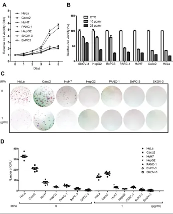
MPA Is Very effective for Inhibiting Cancer Cell Proliferation in Fast-Growing Cell Lines
A first indication as to how MPA affects cancer cells in relation to cell cycle velocity comes from experiments in which we investigated the effects of MPA on cell proliferation and the colony-forming potential of different cancer cell lines. To this end, seven different cancer cell lines derived from various tumor types were compared. Growth curves show substantial vari-ation in the prolifervari-ation rate and colony- forming potential of these cell lines, with, in general, HeLa and Caco2 showing more aggressive behavior as compared with the other cell lines (Figure 1). Challenge with MPA inhib-ited both cell proliferation and colony unit-forming potential of cancer cell lines, but, strikingly, fast-growing cell lines are more affected by MPA treat-ment as compared with slow-growing cell lines (Figure 1), indicating that MPA mainly affects the fast-cycling compartment.
MPA Inhibited Cell Proliferation, Arrested Cell Cycling and Induced Cell Apoptosis in Fast-Growing Cancer Cell Line
To further understand how MPA acts on fast-growing cancer cells, HeLa cells (the most sensitive cell line to MPA from our panel of cancer cell lines) were treated with clinically relevant MPA concentrations (14) and analyzed in more detail for the effects of MPA on cellular expansion, cell cycle and programmed cell death. MPA counteracted HeLa cell proliferation and colony-forming poten-tial in a time- and dose-dependent man-ner (Figure 2A). Indeed, even a relatively low concentration (1 μg/mL) of MPA already substantially impeded colony formation, whereas higher concentra-tions (2–5 μg/mL) completely inhibited harvested and macroscopically analyzed.
Tumor tissues were fixed with 4% para-formaldehyde and embedded in paraffin for evaluation by histology or immuno-histochemistry.
Immunohistochemistry
Paraffin-embedded tumor tissue slides were deparaffined in xylene, rehydrated in graded alcohols, and rinsed in PBS supplemented with 0.05% Tween 20. Slides were boiled in citrate acid buffer (pH 6.0) for 10 min to retrieve antigen. A 3% H2O2 treatment for 20 min at room temperature was used to block endo-genous peroxidase activity. The slides were incubated in 5% milk-containing blocking solution followed by overnight incubation with either a rabbit monoclo-nal antibody against IMPDH2, a rabbit polyclonal antibody against p-Histone H3 or a rabbit polyclonal antibody against cleaved caspase-3, used at a final dilution of 1:500, 1:1000 or 1:3000, respectively, and then counterstained with hematoxylin according to routine procedures. As a negative control, the primary antibody was omitted; positive controls were taken from other slides that had been successfully stained be-fore. IMPDH2, phospho-histone H3 and cleaved caspase-3 staining were scored by two independent expert observers. The numbers of mitotic cells and cleaved caspase-3-positive cells were counted in ten high-power fields. Median numbers of positive cells in each of the ten fields were calculated for each sample of the different groups using a semiquantitative assess-ment. Three categories were used to eval-uate the percentages of apoptotic cells: <10%, mild; 10% to 50%, moderate; >50%, high. The intensity of IMPDH2 staining was presented by categories: + weak; + + moderate; + + + strong.
Statistical Analysis
Statistical analysis was performed by using the nonparametric Mann–Whitney test for paired or non-paired data, or the paired t test using GraphPad InStat software as appropriate. A P value < 0.05 was considered statistically significant. of MPA at concentrations of 5, 10, 15, 20
and 25 μg/mL for 48 h. Vehicle control was performed through the addition of an equal volume of PBS. After 48 h, con-trol and treated cells were trypsinized and washed with PBS and then fixed in cold 70% ethanol overnight at 4°C. The cells were washed twice with PBS and incubated with 20 μg/mL RNase at 37°C for 30 min, and then with 50 μg/mL propidium iodide (PI) at 4°C for 30 min. The samples were analyzed immediately by FACS. Cell cycle was analyzed by ModFit LT 3.0 software.
Analysis of Cell Apoptosis
Cell apoptosis analysis was performed by staining cells with annexin V-FITC and PI. Cells (5 × 10⁵/well) were seeded
into six-well plates and incubated at 37°C in 5% CO2 overnight, then serial dilutions of MPA (5, 10, 15, 20 and 25 μg/mL) were added; whereas for vehicle control, an equal volume of PBS was used. After 48 h, all of the cells were trypsinized and resuspended in annexin- binding buffer and stained with Alexa Fluor 488 annexin V and PI at room temperature for 15 min. Detection of apoptosis was performed by FACS.
Xenograft Assays in nude Mice The xenograft tumor model was performed using nude mice in accor-dance with current prescribed guidelines and under a protocol approved by the Institutional Animal Care and Use Com-mittee of Hangzhou Normal University, China. Mice were bred in an SPF envi-ronment during the whole experimental period. Mice were all female and 4 to 6 wks of age at the time of inoculation, and were subcutaneously inoculated with 5 × 10⁶ of HeLa cells. After 20 h,
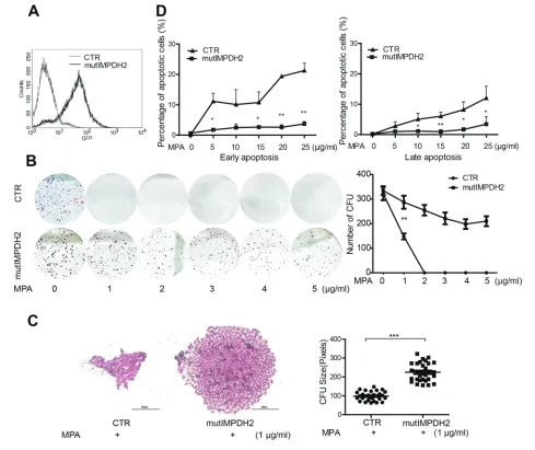
IMPDh2 Is a Relevant Target for MPA in Inhibiting Cancer Cell Growth
The clinical effects of MPA are presumed to be mediated through inhibition of IMPDH enzymatic activity and subsequent inhibition of de novo nucleotide biosynthesis. Although two isoforms of IMPDH exist, the type II isoform (IMPDH2) exhibits a five-fold higher sensitivity to inhibition by MPA as compared with the type I isoform (IMPDH1) (3). IMPDH2 is assumed to be the major target of MPA. Moreover, IMPDH2 is often upregulated in cancer (15), suggesting that IMPDH2 is the relevant target for MPA in the experi-ments described above. To substantiate this notion, we employed a lentiviral vector expressing an experimentally mutated IMPDH2 (mutIMPDH2) fused to GFP (16,17). The product of this con-struct has normal IMP dehydrogenase activity but lacks the binding site for MPA and thus confers MPA resistance. Transduction of this vector resulted in successful expression of this mutated allele in HeLa cells ( Figure 3A). In the CFU assay, forced expression of this mutated IMPDH2 provoked resistance to MPA treatment (Figures 3B,C). Further-more, the mutIMPDH2 cells prevented MPA- induced apoptosis (Figure 3D). These results are consistent with a key role of IMPDH2 in mediating the effects of MPA in our experimentation.
MPA Delayed Tumor Initiation, Inhibited Cancer Cell Proliferation
and Induced Cell Apoptosis In Vivo
Insight into the effects of MPA on tumor cell in vivo was obtained in ex-periments in which nude mice were used for subcutaneous engraftment of the HeLa cell line. Twenty h after in-oculation, mice were injected IP with MPA for 20 consecutive days. In this xenograft model, treatment of MPA (60 mg/kg body weight) significantly (P < 0.05) delayed tumor initiation ( Figure 4A). In the 240-mg/kg body weight of the MPA-treated group, one mouse failed to form a tumor; while tumor formation also tended to be P < 0.01) (Figure 2C). Furthermore,
MPA dose-dependently provoked the G0/G1-phase arrest (Figure 2D). In addition, MPA dose-dependently trig-gered both early and late cell apoptosis (Figure 2E). These data suggest that MPA profoundly interferes with the physi-ology of fast-growing cancer cells and raises questions as to how cancers can escape the effects of MPA.
colony formation. The result reports 322 ± 27 colonies/500 cells were formed in untreated group, but only 148 ± 27 colonies were formed in 1 μg/mL MPA-treated groups (mean ± SEM, n = 6, P < 0.01) (Figure 2B). Accordingly, the size of CFUs was significantly smaller in MPA-treated groups compared with untreated groups (96 ± 5 pixels versus 278 ± 8 pixels, mean ± SEM, n = 30,
existence of Slow-Cycling Cancer Cells Compartment in Fast-Growing Tumors
Substantial evidence indicated that slow-cycling cancer cells can evade therapeutic agents and repropagate the tumor (18). To identify whether there are slow-cycling cancer cells in our experi-mental system, we established a lentivi-ral-based Tet-on label-retaining system that enables us to isolate slow-cycling (P < 0.05) of proliferating cells assessed
by the percentage of p-histone H3 pos-itive cells (Figure 4D). Furthermore, MPA treatment provokes substantial apoptosis in the tumor cell compart-ment, as evidenced from the significant increase in the percentage of cleaved caspase-3-positive cells (Figure 4E). These results show that MPA coun-teracts tumor growth elicited by a fast-growing cancer cell in vivo. delayed in the other mice (Figure 4B).
Thus MPA counteracts growth of exper-imental tumors in this model.
Immunohistochemical staining of tumors harvested from these mice demonstrated significant downreg-ulation (P < 0.05) of IMPDH2 at the protein level following treatment with MPA (Figure 4C). Concomitantly, MPA inhibited tumor cell proliferation, as shown by a significant reduction
cells for further functional characterization. To this end, cells were transduced with two vectors. One vector expresses a reverse Tet transactivator (rtTA), dsRed fluorescent protein and a neomycin- resistance cassette. The other vector expresses a histone-GFP fusion protein driven by a tetracycline response element (TRE) and puromycin-resistance gene (Supplementary Figure S1A). Stable cell lines can be established by cotransducing with these two vectors and the clones can be selected either via drug resistance or by cell sorting based on fluorescence. This constitutes a genotoxic free and cell proliferation-independent approach to identify slow-cycling cells. Upon induction by doxycycline, all the cells are labeled with GFP. After doxycycline withdrawal, dividing cells lose their GFP signal, whereas quiescent or slow-cycling cells retain their GFP expression, which thus serves as a label for LRCs (Figure 5A and Supplementary Figure S1).
HeLa cells engineered with
histone-GFPTet-on were subcutaneously
engrafted in immunodeficient mice ( Figure 5B). Once a small tumor was formed, histone-GFP expression was induced by doxycycline in the drinking water of the animal. Following doxy-cycline withdrawal, mice were killed at different time points. As shown by both FACS and immunohistochemical staining, a small population of LRCs was detected in tumors (Figures 5C,D). Thus we con-cluded that even fast-growing tumors harbor LRC compartment.
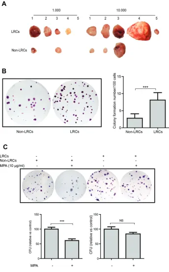
Slow-Cycling Cells Are Superior in Tumor Formation and Display More Resistance to MPA
To characterize LRCs functionally, LRCs and non-LRCs were isolated from HeLa cell-derived tumors using FACS sorter (Figure 5B and Supplementary Figure S3). Surprisingly, ex vivo CFU assay showed that LRCs were signifi-cantly more efficient in colony formation (non-LRCs: 2.8 ± 1.3 colonies/100 cells; LRCs: 8.1 ± 2.2, mean ± SEM, n = 19, P < 0.001) when compared with fast- cycling cells (Figure 6A). Consistently, Figure 6. Grafted label-retaining cells are superior in tumor initiation and more resistant
enzyme in the purine nucleotide synthe-sis pathway, we studied the effects of a modulating purine nucleotide pool on cell growth. Increasing the nucleotide concen-tration by supplementation of exogenous guanosine did not have a major effect on cell growth (Supplementary Figure S4). In addition, we found, surprisingly, that supplementation of exogenous guanosine counteracts the inhibitory effect of MPA only to a minor extent (Supplementary Figure S5). Although the antiproliferative effects of MPA are mainly dependent on targeting IMPDH2, depletion of nucleo-tides could only explain part of its mecha-nism of action.
Interestingly, a recent study has demonstrated a double functionality for IMPDH in Drosophila; in stress condi-tions, it can act as a transcription factor that inhibits cell proliferation (29). Thus, we speculate that IMPDH2 might be a ligand-regulated transcription factor and that MPA might act as a ligand. In-deed, IMPDH2 is located predominantly in cytoplasms in normal conditions of cultured HeLa cells, but efficiently translocated into the nucleus upon MPA treatment (Supplementary Figure S6A). Consistently, a mutated IMPDH2 lacking the binding site of MPA was not able to translate into the nucleus, even with MPA treatment (Supplementary Figure S6B). These results appear to support the pre-vious observation in Drosophila cells and our hypothesis. Furthermore, it is already known that MPA works as a ligand to activate the activity of peroxi-some proliferator-activated receptors, such as PPARγ (30), a critical nuclear receptor on adipocyte differentiation. However, gene silencing of PPARα and PPARγ did not affect the sensitivity of HeLa cells to MPA treatment, excluding their potential involvement (Supplemen-tary Figure S7). The scenario that MPA acts as a ligand for ligand-regulated tran-scription factors to regulate cancer cell growth is certainly interesting and there-fore deserves further investigation.
In summary, this study demonstrated that through inhibiting IMPDH2, MPA was capable of constraining the growth Classically, nucleotide analogs, such as
5-iodo-2’-deoxyuridine and bromode-oxyuridine, are used to identify LRCs. These agents can be used to identify LRCs, but the cells are not able to be isolated for functional study (22). Thus, fluorescent-coupled nucleotide analogs were developed for identification and isolation of LRCs. However, these agents are not competent for in vivo application (23). Although nucleotide analogs were also able to label cells in vivo, subsequent isolation of LRCs from the tissue is often a challenge that hampers further func-tional investigation (22). Another major drawback is that most of these labeling methodologies rely on cell division to label cells. Therefore, the real quiescent cells, in fact, cannot be labeled. Conse-quently, the introduction of modified nucleotides into cells profoundly alters the status of the cells (24). Integrating a lentiviral-based Tet-on cell-labeling sys-tem has circumstanced these limitations. Upon induction of the GFP fluorescent protein, all the cells can be labeled without genotoxic effect. It can be used for identification and isolation of slow- cycling cells both in vitro and in vivo. In this study, we used a histone-GFP fusion protein that localized in the cell nucleus with prolonged half-life (25). Thus, this technique bears broad implications for studying cell cycling. Indeed, we found the existence of a slow-cycling cancer cell compartment within the fast growing tumors formed by HeLa cells. These cells are superior in tumor initiation and more resistant to MPA. We speculate that the existence of slow-cycling cancer cells in different patient populations may affect the ultimate responsiveness of MMF/ MPA treatment (19–21).
IMPDH2 is upregulated in proliferating cells (26), including in various types of cancer cells (27,28), and exhibits a five-fold higher sensitivity to MPA compared with IMPDH1. Mechanistically, the effect of MPA appears through inhibition of its canonical target, IMPDH2. Ectopic expres-sion of a mutated IMPDH2 (mutIMPDH2) largely nullifies the antiproliferative effects of MPA. Since IMPDH is the key LRCs are more efficient in forming
tumors, both with regard to size and number, in immunodeficient mice upon subcutaneous engraftment (Figure 6B).
Subsequently, we evaluated the rela-tive sensitivity of LRCs and non-LRCs to MPA. Both populations were sorted and colony-forming potential was assessed in the presence or absence of MPA. Com-pared with the control groups, treatment with MPA (10 μg/mL) significantly inhibited the colony formation efficiency of non-LRCs but not of LRCs (where only minor effects were seen; Figure 6C). Thus, slow-cycling cancer cells when compared with fast-cycling cancer cells appear more resistant to MPA and may thus constitute the MPA-resilient reservoir in cancers.
DISCUSSIOn
IMPDH is a key enzyme in de novo guanine nucleotide biosynthesis and is thus a target for oncologic disease. MPA works as a potent IMPDH inhibitor that is used as an immunosuppressive drug (3). A phase I trial in patients with advanced multiple myeloma showed a positive correlation between clinical re-sponses and depletion of the intracellular deoxyguanosine triphosphate levels by mycophenolate mofetil (MMF), the prod-rug of MPA. MMF was administered up to a maximum dose of 5 g/day, which is two to three times higher than general use in organ transplantation patients, but was well tolerated in this study (19). In renal transplant patients, a tendency toward a lower risk of malignancy in MMF-treated patients versus non-MMF-treated has been reported in a large, prospectively conducted, observational cohort study (20). However, another clinical study in pancreatic cancer failed to show any beneficial effects (21). We found that only fast-cycling, but not slow-cycling, cancer cells are sensitive to the inhibitory effects of MPA. Thus, dis-secting the heterogeneity of cancer may help to understand the distinct respon-siveness to MPA treatment.
inosine monophosphate dehydrogenase 2 after HIV vector transduction: effects on lymphocytes, monocytes, and CD34+ stem cells. Mol. Ther. 14:236–44.
18. Moore N, Houghton J, Lyle S. (2012) Slow- cycling therapy-resistant cancer cells. Stem Cells Dev. 21:1822–30.
19. Takebe N, et al. (2004) Phase I clinical trial of the inosine monophosphate dehydrogenase inhibitor mycophenolate mofetil (cellcept) in advanced multiple myeloma patients. Clin. Cancer Res. 10:8301–8.
20. Robson R, Cecka JM, Opelz G, Budde M, Sacks S. (2005) Prospective registry-based observa-tional cohort study of the long-term risk of malignancies in renal transplant patients treated with mycophenolate mofetil. Am. J. Transplant. 5:2954–60.
21. Rodriguez-Pascual J, et al. (2013) A preclinical and clinical study of mycophenolate mofetil in pancreatic cancer. Invest New Drugs. 31:14–9. 22. Pan Q, et al. (2013) Identification of lineage- uncommitted, long-lived, label-retaining cells in healthy human esophagus and stomach, and in metaplastic esophagus. Gastroenterology. 144:761–70.
23. Xin HW, et al. (2013) Label-retaining liver cancer cells are relatively resistant to sorafenib. Gut. 62:1777–86.
24. Xin HW, et al. (2012) Tumor-initiating label- retaining cancer cells in human gastrointestinal cancers undergo asymmetric cell division. Stem Cells. 30:591–8.
25. Wang Y, et al. (2012) Identification of quiescent, stem-like cells in the distal female reproductive tract. PLoS One. 7:e40691.
26. Thomas EC, et al. (2012) Different characteristics and nucleotide binding properties of inosine monophosphate dehydrogenase (IMPDH) iso-forms. PLoS One. 7:e51096.
27. Hager PW, Collart FR, Huberman E, Mitchell BS. (1995) Recombinant human inosine monophos-phate dehydrogenase type I and type II proteins. Purification and characterization of inhibitor binding. Biochem. Pharmacol. 49:1323–9. 28. Zimmermann A, Gu JJ, Spychala J, Mitchell BS.
(1996) Inosine monophosphate dehydrogenase expression: transcriptional regulation of the type I and type II genes. Adv. Enzyme Regul. 36:75–84. 29. Kozhevnikova EN, et al. (2012) Metabolic enzyme
IMPDH is also a transcription factor regulated by cellular state. Mol. Cell. 47:133–9.
30. Makoto Ubukata, et al. (2007) Mycophenolic acid as a latent agonist of PPARr. Bioorg. Med. Chem. Lett. 17:4767–70.
Cite this article as: Chen K, et al. (2015) Differential sensitivities of fast- and slow-cycling cancer cells to inosine monophosphate dehydrogenase 2 inhibition by mycophenolic acid. Mol. Med. 21:792–802. ReFeRenCeS
1. Hanahan D, Weinberg RA. (2011) Hallmarks of cancer: the next generation. Cell. 144:646–74. 2. Natsumeda Y, et al. (1990) Two distinct cDNAs
for human IMP dehydrogenase. J. Biol. Chem. 265:5292–5.
3. Carr SF, Papp E, Wu JC, Natsumeda Y. (1993) Characterization of human type I and type II IMP dehydrogenases. J. Biol. Chem. 268:27286–90. 4. Moosavi MA, Yazdanparast R, Sanati MH,
Nejad AS. (2005) 3-Hydrogenkwadaphnin tar-gets inosine 5′-monophosphate dehydrogenase and triggers post-G1 arrest apoptosis in human leukemia cell lines. Int. J. Biochem. Cell. Biol. 37:2366–79.
5. Guidicelli G, et al. (2009) The necrotic signal induced by mycophenolic acid overcomes apoptosis-resistance in tumor cells. PLoS One. 4:e5493.
6. Fellenberg J, Kunz P, Sahr H, Depeweg D. (2010) Overexpression of inosine 5′-monophosphate dehydrogenase type II mediates chemoresistance to human osteosarcoma cells. PLoS One. 5:e12179. 7. Fellenberg J, Bernd L, Delling G, Witte D,
Zahlten-Hinguranage A. (2007) Prognostic significance of drug-regulated genes in high-grade osteosarcoma. Mod. Pathol. 20:1085–94. 8. Inai K, et al. (2000) Differentiation induction in
non-lymphocytic leukemia cells upon treatment with mycophenolate mofetil. Leuk. Res. 24:761–8. 9. Takebe N, et al. (2006) IMP dehydrogenase
inhibitor mycophenolate mofetil induces caspase- dependent apoptosis and cell cycle inhibition in multiple myeloma cells. Mol. Cancer Ther. 5:457–66.
10. Tressler RJ, Garvin LJ, Slate DL. (1994) Anti- tumor activity of mycophenolate mofetil against human and mouse tumors in vivo. Int. J. Cancer 57:568–73.
11. Govindasamy N, Murthy S, Ghanekar Y. (2014) Slow-cycling stem cells in hydra contribute to head regeneration. Biol. Open. 3:1236–44. 12. Schillert A, Trumpp A, Sprick MR. (2013) Label
retaining cells in cancer—the dormant root of evil? Cancer Lett. 341:73–9.
13. Morris RJ, Potten CS. (1994) Slowly cycling ( label-retaining) epidermal cells behave like clono-genic stem cells in vitro. Cell Prolif. 27:279–89. 14. Patel CG, Akhlaghi F. (2006) High-performance
liquid chromatography method for the deter-mination of mycophenolic acid and its acyl and phenol glucuronide metabolites in human plasma. Ther. Drug Monit. 28:116–22. 15. Chen K, et al. (2014) Rationale of personalized
immunosuppressive medication for hepatocellu-lar carcinoma patients after liver transplantation. Liver Transpl. 20:261–9.
16. Liang W, et al. (2012) Sirolimus-based immuno-suppression in liver transplantation for hepato-cellular carcinoma: a meta-analysis. Liver Transpl. 18:62–9.
17. Yam P, et al. (2006) Ex vivo selection and expan-sion of cells based on expresexpan-sion of a mutated of fast-cycling cancer cells. Using a
lentiviral Tet-on cell labeling technique, we identified slow-cycling cancer cells within the fast-growing tumors that are superior in tumor initiation, but more resistant to MPA. Thus, it is very necessary to de-velop regimens that can effective target slow-cycling cancer cells. Combining these regimens with agents targeting fast-cycling cancer cells, such as MPA, may be a viable option in cancer therapy.
COnCLUSIOn
Slow-cycling cancer cells within fast-growing tumors were identified. These cells, compared with fast-cycling cells, were superior in tumor initiation and resistant to IMPDH2 inhibition by MPA. Thus, simultaneous targeting of slow- and fast-cycling cells is necessary to eradicate cancer.
ACKnOWLeDGMenTS
The authors thank Lifeng Ni from the Animal Care at Hangzhou Normal University, Hangzhou, China, for help-ing with the animal experiments. We also thank Riccardo Fodde (Department of Pathology, Erasmus Medical Center Rotterdam, The Netherland) for providing plasmids pLV.EX3D/EF1A-rtTA (M2)-dsRed-Express2 and pLV.EX2D/TRE-eGFP. Our funding included the follow-ing: support from the Daniel den Hoed Foundation for Centennial Fellowship 2014, from the Netherlands Organization for Scientific Research (NWO/ZonMw) for a VENI grant (no. 916-13-032), and from the Dutch Digestive Foundation (MLDS) for a career development grant (no. CDG 1304) to Q Pan. Funding also came from the Zhejiang Provincial Top Key Discipline of Biology (no. 2014A09-C) to K Chen and from the National Nat-ural Science Foundation of China (no. 51272236) to X Kong.
DISCLOSURe