1
Variable Structural Networks at the Active Site of the SARS-CoV and
SARS-CoV2 Main Proteases
Navaneethakrishnan Krishnamoorthy1,2*
1Systems Biology, Sidra Medicine, Doha, Qatar. 2National Heart and Lung Institute, Imperial
College London, London, UK. *Correspondence: [email protected]
Abstract
The novel coronavirus SARS-CoV2 (CoV2) emerged in December 2019. This virus has 88%
genomic similarity with SARS-CoV (CoV), and both viruses largely depend on their main
protease (Mpro) to regulate infection. Mpro thus represents an attractive target for anti-SARS
drug design. The CoV and CoV2 Mpro are 97% identical at the sequence level, with 12 variable
residues, and their X-ray structures appear similar. We thus structurally analysed how these
variable residues affect the intra-molecular interactions between key residues in the CoV2 Mpro
active-site. Compared to CoV Mpro, the 12 divergent residues in CoV2 Mpro exhibit modified
intra-molecular interaction networks that ultimately restructure the molecular
micro-environment. These altered networks also indirectly affect the networks of other active-site
residues at the entrance (T26, M49 and Q192) and near the catalytic region (F140, H163, H164,
M165 and H172) of the Mpro. This suggest CoV2 indirectly (via neighbours) reshape key
molecular networks around the Mpro active-site. It seems that the CoV2 Mpro deceives us with
its apparent structurally identical to the CoV Mpro while this viral system accumulates mass
mutations (12 variable residues) at key positions. Some of these identified CoV2 Mpro networks
at the active-site might guide design of efficient CoV2 Mpro inhibitors.
Keywords: COVID-19; SARS-CoV2; SARS-CoV; variable residues; main protease; structural analysis
2 Introduction
In March 2020, the WHO declared that the outbreak of a novel coronavirus, SARS-CoV2
(CoV2), constituted a pandemic. This virus causes the transmissible disease, severe acute
respiratory syndrome (SARS) [1,2]. Although the source of this virus is still unknown, CoV2
shares 88% genomic similarity with SARS-CoV (CoV) that was identified in 2003 [3,4]. CoV
is highly dependent on the main protease (Mpro, or 3C-Like protease) for replicase polyprotein
processing. By proteolytic cleavage, the Mpro generates functional pp1a and pp1b replicases in
the host system that help to initiate and regulate infection [5]. Mpro is highly conserved among
the coronaviruses, including CoV2; due to its essential role in the viral life cycle, it is
considered as a major target for drug discovery [6-9]. Indeed, several studies have suggested
that inhibitors of the CoV Mpro active site might be repurposed to inhibit CoV2 Mpro [10-13].
The first X-ray structure of the CoV Mpro was released with modified N and C terminals soon
after the 2003 CoV outbreak [7]. Years later, the authentic wild type structure of CoV Mpro
(PDB ID: 2HOB. Fig. 1A) with an anti-coronavirus inhibitor (N3) was reported as a dimer
(protomer A and protomer B) and demonstrated the importance of the original N terminal,
mainly residue S1 (protomer B) [14] for Mpro activity and inhibitor binding. The CoV Mpro has
three functional domains I (1-101), II (102-184) and III (201-306) and a long loop (185-200)
that connects domains I and II (Fig. 1 A-B). The catalytic residues H41 and C145 [15] are
highly conserved among many of the SARS family proteins (MERS-Mpro, HKU5-Mpro,
HKU4-Mpro and SARS-Mpro) [9] and they are also conserved in CoV2 Mpro. The substrate binding
pockets including the active site (T25, T26, H41, M49, F140, N142, G143, S144, C145, H163,
H164, M165, E166, P168, H172, Q189, T190, A191, Q192) are located in-between the cleft
of domains I and II in the highly active wild type protease (PDB ID: 2HOB, [8,14]; these
domains are rich in beta-sheets. The dimeric form is reported to be functional due to key
3
and H163 (from protomer A) that serve to open and close the active site for ligand binding
([7,14,16-18]. Understanding the interactions between these functional residues in the new
CoV2 Mpro is essential.
In February 2020, the first structure of the CoV2 Mpro (PDB ID: 6LU7, unpublished) was
released. At high resolution, the CoV Mpro and CoV2 Mpro X-ray structures look very similar
with only a 0.5 Å structural deviation (Fig. 1C). While sequence alignment between the two
Mpro shows 97% identity (Fig. 1D), there are only 12 variable residues between them (Table 1).
Furthermore, the two Mpro structures accommodate the same ligand (N3) differently (Fig. 1C).
As molecular networks shape protein function, we analysed the impact of these 12 variable
residues on their intra-molecular networks and subsequent functional relevance. Because
functional studies are time consuming during this period of international emergency, we used
a structural systems biology approach to initiate the dissection of these networks.
Materials and Methods
The high-resolution dimeric (protomer A and B) X-ray 3D structures of the CoV Mpro and the
CoV2 Mpro were obtained from the protein data bank (PDB ID: 2HOB and 6LU7, respectively)
to compare the equivalent structures. The structure of CoV2 Mpro was released by the same
team [(Xue, (PDB, February 2020) unpublished] who released the highly active authentic wild
type CoV Mpro structure [14]. Pymol was used for structural analyses and to represent the
molecular structures (www.pymol.org). Sequence alignment was carried out with Clustal
Omega [19]. Dimplot in Ligplot with default parameters for hydrogen bonds and non-bonded
4 Results
The Mpro of CoV2 and CoV differ by 12 residues
A parallel sequence alignment of the CoV2 Mpro and the CoV Mpro confirmed 12 variable
residues at positions 35, 46, 65, 86, 88, 94, 134, 180, 202, 267, 285 and 286 (Fig. 1D and Table
1). The CoV2 Mpro X-ray structure of homodimer seemed to structurally mimic the CoV Mpro
structure in terms of possessing similar domains and a comparable active site (Fig. 1 A-B).
Most (8/12) of the variable residues were found in the Mpro-sheet-rich domains I and II, where
the inhibitor/catalytic site is located; the remaining four residues were found in domain III. By
contrast, the connecting loop possessed no variable residues.
Variable positions 46 and 65 are close to the bottle neck of the binding site (T25, T26, M49
and Q189), while variable positions 86, 88, 134 and 180 are close to the catalytic site. Variable
position 134 also seems critical based on the detection of many functionally important residues
[H172, E166, F140, S1 (B) and the oxyanion loop] in the vicinity. Notably, variable positions
46 and 134 are found in the loop that leads to the catalytic residues H41 and C145, respectively.
These data suggest that despite overall structural similarity with the CoV Mpro, 12 divergent
residues in thenovel CoV2 Mpro might affect the activity of the cartalytic domain.
The impact of the 12 variable CoV2 Mpro residues on neighbouring residue interactions
We next investigated the divergent interacting partners of the 12 variable residues. The
interacting partners and/or interactions of the variable residues differed between the two
proteases (Fig. 2 and Table 1). In CoV2 Mpro, variable position 46 is located near the entrance
of the binding site and shares the same loop with H41, however it is not interacting with M49
but this interaction is there in CoV Mpro. Variation at position 86 (near to the catalytic site) has
a high impact on protease interactions, as this residue manages 10 (CoV2 Mpro) and 12 (CoV
5
H164). Variation at position 134 results in a change from a positively charged (H134) to a
hydrophobic (F134) residue that ultimately expands the network of interacting partners in Cov2
Mpro (unique to F134: P108, F185).
The variable residues in domain III are located in the core region (202, 267) and likely have
roles in the dimer formation (285 and 286). The residue at position 202 in the CoV Mpro has
two unique partners, L250 and P293, and the total number of interactions decrease from nine
in CoV Mpro to seven in CoV2 Mpro. The residue at position 267 is involved in 10 (CoV Mpro)
and 11 (CoV2 Mpro) interactions (unique to CoV: F219 and CoV2: E270 and L220). In the
CoV2 Mpro, the residue at position 286 makes new connections with T280 (protomer B) and
G283 (protomer B), at the interface of the dimer.
Taken together, divergent interactions mediated by these 12 variable residues implies that they
are networking differently in the CoV2 Mpro compared to CoV Mpro. The consequent changes
in the nature of the amino acids (Table 1) at these variable positions might underlie these
alterations to the interaction networks.
The variable residues indirectly alter the interaction networks of the Mpro active site
To understand the consequences of the modified networks on the protease active sites, we
compared the networks established by residues comprising the active site (including the
entrance to the binding site region) between the CoV2 Mpro and CoVMpro (Table 2, Fig. 2 and
Fig. S1). At the entrance, T26 changes its role with its partner T21 (from forming a hydrogen
bond (in CoV Mpro) to forming a hydrophobic interaction (in CoV2 Mpro)). The M49 in CoV
Mpro, used P52 and A46 (variable residue) these interactions were not in CoV2 Mpro. Residue
Q192 at the entrance region forms two new hydrogen bonds with R188 and V186 as a result of
6
In the oxyanion loop, F140 is considered a functional regulator and (Xue et al., 2007); it forms
hydrogen bonds with S1 (B) in CoV Mpro and in CoV2 it makes a new link with S147. H163
interacts with C145 in CoV Mpro, but has lost a hydrogen bond with G146 in CoV2 Mpro. The
neighbouring residue H164 in CoV2 Mpro, has lost its interaction with L86 but gained an
interaction with G174. M165 lies adjacent to the key residue E166 that is necessary to open the
substrate binding site in CoV Mpro [14]. This residue shows two changes in interacting partners
in CoV2 compared to CoV, losing D187 and R188 and gaining F181 and F185. E166 still
makes its typical hydrogen bonds with S1 (B) and H172 in CoV2 Mpro as described in CoV
Mpro [14]. In addition, we identified a hydrogen bond between H172 (as it is one of the essential
regulators in the active site of CoV Mpro) and S1 (B) in CoV2 Mpro, which is not found in the
CoV Mpro. Altogether, a few of the key active site networks are indirectly modified between
CoV Mpro and CoV2 Mpro as a result of direct changes to the neighbouring networks of the 12
variable residues.
Discussion
Our structural analysis of CoV2 Mpro highlights that this new viral system not directly altering
any of the key residues E166, F140, H163, H172 and S1 (B) in the protease active site but
rather changing the neighbouring residues to modify their micro-environment and their
interaction networks (Table 2, Fig. 2 and Fig. S1). Why this indirect approach has been
favoured to alter these key networks at the active site is unclear; it might be to preserve the
original functional role of these residues (as observed in CoV Mpro) while simultaneously
modifying the way they function via their new networks. This concept now warrants detailed
7
outlook; however, specific variations conferred by just 12 variable residues that modify
interactions, especially in the protease’s active site region, are clear at the 3D structural level.
The interactions made by residues in the oxyanion loop (140-145) stabilize the S1 pocket to
control the conformational changes in their micro-environment that differ between the active
and inactive forms of the CoV Mpro [7,14,17]. In CoV2 Mpro, the residue at position 134 changes
from being positive (H134) in CoV Mpro to hydrophobic (F134); this change causes a
modification to its network and it is located on the loop that lead to the oxyanion loop, that
might ultimately serve to regulate the active site, as required. At the active site entrance,
residues M49 and Q189 are essential gatekeepers for substrate binding [13,14]. While A46
interacts with M49 in CoV Mpro, this interaction is lost in CoV2 Mpro (S46) and thus might
indirectly change the role of residues M49 and Q189. The other variable positions 86, 88 and
180 are also located near to the catalytic site and their modified networks might similarly confer
a potential change in their roles in CoV2 Mpro. The networks produced by residues at the
variable positions 285 and 286 in domain III are involved in dimer formation [18]. We now
need to understand how the resulting altered networks in CoV2 Mpro, especially around 286
(seven interactions in CoV Mpro vs. ten in CoV2 Mpro), impact the function of the protease.
Viral systems evolve rapidly at molecular level by mutations for their functional requirements
during the process of natural selection [21,22]. Here, in both the Mpro’s, the catalytic residue
H41 is sandwiched between variable positions 35 and 46 in the same super-secondary structure;
these variable positions have both modified their nature from CoV Mpro to CoV2 Mpro (polar to hydrophobic and hydrophobic to polar, respectively). H41 is conserved across all coronaviruses
[9], thus there could be a functional reason or requirement behind a structural selection or
8
The X-ray structure of CoV Mpro highlighted the role of residue S1 (from protomer B) in
stabilizing the active site by interacting with E166 (A) and F140 (A) and mediating inhibitor
binding [14,16]. In CoV2 Mpro, S1 (B) also seems to stabilize the active site, thus it is essential
to consider a dimeric structure for ligand design. Interestingly, S1 (B) in CoV2 Mpro forms a
unique hydrogen bond with H172: this new interaction might also contribute to its restructuring
process. Going forward, the functional consequences of the 12 variable regions should be
assessed at the conformational level and in terms of the regulation of protease activity.
Functional assays, X-ray analyses like those performed previously [14] and molecular
modelling approaches [17] are all warranted.
The CoV2 Mpro is one of the most targeted novel viral proteins for drug design following the
recent outbreak of COVID 2019. However, in CoV2 Mpro, the internal networks created by
variable residues around the active site compared to well-known CoV Mpro are not under the
focus. This basic study shows their structural networks and thus can open up avenues of
research in to developing effective targets for this novel virus. However, the functions of these
new networks remain to be determined for insight into the mechanism of CoV2 Mpro.
Conflict of Interest Statement: None to declare
Acknowledgement
The author would like to thank Dr. Damien Chaussabel, Director systems biology and
9 Figure 1
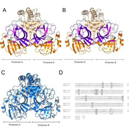
Figure 1. The X-ray structures and sequence alignment of the SARS-CoV Mpro and SARS-CoV2 Mpro. A) The dimeric structure of SARS-CoV Mpro. (B) The dimeric structure of SARS-CoV2 Mpro. (C) The overlapped structures of CoV Mpro (marine blue) and CoV2 Mpro
(grey). The structures coloured as follows (in A&B): orange: domain I (1-101), violet: domain
II (102-184), wheat: domain III (201-306), sky blue: connecting loop (185-200), green sticks:
catalytic residues H41 and C145, magenta: active site pocket and white sticks: inhibitor N3.
(D) Multiple sequence alignment of the CoV Mpros, where (*) indicates conserved residues and
10 Figure 2.
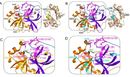
Figure 2: The variable positions and active site of the SARS-CoV Mpro and SARS-CoV2 Mpro. (A) The monomeric structure of SARS-CoV Mpro. (B) The monomeric structure of SARS-CoV2 Mpro. The boxed regions are magnified and showed in smooth-loop cartoons for
clarity in (C) and (D) with colour-matched labels to indicate the positions of the key residues
except S1 (protomer B) (removed for the clarity but indicated with symbol *S1(B)). Here,
colour codes are: yellow sticks: unique residues in CoV Mpro, cyan sticks: unique residues in
CoV2 Mpro, orange: domain I, violet: domain II, wheat: domain III, sky blue: connecting loop,
11 References
1. Huang C, Wang Y, Li X, Ren L, Zhao J, Hu Y, Zhang L, Fan G, Xu J, Gu X, et al. (2020)
Clinical features of patients infected with 2019 novel coronavirus in Wuhan, China. The Lancet
395: 497-506
2. Zhu N, Zhang D, Wang W, Li X, Yang B, Song J, Zhao X, Huang B, Shi W, Lu R, et al.
(2020) A Novel Coronavirus from Patients with Pneumonia in China, 2019. 382: 727-733 3. Ashour HM, Elkhatib WF, Rahman MM, Elshabrawy HA (2020) Insights into the Recent
2019 Novel Coronavirus (SARS-CoV-2) in Light of Past Human Coronavirus Outbreaks.
Pathogens (Basel, Switzerland)9:
4. Xu J, Zhao S, Teng T, Abdalla AE, Zhu W, Xie L, Wang Y, Guo X (2020) Systematic
Comparison of Two Animal-to-Human Transmitted Human Coronaviruses: SARS-CoV-2 and
SARS-CoV. Viruses12:
5. Snijder EJ, Bredenbeek PJ, Dobbe JC, Thiel V, Ziebuhr J, Poon LL, Guan Y, Rozanov M,
Spaan WJ, Gorbalenya AE (2003) Unique and conserved features of genome and proteome of
SARS-coronavirus, an early split-off from the coronavirus group 2 lineage. Journal of
molecular biology331: 991-1004
6. Anand K, Ziebuhr J, Wadhwani P, Mesters JR, Hilgenfeld R (2003) Coronavirus main
proteinase (3CLpro) structure: basis for design of anti-SARS drugs. Science (New York, N.Y.)
300: 1763-1767
7. Yang H, Yang M, Ding Y, Liu Y, Lou Z, Zhou Z, Sun L, Mo L, Ye S, Pang H, et al. (2003)
The crystal structures of severe acute respiratory syndrome virus main protease and its complex
with an inhibitor. Proceedings of the National Academy of Sciences of the United States of
12
8. Yang H, Xie W, Xue X, Yang K, Ma J, Liang W, Zhao Q, Zhou Z, Pei D, Ziebuhr J, et al.
(2005) Design of wide-spectrum inhibitors targeting coronavirus main proteases. PLoS biology
3: e324
9. Tomar S, Johnston ML, St John SE, Osswald HL, Nyalapatla PR, Paul LN, Ghosh AK,
Denison MR, Mesecar AD (2015) Ligand-induced Dimerization of Middle East Respiratory
Syndrome (MERS) Coronavirus nsp5 Protease (3CLpro): IMPLICATIONS FOR nsp5
REGULATION AND THE DEVELOPMENT OF ANTIVIRALS. The Journal of biological
chemistry290: 19403-19422
10. Liu C, Zhou Q, Li Y, Garner LV, Watkins SP, Carter LJ, Smoot J, Gregg AC, Daniels AD,
Jervey S, et al. (2020) Research and Development on Therapeutic Agents and Vaccines for
COVID-19 and Related Human Coronavirus Diseases. ACS Central Science,
10.1021/acscentsci.0c00272
11. Chen Y, Yiu C, Wong K (2020) Prediction of the SARS-CoV-2 (2019-nCoV) 3C-like
protease (3CLpro) structure: virtual screening reveals velpatasvir, ledipasvir, and other drug
repurposing candidates [version 1; peer review: 2 approved]. 9:
12. Xu Z, Peng C, Shi Y, Zhu Z, Mu K, Wang X, Zhu W (2020) Nelfinavir was predicted to
be a potential inhibitor of 2019-nCov main protease by an integrative approach combining
homology modelling, molecular docking and binding free energy calculation.
10.1101/2020.01.27.921627 %J bioRxiv2020.2001.2027.921627
13. Ton A-T, Gentile F, Hsing M, Ban F, Cherkasov A (2020) Rapid Identification of Potential
Inhibitors of SARS-CoV-2 Main Protease by Deep Docking of 1.3 Billion Compounds. n/a: 14. Xue X, Yang H, Shen W, Zhao Q, Li J, Yang K, Chen C, Jin Y, Bartlam M, Rao Z (2007)
Production of authentic SARS-CoV M(pro) with enhanced activity: application as a novel
13
15. Ziebuhr J, Snijder EJ, Gorbalenya AE (2000) Virus-encoded proteinases and proteolytic
processing in the Nidovirales. The Journal of general virology81: 853-879
16. Shi J, Sivaraman J, Song J (2008) Mechanism for controlling the dimer-monomer switch
and coupling dimerization to catalysis of the severe acute respiratory syndrome coronavirus
3C-like protease. Journal of virology82: 4620-4629
17. Tan J, Verschueren KH, Anand K, Shen J, Yang M, Xu Y, Rao Z, Bigalke J, Heisen B,
Mesters JR, et al. (2005) pH-dependent conformational flexibility of the SARS-CoV main
proteinase (M(pro)) dimer: molecular dynamics simulations and multiple X-ray structure
analyses. Journal of molecular biology354: 25-40
18. Lim L, Shi J, Mu Y, Song J (2014) Dynamically-driven enhancement of the catalytic
machinery of the SARS 3C-like protease by the S284-T285-I286/A mutations on the extra
domain. PloS one9: e101941
19. Sievers F, Higgins DG (2014) Clustal Omega, accurate alignment of very large numbers of
sequences. Methods in molecular biology (Clifton, N.J.)1079: 105-116
20. Laskowski RA, Swindells MB (2011) LigPlot+: multiple ligand-protein interaction
diagrams for drug discovery. Journal of chemical information and modeling51: 2778-2786 21. Forni D, Cagliani R, Clerici M, Sironi M (2017) Molecular Evolution of Human
Coronavirus Genomes. Trends in microbiology25: 35-48
22. Dolan PT, Whitfield ZJ, Andino R (2018) Mechanisms and Concepts in RNA Virus
14
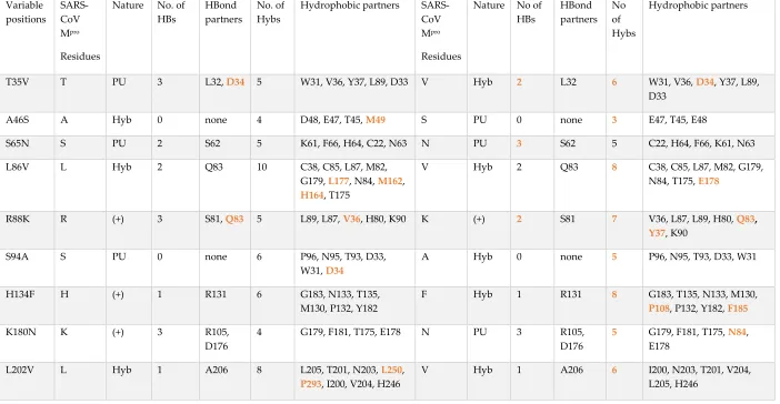
Table 1. The networks of the 12 variable residues in the SARS-CoV Mpro and SARS-CoV2 Mpro
Variable positions SARS-CoV Mpro Residues
Nature No. of
HBs
HBond partners
No. of Hybs
Hydrophobic partners
SARS-CoV
Mpro
Residues
Nature No of HBs HBond partners No of Hybs Hydrophobic partners
T35V T PU 3 L32, D34 5 W31, V36, Y37, L89, D33 V Hyb 2 L32 6 W31, V36, D34, Y37, L89,
D33
A46S A Hyb 0 none 4 D48, E47, T45, M49 S PU 0 none 3 E47, T45, E48
S65N S PU 2 S62 5 K61, F66, H64, C22, N63 N PU 3 S62 5 C22, H64, F66, K61, N63
L86V L Hyb 2 Q83 10 C38, C85, L87, M82,
G179, L177, N84, M162,
H164, T175
V Hyb 2 Q83 8 C38, C85, L87, M82, G179,
N84, T175, E178
R88K R (+) 3 S81, Q83 5 L89, L87, V36, H80, K90 K (+) 2 S81 7 V36, L87, L89, H80, Q83,
Y37, K90
S94A S PU 0 none 6 P96, N95, T93, D33,
W31, D34
A Hyb 0 none 5 P96, N95, T93, D33, W31
H134F H (+) 1 R131 6 G183, N133, T135,
M130, P132, Y182
F Hyb 1 R131 8 G183, T135, N133, M130,
P108, P132, Y182, F185
K180N K (+) 3 R105,
D176
4 G179, F181, T175, E178 N PU 3 R105,
D176
5 G179, F181, T175, N84,
E178
L202V L Hyb 1 A206 8 L205, T201, N203, L250,
P293, I200, V204, H246
V Hyb 1 A206 6 I200, N203, T201, V204,
15
A267S A Hyb 3 L271,
E270, D263
7 A266, L268, M264, F219,
N221, C265, K269
S PU 3 F219,
L271, D263
8 A266, L268, M264, E270,
N221, L220, C265, K269
T285A T PU 0 5 T280, S284, I286, M276,
I286 (B)
A Hyb 0 none 5 S284, L286, M276, A285
(B), L286 (B)
I286L I Hyb 2 S284 5 W207, L287, T285, E288,
T285 (B)
L Hyb 2 S284 8 M276, L287, A285, E288,
W207, T280 (B), G283 (B),
A285 (B)
Keys: HBs: hydrogen bonds, Hybs: hydrophobic interactions, Hyb: hydrophobic, PU: polar and un-charged, (+) positively charged and Bold
16
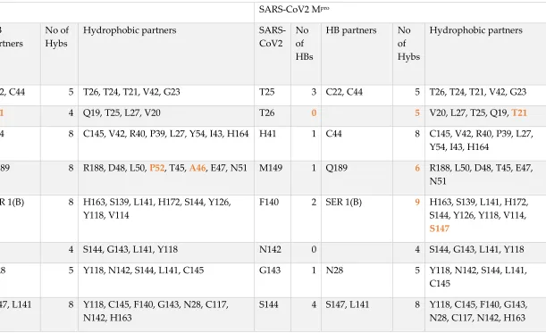
Table 2. The networks at the active site of the SARS-CoV Mpro and SARS-CoV2 Mpro
SARS-CoV Mpro SARS-CoV2 Mpro
SARS-CoV No of HBs HB partners No of Hybs
Hydrophobic partners
SARS-CoV2 No of HBs
HB partners No
of Hybs
Hydrophobic partners
T25 3 C22, C44 5 T26, T24, T21, V42, G23 T25 3 C22, C44 5 T26, T24, T21, V42, G23
T26 1 T21 4 Q19, T25, L27, V20 T26 0 5 V20, L27, T25, Q19, T21
H41 1 C44 8 C145, V42, R40, P39, L27, Y54, I43, H164 H41 1 C44 8 C145, V42, R40, P39, L27,
Y54, I43, H164
M49 1 Q189 8 R188, D48, L50, P52, T45, A46, E47, N51 M149 1 Q189 6 R188, L50, D48, T45, E47,
N51
F140 2 SER 1(B) 8 H163, S139, L141, H172, S144, Y126,
Y118, V114
F140 2 SER 1(B) 9 H163, S139, L141, H172,
S144, Y126, Y118, V114,
S147
N142 0 4 S144, G143, L141, Y118 N142 0 4 S144, G143, L141, Y118
G143 1 N28 5 Y118, N142, S144, L141, C145 G143 1 N28 5 Y118, N142, S144, L141,
C145
S144 4 S147, L141 8 Y118, C145, F140, G143, N28, C117,
N142, H163
S144 4 S147, L141 8 Y118, C145, F140, G143,
17
C145 3 H164, N28 7 G146, S144, H41, H163, L27, G143, S147 C145 3 H164, N28 7 G146, S144, H41, H163,
L27, G143, S147
H163 3 S147, Y161,
G146
8 H164, F140, M162, C145, M165, H172, P39, S144
H163 2 Y161, S147 8 H164, F140, M162, C145,
M165, H172, P39, S144
H164 3 C145,
T175, A173
8 H163, M165, P39, L86, H41, C85, M162,
F181
H164 3 C145, T175,
A173
8 H163, M165, P39, H41,
C85, M162, F181, G174
M165 2 A173 7 H163, E166, H164, H172, V186, D187,
R188
M165 2 A173 7 H163, E166, H164, H172,
V186, F181,F185
E166 2 S 1(B),
H172
3 M165, L167, V171 E166 2 S 1(B), H172 3 M165, L167, V171
P168 0 4 G170, T169, L167, Q192 P168 0 4 G170, T169, L167, Q192
H172 4 G138, I136,
E166
8 V171, F140, A173, T135, M165, H163,
G170, S1(B)
H172 5 G138, I136,
E166, S1(B)
7 V171, F140, A173, T135,
M165, H163, G170,
Q189 1 M149 3 L50, T190, R188 Q189 1 M149 3 L50, T190, R188
T190 1 R188 4 Q192, Q189, A191, L50 T190 1 R188 4 Q192, Q189, A191, L50
A191 0 2 T190, Q192 A191 0 2 T190, Q192
Q192 0 6 V186, A193, A191, F185, T190, P168 Q192 3 R188, V186 5 A193, A191, F185, T190,
P168
Keys: HBs: hydrogen bonds, Hybs: hydrophobic interactions, Hyb: hydrophobic and Bold fonts: variables partners or interactions in the