Tibial eminence fractures occur as a result of high amounts of tension placed upon the anterior cruciate ligament (ACL). The incidence of these fractures is higher among adolescent girls due to their inherent skeletal immaturity. In such an injury, direct trauma causes an avulsion fracture occurring at the tibial eminence while the ACL is spared. Imaging is used to confirm the diagnosis of a tibial eminence fracture and regardless of the extent of injury, rehabilitation is crucial for a full recovery. The following is a case study of a 17-year-old girl who was involved in a motor vehicle accident. In the accident, she sustained a left lateral tibial eminence fracture, along with soft tissue injuries at the cervical and lumbar spine. Her treatment included passive and active range of motion (ROM), strength training, physical modalities, and proprioceptive training of the injured areas. An improvement was noted post-treatment and after a 5-month follow-up according to subjective reports and objective assessments (ROM and girth measurements).
(JCCA 2007; 51(2):99–105)
k e y wo r d s: tibial eminence, fracture, rehabilitation.
Introduction
Although tibial eminence fractures can occur at any age, most occur between the ages of 8 and 14 years.1
Typical-ly, tibial eminence fractures are associated with falls from
bicycles, where an excessive force is exerted on the ACL causing a bony avulsion instead of an acute ACL tear.2
The ACL inserts into the anterior attachment of the later-al and medilater-al menisci in the recess anterior to the medilater-al
* Simply Align Rehabilitation, 4129 Lawrence Avenue, East, Scarborough, Ontario M1E 2S2. Tel: 416-438-3230 email: [email protected]
© JCCA 2007.
Une fracture de l’éminence du tibia se produit quand une tension excessive est placée sur le ligament croisé antérieur du genou. L’incidence de ces fractures est plus fréquente chez les jeunes adolescentes en raison de l’immaturité inhérente du squelette. Lors d’une telle blessure, un trauma direct cause une avulsion-fracture qui se loge dans l’éminence du tibia alors que le ligament croisé antérieur du genou est épargné. On utilise l’imagerie médicale pour confirmer le diagnostic de la fracture de l’éminence du tibia et, peu importe la gravité de la blessure, la réadaptation est essentielle pour un rétablissement complet. Voilà une étude de cas d’une jeune fille de 17 ans impliquée dans un accident de motocyclette. Dans l’accident, elle a subi une fracture de l’éminence du tibia du côté gauche, en plus de
meurtrissures aux tissus mous au niveau cervical et lombaire. Son traitement a consisté en amplitude de mouvements passifs et actifs, l’entraînement musculaire, la modalité physique, un entraînement proprioceptif des parties blessées. On a observé une amélioration après les traitements et après un suivi à cinq mois selon les rapports subjectifs et les évaluations objectives (amplitudes de mouvement et mesures des circonférences).
(JACC 2007; 51(2):99–105)
tibial spine, also known as the anterior intercondylar area. The anterior attachment of the lateral meniscus is located posteriorly to the ACL insertion1 (Figure 1).
One of the most widely used classification methods for a tibial eminence fracture is Meyers and McKeever’s cat-egorization, which delineates different displacement lev-els of avulsion as well as different management strategies (Figure 2).
Type I is the minimal displacement of an avulsed frag-ment. This type of injury is treated conservatively by closed reduction, where the tibia is set into place without a surgical incision. The knee is then immobilized in a long-leg cast or a fracture brace set at 10 to 20 degrees of knee flexion. Fixed flexion is recommended due to the fact that full extension may place excessive tension on the ACL and popliteal artery.1 Immobilization is then
recommended for approximately 6 weeks depending on the age of the patient, healing rate, and radiological find-ings.1 Type II classification is the displacement of half or
the anterior third of the ACL insertion causing a posteri-or hinge. Type III is the complete separation of the avul-sion site. Type II and III can be treated by closed reduction or an open reduction. In an open reduction in-cisions are made and wires, pins or screws are used to help immobilize the avulsion site. Orthopedic testing such as positive Lachman’s and anterior Drawer test, in-dicating ACL instability,3 along with appropriate
imag-ing confirm the diagnosis and success of reduction. Imaging typically entails a full set of knee radiographs including anterior-posterior and lateral views as well as magnetic resonance imaging (MRI) confirming the diag-nosis, and proper reduction.1 The selection of
appropri-ate imaging is important as partial ACL ruptures with
associated increases in laxity are common sequelae of tibial eminence fractures.1,4
Tibial eminence fractures have excellent prognoses.1
Prolonged immobilization may lead however, to arthrofi-brosis and a permanent loss of full extension.1 Formal
re-habilitation is crucial as it encourages a faster recovery and prevents the development of secondary complica-tions.1 During immobilization, the patient is given
axil-lary crutches along with strict rules in regards to their weight bearing status. ROM, and strengthening of quadri-ceps and hamstrings and proprioceptive training are uti-lized for rehabilitation of the knee post immobilization.
Case study
History
Written consent was obtained from this patient to report the following findings. A 17-year-old female student am-bulating across a street was struck by a motor vehicle ac-cident. The precise mechanism of injury is unknown due to the patient’s loss of consciousness upon impact. The patient was then sent to a nearby hospital. While in the hospital, radiographs of her left knee revealed a Type I tibial eminence fracture. The knee specialist performed a closed reduction procedure and placed the patient’s left lower extremity into a long-leg cast. The orthopedic sur-geon also recommended weight bearing as tolerated (WBAT), ROM and strengthening exercises for the in-jured areas. The patient was then referred to a chiroprac-tic rehabilitation clinic ten days after her accident by her
Figure 1 Dashed area depicts avulsion fracture of ACL. LM, lateral meniscus; MM, medial meniscus; PCL, cut posterior
cruciate ligament. Figure 2 Meyers and McKeever classification of tibial
instructed by her medical doctor to take Advil if her pain increased. Although the purpose of this paper is related to her tibial eminence fracture, minor references are made in regards to the patient’s cervical and lumbar pain. This is done since at the time of injury, the patient’s lumbar pain was the chief complaint.
Findings
During the examination, the patient’s left knee was in a long-leg cast. She was ambulating WBAT, with axillary crutches. Her cervical spine ROM was restricted in later-al bending and rotation by five degrees.5 The patient had
decreased external rotation of both shoulders with associ-ated pain. According to the orthopedic examination of the patient possible soft tissue injuries were revealed for the cervical and lumbar regions.3 Neurological testing with
respect to dermatomes, myotomes, and deep tendon re-flexes were unremarkable.
Treatment
The patient was treated initially for her lumbar and cervi-cal soft tissue injuries. As her knee was in a long-leg cast, lower extremity exercises could not be performed and were delayed. Six weeks later, the cast was removed and the patient began ambulation training with crutches. Her status in this phase of rehab was WBAT. Measurements of the patient’s thigh and calf musculature after the re-moval of the cast revealed a 2 cm reduction of diameter on her left leg compared to the right. Left knee flexion was measured at 105/135 degrees.6 Along with
ambula-tion training, her treatment regimen included the use of physical modalities such as TENS and heat for pain re-duction,7,8 as well as PROM exercises and myofascial
re-lease to her knee. Myofascial rere-lease was performed on the quadriceps, hamstring, adductor muscles and IT band of the injured knee. To resolve soft tissue adhesions a heat pack was applied for 15 minutes followed by manual myofascial release of restricted areas identified during
left knee pain following the removal of the cast. For this reason, manipulation, ROM exercises, deep neck flexor and core stability exercises were utilized throughout the treatment duration.9,10 Her treatment frequency was
in-creased to three times a week. As the rehabilitation of tib-ial eminence fractures follow similarly to ACL protocols,11 other activities such as bicycling, leg presses,
elastic tubing exercises, and jogging were also indicat-ed.12 Passive knee flexion and extension with
mobiliza-tions were utilized to increase knee ROM.13 Initially
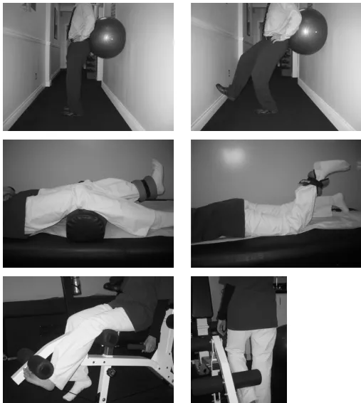
closed kinetic chain exercises, such as wall squat and sin-gle leg wall squats, with the knee in sub-terminal exten-sion were explored (Figure 3). Quadriceps, hamstring and calf raise exercises were introduced later in the exer-cise program to stabilize and strengthen the knee.13,14
Quadriceps and hamstring exercises were performed with a 5 lb weight in a lying position initially, progressing to a 10 lb weight in a standing position (Figure 3).
A stationary bike was used as part of her rehabilita-tion.12 Recent studies have shown the importance of
pro-prioceptive training in an ACL rehabilitation.11,12,13,15 To
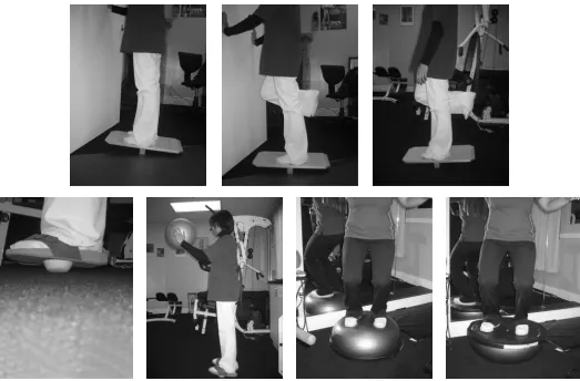
apply this concept, the patient was instructed to perform two-legged dorsi and plantar flexion motions on a rocker board using the wall for support. Progression was made in subsequent visits by performing the same exercise with the left leg only. In the following weeks, the rocker board was replaced by a wobble board and the patient was in-structed to perform all the previous exercises without us-ing the wall for support. In the latter part of her rehab program, balance sandals requiring higher levels of bal-ance and stability were used (Figure 4). To improve the patient’s proprioceptive response, tossing a ball between the therapist and the patient was established in conjunc-tion with the balance sandals (Figure 4).
Follow-up
provement in her knee, cervical and lumbar pain accord-ing to pre (8/10) and post (1–2/10) VAS scores. Despite the dramatic improvement, the patient still complained of sharp, stabbing, “deep” knee pain after prolonged stand-ing and walkstand-ing. Objectively, the patient had full ROM in the cervical and lumbar spines and in her left knee.5,6
Post-treatment evaluation revealed identical thigh girth measurements. Calf measurements differed only by 1 cm. Cervical and lumbar orthopedic special tests were nega-tive.5 Minor laxity was noted using the anterior drawer
test, while all other special tests of the knee were unre-markable.5 The patient was instructed to continue with
her conservative treatments at a frequency of once a week, as well as home exercises. The patient was also
ad-vised to see her specialist due to minor anterior instability as well as the persistent knee pain. An MRI was then or-dered revealing a partial tear in the mid-ACL, a partial tear of the proximal MCL, as well as displaced tear of the lateral meniscus. Following this discovery, the patient was then scheduled for an arthroscopic knee evaluation. This scheduled procedure has not yet been performed. According to the orthopedic surgeon, the patient’s prog-nosis is good since the ACL was only partially torn and therefore may not require reconstruction.
Discussion
Tibial eminence fractures and ACL injuries, in skeletally immature patients, are usually seen in sports medicine
and pediatric orthopedic practices.16 The literature on
in-ternet-based search engines such as EBSCO, PUBMED and OVID did not reveal any studies relating to the treat-ment of Type I tibial eminence fracture rehabilitation pro-tocols. Furthermore, aside from the work performed by Rosenburg et al., there were no other studies indicating a step-by-step rehabilitation protocol for tibial spine avul-sion fractures.12 It is important to realize that although a
total tear of the ACL is spared in a tibial eminence avul-sion, ACL sprains are still very common.1,17 Closed
ki-netic chain exercises without full extension are highly recommended for ACL injuries.13 As mentioned
previ-ously, the avoidance of terminal knee extension during the rehabilitation of ACL sprains is advised as this mo-tion increases the tension placed on the ACL.13,14
Howev-er, if a medial meniscus injury has occurred without any damage to the ACL, introduction of open kinetic chain exercises takes precedence.13 MRI has proven to be
accu-rate for the diagnosis of intra- and peri-articular patholo-gy, especially for meniscal patholopatholo-gy, accounting for 86% of the indications for arthroscopy, and ligamentous injuries. MRI, when used in all patients with high clinical suspicion of intra-articular knee pathology instead of di-rect arthroscopy, can avoid 35% of arthroscopies with sensitivity of 87.3% and specificity of 88.4%.18 MRI can
also be up to 95% accurate in identifying ACL tears.19
Therefore, advanced imaging in conjunction with regular radiographs, and orthopedic testing may be a preferred approach to completely diagnose ACL and meniscus in-juries in a tibial avulsion fracture. A recent study by Ishi-bashi et al. also agrees with the above suggestion of performing advanced imaging on tibial spine fracture pa-tients.4 In this study out of 25 patients with tibial spine
fractures, 15 adults and 10 children, MRI that was not seen on original radiographs confirmed additional liga-ment injuries. This study suggests that because tibial spine fractures in adults may be accompanied by con-comitant injuries requiring surgical treatment, magnetic resonance imaging is recommended.4
Proprioception is also crucial in rehabilitation of most knee injuries.11,12,13,15 Such exercises require little to no
equipment in a chiropractic rehabilitation setting. Other closed kinetic chain exercises that could have been im-plemented are step-ups, step-downs and 1/4 squats.14 An
affordable and multifunctional half ball could also be used for balance and core stability training (Figure 4).
Proprioceptive neuromuscular facilitation (PNF) patterns are also suggested to strengthen the rotational component of the knee motion.13
A shortcoming of this case was that there was no knee questionnaire aside from the VAS that was completed at the time of evaluation and post-treatment. The Cincinnati knee rating system is a possible questionnaire that could be used for future knee patients.3 The Lower Extremity
Functional Scale (LEFS) can also be used as a functional outcome measure with internal consistency of 0.93 to 0.96 and Test-Retest of 0.94.20 In addition, there was also
no ligamentous stability testing after removal of the cast. This may have helped with earlier identification of the ACL tear. The importance of advanced imaging such as MRI, was apparent in this case since ACL, MCL, and lat-eral meniscus tears were not diagnosed originally. As the rehabilitation protocols for ACL and meniscus tears are different, future studies may indicate the need for ad-vanced imaging in all type I tibial eminence fracture as a gold standard.
Conclusion
The rehabilitation of tibial eminence fractures may be complicated, due to the high likelihood of associated un-derlying injuries to other structures of the knee. This par-ticular case study is a good example of a patient with a lateral tibial eminence fracture whose partial ACL and lateral meniscus tear was left undiagnosed. Nevertheless, the original fracture was healed and other soft tissue inju-ries such as cervical and lumbar sprains and strain were also improved through treatment. Regardless of the diag-nosis, low-tech rehabilitation of the knee could be ap-plied in a chiropractic rehabilitation setting. However, future studies are warranted in order to determine proper rehabilitation protocols for uncomplicated and compli-cated tibial eminence fractures.
References
1 Accousti WK, Willis RB. Tibial eminence fractures. Orthop Clin N Am 2003; 34(3):365–375.
2 Ahmad CS, Shubin Stein BE, Jeshuran W, Nercessian OA, Henry JH. Anterior cruciate ligament function after tibial eminence fracture in skeletally mature patient. Am J Sports Med 2001; 29(3):339–345.
7 Al-Smadi J, Warke K, Wilson I, Cramp AFL, Nobel G, Walsh DM, Lowe-Strong AS. A pilot investigation of the hypalgesic effects of transcutaneous electrical nerve stimulation upon low back pain in people with multiple sclerosis. Clinical Rehabilitation 2003; 17(7):742–749. 8 Philadelphia Panel Evidenced-Based Clinical Practice
Guidelines on Selected Rehabilitation Interventions for the knee. Physical Therapy 2001; 10:1629–1640.
9 McGill SM. Lower back disorders: evidence-based prevention and rehabilitation. Champaign, IL. Human Kinetics, 2002.
10 Liebenson C. Rehabilitiation of the spine. 2nd edition. Baltimore. Lippincott Williams & Wilkins, 1996.
11 Nicholas JA, Heshman EB. The lower extremity & spine in sports medicine. 2nd edition. Mosby 1995.
12 Griffin LY. Rehabilitation of the injured knee. 2nd edition. Mosby 1995.
16 Fehnel DJ, Johnson R. Anterior cruciate injuries in the skeletally immature athlete. Sports Med 2000; 29(1):51–63.
17 Willis RB, Blokker C, Still TM, Paterson DC, Galpin RD. Long-term follow-up of anterior tibial eminence fracture. J Pediatric Orthopedics 1993; 13:361–4.
18 Vincken P, Braak BP, Van Erkel AR, Coerkamp EG, De Rooy TPW, Mallens MC, Bloem JL. Magnetic resonance imaging of the knee: a review. Imaging Decisions 2006; 10(1):24–30.
19 Koon D, Bassett F. Anterior cruciate ligament rupture. Southern Med J 2004; 97(8):755–756.
20 Finch E, Brooks D, Stratford PW, Mayo NE. Physical Rehabilitation Outcome Measures. 2nd edition. Hamilton, Ontario. BC Decker Inc., 2002.