Article
1
Z Probe, an Efficient Tool for Characterizing Long
2
Non-Coding RNA in FFPE Tissues
3
Manish Tripathi
1#*, Chidi Zacheaus
1#, Kyle Doxtater
1, Fatemeh Keramatnia
1, Lani Gao
2,
4
Meena Jaggi
1, Murali Yallapu, Subhash Chauhan
1*
5
1 Department of Pharmaceutical Sciences, University of Tennessee Health Science Center, Memphis, TN,
6
USA.
7
2 University of Tennessee at Chattanooga, TN, USA.
8
9
#Contributed equally to this article
10
* Correspondence: [email protected]; [email protected]; Tel.: +901-448-7313
11
12
Abstract:
Formalin-Fixed Paraffin Embedded (FFPE) tissues are a valuable resource in studying
13
different markers and mechanistic molecules (protein, DNA and RNA) in order to understand the
14
etiology of different cancers as well as many other diseases. Degradation and modification of RNA
15
is the major challenge in utilizing FFPE tissue samples in medical research. Recently, non-protein
16
coding transcripts long non-coding RNAs (lncRNAs), have gained significant attention due to their
17
important biological actions and potential involvement in cancer. There is no validated method
18
except qRTPCR or RNAseq to evaluate and study lncRNA expression. We have standardized and are
19
reporting a sensitive Z probe based
in situ
hybridization method to identify, localize and quantitate
20
lncRNA in FFPE tissues. This assay is sensitive to single transcript and localizes lncRNA in individual
21
cells within tumor. We have characterized a tumor suppressor lncRNA-NRON (non coding repressor
22
of NFAT), which is scarcely expressed, a moderately expressed oncogeneic lncRNA UCA1 (urothelial
23
cancer associated 1), and a highly studied and expressed lncRNA MALAT1 (metastasis associated
24
lung adenocarcinoma transcript1) in different cancers. High MALAT1 staining was found in
25
colorectal, breast and pancreatic cancer. MALAT1 expression increased with the progression of the
26
stage in colorectal cancer and invasiveness in breast cancer.
27
28
Keywords:
long noncoding RNA; MALAT1; UCA1; NRON; Z probe; colorectal cancer; pancreatic
29
cancer; breast cancer
30
1. Introduction
31
LncRNA a class of noncoding RNA has gained significant traction with exponential publications
32
and interests from the scientific community. The role of miRNA in regulation of gene expression has
33
been extensively studied in the last decade since its discovery. LncRNA provides a novel way of
34
regulating gene expression and function at all levels of DNA, RNA or proteins. In 2005 when 512
35
known lncRNA were systematically studied [1], It was not anticipated that the number will grow
36
exponentially to 19,175 potentially functional lncRNAs in human genome. Many of these potential
37
lncRNA identified by FANTOM5 analysis of CAGE (cap analysis of gene expression) data and
38
overlapping expression qualitative trait loci (eQTL), have not been functionally described, it is
39
anticipated that this number of lncRNA will increase significantly [2]. Many of the lncRNA are been
40
characterized by revisiting the array datasets on publicly available resources [3-7]. LncRNA, which
41
are now at the center of various physiological and pathological processes have been linked to various
42
cellular pathways including progression of different cancers and various diseases. It is imperative to
43
invest efforts in understanding the roles of these new class of regulators and further elucidate the
44
mechanism.
45
Immunohistochemistry(IHC) for decades has been the primary diagnostic method of choice for
46
identifying important biomarkers at the protein level in cancer and other diseases. The ability of IHC
47
to detect important protein receptors and membrane bound proteins makes it an attractive diagnostic
48
method. The information gained by IHC helps in the designing of a better therapeutic regimen for
49
the treatment of cancer and other diseases [8]. RNA
in situ
hybridization has been used widely to
50
analyze mRNA in different studies [9-11]. Quantitative RT-PCR, RNA sequencing and microarrays
51
are the current gold standard for detecting lncRNA in cells and tissues. The problem with this method
52
is its inability to differentiate different cell populations within the tissue and the compartmental
53
location of biomarker, which are important information for better targeted therapy [12]. The
54
limitation also lies in low copy numbers and lack of localization information while using these
55
techniques. Most lncRNA studies are based on bioinformatics analysis and correlation studies from
56
the data available from public resources [2,6,7]. A major problem in understanding lncRNA’s role in
57
cancer progression and other diseases is a lack of an efficient tool to characterize them. A new Z probe
58
based technology was reported first time to investigate into the levels of program death ligand-1 and
59
its receptor PD1 mRNA expression in tissues cohorts of non-small cell lung cancer. [13]. If used as a
60
corroborative diagnostic tool, this method may provide more confidence as the validation step to
61
identify the same cancer biomarkers observed with IHC. In the interest of having a specific, sensitive
62
and reproducible lncRNA assay, we analyzed and standardized Z probe based chromogenic method
63
for further diagnostic detection of lncRNAs as cancer biomarkers. This method with rigorous steps,
64
sensitivity and specificity, can improve patient lives through identification of localized gene
65
expression within individual cancer cells as well as expression changes in different cell populations
66
of cancer tissues.
67
68
2. Results
69
2.1. Schematic and Controls
70
Schematic work flow of RNAScope assay using the Z probes is represented in Figure 1. Each Z
71
probe has a18-25 base complimentary sequence to the target lncRNA and a 14 base sequence
72
complementary to the pre-amplifier. Three double ZZ probes are sufficient for a signal, but this assay
73
utilizes 20 double ZZ probes, which covers around 1kb of the target transcript.
74
FFPE sectioned HeLa cells from ACD were used first to validate the assay as shown in Figure
75
2(c). PPIB (Peptidylprolyl isomerase B), a human gene and DapB (dihydrodipicolinate reductase), a
76
bacterial (
E.coli
) gene, were used as positive control and negative control probe respectively. DapB
77
did not show any signal in pancreatic and colorectal cancer tissues, but PPIB stained nicely on the
78
tissues (pancreatic and colorectal cancer) as well as on the FFPE sectioned HeLa cells. This result
79
demonstrated the sensitivity and specificity of the assay and that FFPE tissues can be tried with the
80
experimental probe staining with similar results as with the embedded cells.
81
2.2. Validation of assay
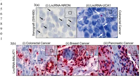
84
In order to validate the assay method in identifying lncRNAs on FFPE tissues we selected a low,
85
moderately and highly expressed lncRNA. LncRNA NRON, is scarcely expressed and is very hard
86
to quantitate even by qRTPCR. Figure 3(a)(i) shows NRON signal in xenograft FFPE tissue, similarly
87
moderately expressed oncogenic lncRNA UCA1 was very specifically located in colorectal cancer
88
tissue in the epithelial population (Figure 3 (a)(ii)), highlighting the sensitivity of the assay method.
89
Further, we chose highly expressed lncRNA MALAT1 for staining in colorectal, breast and pancreatic
90
cancer. MALAT1 showed a very prominent staining in these three cancer tissues, mostly in epithelial
91
cell population.
92
2.3. Quantitative measure of Progression and Invasiveness
93
Prominent lncRNA MALAT1 staining encouraged us to utilize this assay to analyze MALAT1
94
as a cancer progression and invasiveness marker. Interestingly lncRNA MALAT1 stained very well
95
rwith respect to the the progression of colorectal cancer. Figure 4(a) represents MALAT1 staining in
96
different stages of CRC with mean area intensity represented below in different stages. Figure 4(b)
97
represents the quantitative stain intensity in different stages of CRC tissues. The MALAT1 stain
98
intensity is significantly correlated among different stage (except stage III and stage IV) progression.
99
Additionally, we observe higher MALAT1 expression in epithelial cells than in stroma cells. This
100
might indicate a preferential expression within cell types and perhaps different regulatory
101
mechanism.
102
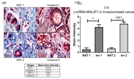
Finally, to assess whether the assay method can quantitatively measure the invasiveness, we
103
used a tumor microarray (TMA) with normal to adjacent tumor (NAT) and matched invasive breast
104
cancer tissues and reassuringly, invasive breast cancer tissues have a higher MALAT1 staining (as
105
shown in Figure 5(a), mean area intensity is higher in both invasive tissues as compared to NAT.
106
Figure 5(b) shows a significant difference in the stain intensity between NAT and invasive tissues
107
indicating lncRNA MALAT1 as a marker of invasive breast cancer.
108
2.2. Figures, Tables and Schemes
110
Figure 1: A schematic representation of Z probe based RNAScope assay for lncRNA analysis. Starting with Z
111
probes hybridizing with target sequence creating a double ZZs up to 20 groups side by side. Preamplifier then
112
binds to the complementary sequence on the 28 base tail (top of the ZZ). Pre-amplifiers contain multiple binding
113
sites for amplifiers to bind to and the amplifiers also have multiple binding sites for labeled probes to bind. Upon
114
chromogenic stain, the label probes fluoresce red color.
115
116
117
Figure 2: Validation and optimization of the Z-probe staining. Paraffin embedded and sectioned, pancreatic
118
cancer tissue, colorectal tissue and HeLa cell pellet were stained with a negative control probe DapB (full form)
119
and positive control probe PPIB (full form). The PPIB stained well with pancreatic, colorectal and HeLa cells
120
(bottom panel (a), (b) & (c). 20x (inset) and 80x magnification using caseview software, scanned and analyzed
121
on Pannoramic 250 Flash III. Arrows point at specific staining.
123
124
125
126
127
128
129
130
131
Figure 3: Paraffin embedded different human cancer tissues Z probe stained for different lncRNAs. (a) Tumor
132
suppressor lncRNA-NRON (very low expressed) (i), and oncogeneic lncRNA-UCA (moderately expressed) (ii),
133
stained in colorectal cancer tissue. (b) LncRNA–MALAT1 stained using specific Z-probes in paraffin embedded
134
(i) colorectal cancer, (ii) breast cancer and (iii) pancreatic cancer tissues. 20x (inset) and 80x magnification using
135
caseview software, scanned and analyzed on Pannoramic 250 Flash III. Arrows point at specific lncRNA signal.
136
137
138
139
140
141
142
143
Figure 4:LncRNA-MALAT in different stages of CRC. Paraffin embedded different stages of colorectal cancer
146
tissues were stained for lncRNA-MALAT1 and quantitated for the staining intensity. (a) Stages 1-IV CRC tissues
147
show a differential stain for lncRNA-MALAT1. Stain intensity correlates to the progression. (b) Quantitation of
148
the lncRNA-MALAT1 staining intensity was performed using Image J software. 10x (inset) and 80x
149
magnification using caseview software, scanned and analyzed on Pannoramic 250 Flash III. Statistical analysis:
150
One way ANOVA, Tukey’ multiple comparison test. Compare the mean of each column with mean of other
151
column. Arrows point at specific lncRNA signal.
152
153
154
155
156
157
158
159
160
161
Figure 5:LncRNA-MALAT1 expression in breast cancer. (a) Matched breast cancer tissues (NAT vs Invasive )
162
from two different patients, 1 & 2 were stained for lncRNA-MALAT1 using Z probe. The invasive breast cancer
163
tissues have higher staining for lncRNA-MALAT1 as compared to normal adjacent tumor. (b) Quantitation of
164
the lncRNA-MALAT1 staining intensity was performed using Image J software.10x (inset) and 80x magnification
165
using caseview software, scanned and analyzed on Pannoramic 250 Flash III. Statistical analysis: Unpaired t test,
166
** p<0.01, *** p<0.001.
167
3. Discussion
168
IHC and RT-QPCR has long been the gold standard for investigating protein and RNA
169
biomarkers respectively [8,14-16]. Lack of specificity, sensitivity and reproducibility of these methods
170
continue to limit our interpretation of data collected. This in turn makes clinical diagnosis
171
challenging. While IHC is beneficial in identifying protein biomarkers, the assay is ineffective in
172
detecting lncRNA in FFPE tissues, which is the limitation of the assay design. As our understanding
173
of long non-coding RNAs and their role in cancer development and progression augments, a more
174
specific, sensitive and reproducible tool is warranted. The strength of Z probe assay lies on the fact
175
that it utilizes approximately 1kb of the target transcript and it employs 20 double ZZ binding probes
176
hybridization assay [9-11]. Differential expression pattern of HOTAIR, H19, KCNQ101T, MEG3,
178
MALAT1, and ZFAS1 via RNAScope on breast cancer patient samples has been shown recently.[12].
179
Use of the appropriate positive and negative controls strengthen the confidence on the results. PPIB
180
positive signal on xenograft HeLa cell and human cancer tissues simultaneously validated the
181
procedure.
182
Tumor suppressor lncRNA-NRON was one of the first lncRNA identified from an unbiased
183
screen [1]. NRON is noncoding repressor of NFAT, and it is a tumor suppressor [17] . NRON is very
184
lowly expressed and its expression was difficult to visualize in human tumor FFPE tissue, so SW480
185
(colorectal cancer cell line) Xenograft FFPE tissue was used. Moderately expressed oncogenic
186
lncRNA-UCA1 has been studied for different pathways and interactions, resulting in cancer
187
progression [18-20]. The assay in study was able to show specific epithelial cell population expressing
188
UCA1 as red dot. We were also able to show that MALAT1 is aberrantly expressed in TMAs of three
189
cancer groups (Colorectal, Breast, and Pancreatic), suggesting that lncRNA MALAT1 is associated
190
with multiple cancer and its use as a therapeutic target should be investigated further. Though
191
MALAT1 has been studied in different cancers as a poor prognostic [21-24], aggressive and metastatic
192
marker [21,25,26], most of the published data on MALAT1 lack visual representation of MALAT1
193
detection. All the studies known so far have used RT-QPCR, RNAseq or curated database analysis
194
for their analysis. This is the first evidence of MALAT1 expression in FFPE patient tissues of breast,
195
colorectal and pancreatic cancer by chromogenic staining, suggesting that lncRNA MALAT1 is
196
associated with multiple cancers and its use as a therapeutic target should be investigated further.
197
We also showed progressive expression of MALAT1 across the different stages of colorectal cancer
198
and were able to correlate the stages with the progression of CRC. To our confidence the stain
199
intensity of different ROI in the CRC tissues was statistically significant except for Stage III and stage
200
IV comparison. More stage III and Stage IV CRC tissues might help to increase the statistical
201
significance. Invasive breast tissues have been shown to have higher MALAT1 expression [21,26-28],
202
but for the first time this study gives a visual evidence. Although we were only able to show UCA1
203
expression in CRC TMA, staining in TMA of other cancer group will be investigated to determine if
204
there is differential expression of UCA1 between cancer types.
205
As we continue to investigate the role of long non-coding RNAs in cancer development and
206
metastasis, this assay method will help provide visual representation of this class of RNA molecules.
207
Collectively, this data suggest that Z probe can be an efficient tool to characterize long non-coding
208
RNA molecules in FFPE tissues and cells. This probe provides high confidence with respect to
209
specificity, sensitivity and reproducibility. This assay alleviates all areas of concern by
210
simultaneously detecting lncRNA molecule of interest while suppressing background noise with
211
more novel Z probe pair design and signal amplifiers. In both laboratory and clinical settings, this
212
assay can be utilized to corroborate the results of both IHC and RT-QPCR strengthening our
213
interpretation of cancer behavior especially in this new field of long non-coding RNAs.
214
4. Materials and Methods
215
Breast cancer TMA # BR243w was purchased from Biomax, human colorectal cancer tissues (FFPE),
216
pancreatic cancer tissue (FFPE) sectioned were procured from department of pathology, UTHSC as per UTHSC
217
IRB guidelines. Colorectal cancer cell line SW480 xenograft (FFPE) were sectioned using Microtome (Leica
218
Biosystems). FFPE HeLa slides were procured from ACD (#310045) to use as positive and negative controls.
Chromogenic staining in FFPE tissues: Formalin fixed (10% neutral buffered formalin, 16-32 hrs) and
220
alcohol treated (70% alcohol, 24-48 hrs) paraffin embedded tissues, sectioned (5-7 M) and mounted on
221
Superfrost slides (Fisherscientific) were used for lncRNA Z-probe staining. RNAScope 2.5 HD Detection Kit
222
(RED) (#322360, ACD), was used as per manufacturer suggestions with modifications. After baking FFPE slides
223
on side warmer for 1 hr at 60 oC, they were deparaffinized in fresh xylene twice for 5 min, dehydrated in 100%
224
alcohol twice for 1 min. and treated with hydrogen peroxide (#322330, ACD) for 10 min at room temperature.
225
Maxed epitopes were retrieved by incubating slides in boiling target retrieval buffer (#322000, ACD) for 25 min
226
(this is a critical step as different tissues require different retrieval times) followed by protease plus (#322340,
227
ACD) treatment for 30 min in the HybEZ (#240200, ACD) incubator preset at 40 oC. Z probes (HsNRON #508481,
228
HsUCA1 #417521, HsMALAT1 #400811) warmed at 40 oC for 10 min, and amplifiers (#322360, ACD) were
229
normalized to room temperature before hybridization steps. After aspirating protease plus, probes were added
230
to respective tissues and incubated at 40 oC for 2 hrs in HybEZ incubator. Hybridized probes bound to
231
complementary bases were amplified through sequential amplification steps from amp-1 to amp-6 followed by
232
incubation at 40 oC in the incubator with washing (#310091, ACD) between each amplification steps. Amplifiers
233
tagged with chromogenic labeled dye probes (mix of RED-B and RED-A at 60:1 ratio) after 10 min incubation
234
gives a red signal. Slides were counterstained with 50% hematoxylin for 2 minutes at room temperature and
235
washed with 0.02% ammonia water followed by distilled water. Slides finally dried at 60 oC for 15 min on a slide
236
warmer, Xylene dipped and mounted with Ecomount (#EM897L Biocare Medical). FFPE mammalian cells
237
sectioned on slides can also be stained using the same method.
238
Quantitation (ImageJ) Analysis: The stained slides were scanned on Pannoramic 250 Flash III. Images
239
were visualized using Caseviewer software (3DHistech). Four different field (ROI: Region of Interest) of the 10x
240
image were analyzed for the total intensity of the red stain, using Image J (nih.gov), representing the
241
corresponding lncRNA staining. Color threshold was adjusted to identify only the red color.
242
Statistical analysis: Statistical analysis were performed using GraphPad Prism 7. P<0.05 was considered
243
significant.
244
245
Author Contributions: Conceptualization, M.K.T, CZ, and SCC; Methodology, CZ and MKT; Software, CZ,
246
MKT; Formal Analysis, CZ and MKT; Investigation, CZ and MKT; Resources, MKT, LG, MMY, MJ and SCC;
247
Writing-Original Draft Preparation, CZ and MKT; Writing-Review & Editing, CZ, MKT, KD, FK, MJ, MMY and
248
SCC; Visualization, CZ, KD, FK, and MKT; Supervision, MKT and SCC; Project Administration, MKT and SCC;
249
Funding Acquisition, MKT, LG, MMY, MJ and SCC.
250
Funding: The research was partially supported by the National Institutes of Health Grant (R01CA204552,
251
R01CA210192 and R01CA206069) to S.C.C. UTHSC-CORNET and UTHSC-Clinical CORNET award to M.K.T.
252
This work was also partially supported by UTHSC-College of Pharmacy-Dean’s Seed Grant and UTHSC New
253
Grant Mechanism Award to S.C.C., M.M.Y. and M.J.
254
Acknowledgments: The authors would like to thank Dr. Marie Chisholm-Burns, Dean COP UTHSC, Dr. Lisa
255
Youngentob, Director R&D and Dr. Steven R. Goodman, Vice Chancellor of Research for suppor through
256
CORNET program. Department of Pathology data acquisition services are also acknowledged.
257
Conflicts of Interest: “The authors declare no conflict of interest.”
References
260
1. Willingham, A.T.; Orth, A.P.; Batalov, S.; Peters, E.C.; Wen, B.G.; Aza-Blanc, P.; Hogenesch, J.B.; Schultz,
261
P.G. A strategy for probing the function of noncoding rnas finds a repressor of nfat. Science (New York,
262
N.Y.) 2005, 309, 1570-1573.
263
2. Hon, C.C.; Ramilowski, J.A.; Harshbarger, J.; Bertin, N.; Rackham, O.J.; Gough, J.; Denisenko, E.;
264
Schmeier, S.; Poulsen, T.M.; Severin, J., et al. An atlas of human long non-coding rnas with accurate 5'
265
ends. Nature 2017, 543, 199-204.
266
3. Ma, X.; Zhang, W.; Zhang, R.; Li, J.; Li, S.; Ma, Y.; Jin, W.; Wang, K. Overexpressed long noncoding rna
267
crnde with distinct alternatively spliced isoforms in multiple cancers. Frontiers of medicine 2018.
268
4. Gutschner, T.; Richtig, G.; Haemmerle, M.; Pichler, M. From biomarkers to therapeutic targets-the
269
promises and perils of long non-coding rnas in cancer. Cancer metastasis reviews 2018, 37, 83-105.
270
5. Shen, J.; Hodges, T.R.; Song, R.; Gong, Y.; Calin, G.A.; Heimberger, A.B.; Zhao, H. Serum hotair and
271
gas5 levels as predictors of survival in patients with glioblastoma. Molecular carcinogenesis 2018, 57,
137-272
141.
273
6. Song, W.; Wang, K.; Zhang, R.; Zou, S. [long noncoding rna malat1: A potential novel prognostic
274
biomarkers in cancers based on meta-analysis]. Zhong nan da xue xue bao. Yi xue ban = Journal of Central
275
South University. Medical sciences 2016, 41, 1163-1167.
276
7. Kong, H.; Wu, Y.; Zhu, M.; Zhai, C.; Qian, J.; Gao, X.; Wang, S.; Hou, Y.; Lu, S.; Zhu, H. Long non-coding
277
rnas: Novel prognostic biomarkers for liver metastases in patients with early stage colorectal cancer.
278
Oncotarget 2016, 7, 50428-50436.
279
8. Matos, L.L.; Trufelli, D.C.; de Matos, M.G.; da Silva Pinhal, M.A. Immunohistochemistry as an
280
important tool in biomarkers detection and clinical practice. Biomarker insights 2010, 5, 9-20.
281
9. Thomsen, R.; Nielsen, P.S.; Jensen, T.H. Dramatically improved rna in situ hybridization signals using
282
lna-modified probes. RNA (New York, N.Y.) 2005, 11, 1745-1748.
283
10. Weiszmann, R.; Hammonds, A.S.; Celniker, S.E. Determination of gene expression patterns using
high-284
throughput rna in situ hybridization to whole-mount drosophila embryos. Nature protocols 2009, 4,
605-285
618.
286
11. Thisse, C.; Thisse, B. High-resolution in situ hybridization to whole-mount zebrafish embryos. Nature
287
protocols 2008, 3, 59-69.
288
12. Zhang, Z.; Weaver, D.L.; Olsen, D.; deKay, J.; Peng, Z.; Ashikaga, T.; Evans, M.F. Long non-coding rna
289
chromogenic in situ hybridisation signal pattern correlation with breast tumour pathology. Journal of
290
clinical pathology 2016, 69, 76-81.
291
13. Velcheti, V.; Schalper, K.A.; Carvajal, D.E.; Anagnostou, V.K.; Syrigos, K.N.; Sznol, M.; Herbst, R.S.;
292
Gettinger, S.N.; Chen, L.; Rimm, D.L. Programmed death ligand-1 expression in non-small cell lung
293
cancer. Laboratory investigation; a journal of technical methods and pathology 2014, 94, 107-116.
294
14. Jacob, F.; Guertler, R.; Naim, S.; Nixdorf, S.; Fedier, A.; Hacker, N.F.; Heinzelmann-Schwarz, V. Careful
295
selection of reference genes is required for reliable performance of rt-qpcr in human normal and cancer
296
cell lines. PloS one 2013, 8, e59180.
297
15. Taylor, S.; Wakem, M.; Dijkman, G.; Alsarraj, M.; Nguyen, M. A practical approach to
rt-qpcr-298
publishing data that conform to the miqe guidelines. Methods (San Diego, Calif.) 2010, 50, S1-5.
299
16. Fleige, S.; Pfaffl, M.W. Rna integrity and the effect on the real-time qrt-pcr performance. Molecular
300
aspects of medicine 2006, 27, 126-139.
17. Sharma, S.; Findlay, G.M.; Bandukwala, H.S.; Oberdoerffer, S.; Baust, B.; Li, Z.; Schmidt, V.; Hogan,
302
P.G.; Sacks, D.B.; Rao, A. Dephosphorylation of the nuclear factor of activated t cells (nfat) transcription
303
factor is regulated by an rna-protein scaffold complex. Proceedings of the National Academy of Sciences of
304
the United States of America 2011, 108, 11381-11386.
305
18. Li, C.; Liang, G.; Yang, S.; Sui, J.; Yao, W.; Shen, X.; Zhang, Y.; Peng, H.; Hong, W.; Xu, S., et al.
306
Dysregulated lncrna-uca1 contributes to the progression of gastric cancer through regulation of the
307
pi3k-akt-mtor signaling pathway. Oncotarget 2017, 8, 93476-93491.
308
19. Wang, Z.Q.; Cai, Q.; Hu, L.; He, C.Y.; Li, J.F.; Quan, Z.W.; Liu, B.Y.; Li, C.; Zhu, Z.G. Long noncoding
309
rna uca1 induced by sp1 promotes cell proliferation via recruiting ezh2 and activating akt pathway in
310
gastric cancer. Cell death & disease 2017, 8, e2839.
311
20. Huang, J.; Zhou, N.; Watabe, K.; Lu, Z.; Wu, F.; Xu, M.; Mo, Y.Y. Long non-coding rna uca1 promotes
312
breast tumor growth by suppression of p27 (kip1). Cell death & disease 2014, 5, e1008.
313
21. Jadaliha, M.; Zong, X.; Malakar, P.; Ray, T.; Singh, D.K.; Freier, S.M.; Jensen, T.; Prasanth, S.G.; Karni,
314
R.; Ray, P.S., et al. Functional and prognostic significance of long non-coding rna malat1 as a metastasis
315
driver in er negative lymph node negative breast cancer. Oncotarget 2016, 7, 40418-40436.
316
22. Li, C.; Cui, Y.; Liu, L.F.; Ren, W.B.; Li, Q.Q.; Zhou, X.; Li, Y.L.; Li, Y.; Bai, X.Y.; Zu, X.B. High expression
317
of long noncoding rna malat1 indicates a poor prognosis and promotes clinical progression and
318
metastasis in bladder cancer. Clinical genitourinary cancer 2017, 15, 570-576.
319
23. Zheng, H.T.; Shi, D.B.; Wang, Y.W.; Li, X.X.; Xu, Y.; Tripathi, P.; Gu, W.L.; Cai, G.X.; Cai, S.J. High
320
expression of lncrna malat1 suggests a biomarker of poor prognosis in colorectal cancer. International
321
journal of clinical and experimental pathology 2014, 7, 3174-3181.
322
24. Handa, H.; Kuroda, Y.; Kimura, K.; Masuda, Y.; Hattori, H.; Alkebsi, L.; Matsumoto, M.; Kasamatsu, T.;
323
Kobayashi, N.; Tahara, K.I., et al. Long non-coding rna malat1 is an inducible stress response gene
324
associated with extramedullary spread and poor prognosis of multiple myeloma. British journal of
325
haematology 2017, 179, 449-460.
326
25. Li, Q.; Pan, X.; Wang, X.; Jiao, X.; Zheng, J.; Li, Z.; Huo, Y. Long noncoding rna malat1 promotes cell
327
proliferation through suppressing mir-205 and promoting smad4 expression in osteosarcoma.
328
Oncotarget 2017, 8, 106648-106660.
329
26. Xiping, Z.; Bo, C.; Shifeng, Y.; Feijiang, Y.; Hongjian, Y.; Qihui, C.; Binbin, T. Roles of malat1 in
330
development and migration of triple negative and her-2 positive breast cancer. Oncotarget 2018, 9,
2255-331
2267.
332
27. Huang, N.S.; Chi, Y.Y.; Xue, J.Y.; Liu, M.Y.; Huang, S.; Mo, M.; Zhou, S.L.; Wu, J. Long non-coding rna
333
metastasis associated in lung adenocarcinoma transcript 1 (malat1) interacts with estrogen receptor and
334
predicted poor survival in breast cancer. Oncotarget 2016, 7, 37957-37965.
335
28. Soudyab, M.; Iranpour, M.; Ghafouri-Fard, S. The role of long non-coding rnas in breast cancer. Archives
336
of Iranian medicine 2016, 19, 508-517.