Repurposing azithromycin and rifampicin against Gram-negative pathogens by combination with 1
peptide potentiators 2
3
Kristin R.Baker a, b #, Bimal Jana a, b, Anna Mette Hansend, Karina Juul Vissing c, Hanne MørckNielsen c, 4
Henrik Franzyk d #, Luca Guardabassi a, b. 5
6
a Department of Veterinary and Animal Sciences, Faculty of Health and Medical Sciences, University of 7
Copenhagen, Frederiksberg, Denmark 8
b Department of Biomedical Sciences, Ross University School of Veterinary Medicine, Basseterre, St 9
Kitts, West Indies 10
c Department of Pharmacy, University of Copenhagen, Copenhagen, Denmark 11
d Department of Drug Design and Pharmacology, University of Copenhagen, Copenhagen, Denmark 12
13
Running title: Peptide-induced antibiotic susceptibility 14
15
# Corresponding author mailing address: Kristin R. Baker, Ross University School of Veterinary 16
Medicine, Main Road, Basseterre, St. Kitts, Tel: +1 869-465-4161 Ext. 401-1211, [email protected] 17
and Henrik Franzyk, University of Copenhagen, Jagtvej 162, DK-2100, Copenhagen, Tel: +45 3533 6255, 18
Abstract 21
Gram-negative pathogens are intrinsically resistant to several antibiotics that are not able to penetrate the 22
envelope barrier. The objective of this study was to identify peptides that at low concentrations induce 23
susceptibility to these antibiotics in multidrug-resistant (MDR) Gram-negative strains of clinical relevance. 24
A pairwise screening of 34 diverse peptides and four antibiotics (erythromycin, linezolid, rifampicin and 25
vancomycin) with primary activity against Gram-positive bacteria identified four peptides that at sub-26
micromolar concentrations conferred susceptibility to rifampicin or erythromycin in Escherichia coli 27
ATCC 25922. The identified peptides exhibited synergy with azithromycin and potentiated clindamycin in 28
MDR E. coli ST131 and Klebsiella pneumoniae ST258. The low cytotoxicity toward eukaryotic cells (IC50 29
>50 µM) observed for two peptides (KLWKKWKKWLK-NH2 and GKWKKILGKLIR-NH2) prompted 30
synthesis and evaluation of the corresponding all-D analogs (D1 and D2), which retained similar synergistic 31
antibacterial profiles. Low concentrations of D1 and D2 in combination with azithromycin and rifampicin 32
inhibited growth of most clinical E. coli, K. pneumoniae and Acinetobacter baumannii strains tested. Our 33
data demonstrate that combinatorial screening at low concentrations constitutes an efficient approach to 34
identify clinically relevant peptide-antibiotic combinations. In vivo PK/PD and toxicity studies are needed 35
to further validate the use of the peptides identified by this study for repurposing azithromycin and 36
rifampicin against Gram-negative pathogens. 37
38
Keywords 39
Antimicrobial peptides, Gram-negative bacteria, multidrug resistance, antibiotic adjuvant, combination 40
therapy, antibiotic potentiation 41
42
1. Introduction 44
As a consequence of the worldwide spread of multidrug-resistant (MDR) Gram-negative clones, the 45
World Health Organization has ranked the development of new therapeutics to treat infections caused by 46
Enterobacteriaceae, Acinetobacter baumannii and Pseudomonas aeruginosa as a critical priority [1]. 47
Intrinsic antibiotic resistance considerably limits the therapeutic options against these pathogens, since 48
several classes of the available antibiotics cannot effectively penetrate the envelope barrier [2]. 49
Combination therapy represents an attractive approach for treating MDR infections as it typically reduces 50
the required dose of the individual components and limits the risk for emergence of resistance [4,5]. 51
Antimicrobial peptides that increase therapeutic potency and expand the spectrum of antibiotics to include 52
Gram-negative pathogens have potential use in combination therapy [4,5,6]. Although many reports have 53
demonstrated synergistic peptide-antibiotic interactions, the clinical potential of such findings have rarely 54
been studied systematically. 55
The objective of the present study was to identify peptides that at low non-toxic concentrations render 56
MDR Gram-negative pathogens susceptible to antibiotics to which they are intrinsically resistant. 57
Following a systematic approach, we designed a pairwise screen based on antibacterial activity of low 58
concentrations of a diverse set of peptides in combination with four antibiotics with primary activity against 59
Gram-positive bacteria. Subsequently, peptide-induced antibiotic susceptibility was confirmed, and 60
cytotoxicity was then assessed for the top four antibiotic-potentiating peptides. This resulted in 61
identification of two lead peptides that displayed low cytotoxicity to different eukaryotic cell types and 62
potentiated azithromycin and rifampicin against several Gram-negative species of clinical relevance. 63
64
2. Materials and methods 65
2.1 Media, antibiotics, bacterial strains and peptide synthesis 66
Bacteria were cultured on Luria-Bertani broth, cation-adjusted Mueller-Hinton agar (MHA) and 67
included Escherichia coli, Klebsiella pneumoniae, Pseudomonas aeruginosa, and Acinetobacter 69
baumannii. MDR strains E. coli ST131 and K. pneumoniae ST258 are clinical isolates from urinary tract 70
[7] and wound infections [8], respectively. A panel of β-lactamase resistant clinical isolates of were 71
provided by Laurent Poirel. Starting materials and solvents for peptide synthesis were purchased from 72
commercial suppliers (Iris Biotech, Sigma-Aldrich and VWR). All peptides and all-D analogs were 73
synthesized and analyzed as previously reported [9]. The peptide stock solutions were made in deionized 74
water, followed by dilution in MHBII. 75
76
2.2 Antimicrobial susceptibility testing 77
Bacterial susceptibility to compounds was determined by microbroth dilution according to CLSI 78
guidelines [10]. In the screen, antibiotics and peptides were combined at fixed concentrations corresponding 79
to the antibiotics’ CLSI susceptibility breakpoint for Staphylococcus species [11]. Antibiotic minimum 80
inhibitory concentrations (MICs) in presence of peptide concentrations (0.5 µM or 1 µM) were determined 81
as above. For growth curve assays, the MIC plates were prepared as above, and then plates were incubated 82
for 24 h at 37 °C with continuous shaking. Optical density (OD) at 600 nm was recorded in 10 min intervals. 83
84
2.3 Checkerboard assay 85
Synergy of peptide-antibiotic combinations was measured by using a two-dimensional checkerboard 86
assay [12] and CLSI guidelines [10]. The fractional inhibitory concentration index (FICI) was calculated 87
and interpreted as previously reported [13]. 88
89
2.4 Cellular viability and IC50 90
Cell viability was determined in ATCC NIH 3T3 fibroblasts and HepG2 hepatocytes by using the 91
MTS/PMS assay as previously reported [14]. Peptide concentrations ranged from 0.1 to 500 µM. The 92
defined as the absorbance values obtained after incubation of cells with SDS (0.2%, w/v in medium) and 94
with medium, respectively. 95
𝑅𝑒𝑙𝑎𝑡𝑖𝑣𝑒 𝑣𝑖𝑎𝑏𝑖𝑙𝑖𝑡𝑦 (%) =(𝐴𝑏𝑠𝑠𝑎𝑚𝑝𝑙𝑒−𝐴𝑏𝑠𝑝𝑜𝑠)
(𝐴𝑏𝑠𝑛𝑒𝑔−𝐴𝑏𝑠𝑝𝑜𝑠) × 100% (1)
96
IC50 values were calculated using GraphPad Prism 7 (GraphPad Software, La Jolla, CA, USA) by 97
fitting the relative viability of the cells to the concentration of the test compound using equation 2: 98
𝑅𝑒𝑙𝑎𝑡𝑖𝑣𝑒 𝑣𝑖𝑎𝑏𝑖𝑙𝑖𝑡𝑦 (%) = 𝑇𝑜𝑝−𝐵𝑜𝑡𝑡𝑜𝑚
1+10(𝐿𝑜𝑔𝐼𝐶50−𝐿𝑜𝑔[𝑝𝑒𝑝𝑡𝑖𝑑𝑒]) ×𝐻𝑖𝑙𝑙 𝑠𝑙𝑜𝑝𝑒 (2)
99
With top and bottom values constrained to 100% and 0%, representing the mean of the highest and 100
of the lowest observed values, respectively. Data were collected from technical triplicates. 101
102
2.5 Time-kill assay 103
Time-kill kinetics assays were performed in K. pneumoniae ATCC 13883 and A. baumannii ATCC 104
19606. Briefly, ~106 CFU/mL logarithmic-phase cells were transferred to 15-mL round-bottom tubes and 105
incubated for 24 h at 37 °C with aeration in the presence or absence of antibiotic, peptide or their 106
combination. At each time point, 100 µL cells were serially diluted in sterile 0.9% NaCl and 10 µL aliquots 107
were plated on MHA in triplicate. The CFU/mL from each condition was calculated following 18-24 h 108
incubation at 37 °C. The detection limit was 102 CFU/mL. All time-kill curves represent the average and 109
standard deviation from biological duplicates. Synergy was defined as a ≥2-log10 CFU/mL decrease for the 110
antibiotic-peptide combination relative to the individual compounds. 111
112
3. Results and Discussion 113
To identify peptide-induced antibiotic susceptibility, a pairwise combinatorial screening of 34 peptides 114
and four antibiotics with poor activity against Gram-negative bacteria (rifampicin, erythromycin, 115
vancomycin, and linezolid) was performed by assaying growth inhibition of E. coli ATCC 25922. Since 116
concentrations (1 or 0.5 µM) and at clinically relevant antibiotic concentrations [11] would identify the 118
most potent antibiotic potentiators, thus expediting the discovery of peptides with potential clinical utility. 119
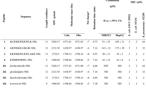
Three peptides (1, 2, and 3) exhibited growth inhibition in combination with rifampicin or erythromycin 120
(Figure S1). A fourth peptide (4) was selected for further analysis due to its ability to enhance susceptibility 121
to both rifampicin and erythromycin at 0.5 µM (Figure S1). All four peptides had a low MIC of 2 µM 122
against E. coli ATCC 25922. These peptides were all short (9 to 13 residues), highly cationic, and possessed 123
similar hydrophobicity as estimated from their retention in reversed-phase analytical HPLC (Table 1) [15– 124
17]. None of the 34 peptides induced susceptibility to linezolid or vancomycin. 125
The ability of the four identified peptides to induce antibiotic susceptibility in two epidemic MDR 126
clones with high clinical relevance (i.e., E. coli ST131 and K. pneumoniae ST258) was evaluated by 127
determining the MICs of rifampicin, erythromycin, clindamycin and azithromycin in combination with low 128
concentrations (≤ 1 µM) of peptide. In the presence of sub-inhibitory concentrations of peptide (Table 1), 129
the MICs of the antibiotics were reduced considerably, resulting in synergistic peptide-antibiotic 130
combinations with estimated FICIs ranging from 0.02 for rifampicin to 0.38 for azithromycin (Tables 2 and 131
S1). The reductions in antibiotic MICs ranged from 8-fold, for azithromycin in combination with peptides 132
1 and 4, to ≥250-fold, for rifampicin in the presence of peptides 2 and 3. All four peptides reduced the MICs 133
of rifampicin and azithromycin to below susceptibility breakpoints [11] in both strains. For clindamycin, 134
the most favorable interactions were observed for peptides 2 and 3 in E. coli ST131 with a reduction of the 135
MICs below the resistance breakpoint [11]. In K. pneumoniae ST258, the clindamycin MICs remained 136
above the resistance breakpoint despite of ≥32-fold reduction of the MICs. Susceptibility to erythromycin 137
was not achieved, most likely due to the high MICs of this macrolide in the two strains (256 and 512 µg/mL, 138
respectively). Consequently, azithromycin was chosen as the representative macrolide for further analyses. 139
For the above combinations that reduced the antibiotic MICs below the resistance breakpoints, synergy was 140
As a preliminary evaluation of the toxicity, and thus potential for clinical application, we determined 142
the cytotoxicity for peptides 1-4 in two relevant eukaryotic cell lines (Table 1). Peptides 1 and 2 exhibited 143
a low cytotoxicity with IC50 values above 50 µM in mouse fibroblasts (NIH 3T3) and ≥100 µM in human 144
hepatocytes (Hep G2), while peptides 3 and 4 reduced cell viability with IC50 values of 19-43 µM (Table 145
1). Regardless, the peptide-antibiotic combinations were non-toxic at synergistic concentrations (Figure 146
S2). 147
All-D analogs of the four selected peptides (denoted as D-peptides D1-D4 hereafter), were synthesized 148
and tested for their ability to induce susceptibility of MDR Gram-negative pathogens to azithromycin, 149
rifampicin and clindamycin. The all-D analogs retained the MICs of the corresponding L-forms (Table 1), 150
and exhibited synergy with the antibiotics in MDR E. coli ST131 and K. pneumoniae ST258 (Table S2). 151
Based on their activity profiles, both forms would be expected to retain similar toxicity profiles while the 152
D-peptides are expected to have greater proteolytic stability [18]. As peptides 1 and 2 alone had significantly 153
lower cytotoxicity compared to peptides 3 and 4, it is likely that D1 and D2 will retain better safety profiles 154
as compared to D3 or D4; hence the first two D-peptides were studied further. 155
We further tested the activity of D1 and D2 in combination with the same three antibiotics 156
(azithromycin, rifampicin and clindamycin) by using a collection of reference and clinical isolates of E. 157
coli, K. pneumoniae, A. baumannii and P. aeruginosa. Overall, the MICs of rifampicin and azithromycin 158
were reduced to below their respective susceptibility breakpoints in three reference strains when co-exposed 159
to sub-MIC concentrations of peptides D1 and D2, while the MIC of azithromycin was reduced to 2-fold 160
above the susceptibility breakpoint in P. aeruginosa (Table S3). Similarly, the MICs of clindamycin were 161
below the resistance breakpoint for K. pneumoniae and A. baumannii. In P. aeruginosa, the peptide-162
antibiotic combinations were overall not synergistic, and only borderline synergy was observed for 163
combinations with rifampicin (Table S3). Overall, the antibacterial activity of the D-peptide-antibiotic 164
S4). Most (88%) and ≥50% of the isolates, except for P. aeruginosa, were inhibited by the D-peptides in 166
combination with rifampicin and azithromycin, respectively (Table S4). 167
The above synergistic combinations were further investigated in growth curve assays, which showed 168
that neither peptide nor antibiotic individually inhibited growth of E. coli or K. pneumoniae at the 169
concentration present in the synergistic combination (Figure S3 A-J). However, A. baumannii growth was 170
retarded in the presence of each antibiotic or peptide D2 alone (Figure S3, K-P). 171
Peptide D2 was studied further to understand the bactericidal kinetics of D2-antibiotic combinations. 172
This peptide was chosen based on its high potency in synergistic combinations. Time-kill experiments with 173
D2 in combination with antibiotics and alone were performed with reference strains of A. baumannii and 174
K. pneumoniae which served as the representative of Enterobacteriaceae. All D2-antibiotic combinations 175
exerted synergistic bactericidal effects in the time-kill assay (Figure 1, A-F). Moreover, at sub-MIC 176
concentrations of D2 (i.e., ≤2 µM), all antibiotic concentrations were below their respective susceptibility 177
breakpoints, except for clindamycin in K. pneumoniae. 178
Time-kill kinetics of the antibiotics, D2, and their combinations were compared to examine whether 179
the D2-antibiotic combinations were able to enhance the rate and efficiency of killing relative to either 180
component individually. In both species, faster killing kinetics were achieved for the D2 combinations with 181
clindamycin and rifampicin than for either antibiotic alone (Figure S4B-C and E-F). The D2-azithromycin 182
combination also exhibited faster killing kinetics than azithromycin alone in A. baumannii (Figure S4D), 183
while the combination displayed similar kinetics in K. pneumoniae (Figure S4A). However, D2 did not 184
exhibit efficient killing in K. pneumoniae (Figure S5)even at concentrations 8-fold above the MIC (Table 185
S5). 186
The approach developed in this study, which combines combinatorial screening at low compound 187
concentrations with cytotoxicity testing, can be used to expedite discovery of clinically relevant peptide-188
antibiotic combinations. This approach enabled rapid identification of two peptides (1 and 2) that at low 189
rifampicin in multiple Gram-negative species of clinical relevance, including epidemic MDR clones. 191
Furthermore, the all-D peptide analogs induced susceptibility to rifampicin and azithromycin and reduced 192
the MICs of clindamycin by more than 500-fold. These findings may help mitigate the lack of novel 193
antibiotics effective against Gram-negative species by opening new avenues to repurpose these antibiotics 194
for treatment of infections caused by Gram-negative MDR pathogens. 195
Peptides 1 and 2 as well as their all-D analogs (D1 and D2) exhibited substantial synergy with 196
rifampicin, azithromycin, and clindamycin in K. pneumoniae and A. baumannii (Tables S2 and S3) at low 197
(≤1 µM) non-toxic peptide concentrations. The present study constitutes the first report on antibiotic 198
synergy of these peptides, while their antimicrobial activity, cytotoxic and haemolytic properties were 199
reported previously [19,20]. Notably, according to these studies peptides 1 and 2 do not exhibit haemolytic 200
activity at concentrations ≥200 µM. The use of the analogue D2 appears to be particularly promising for 201
antibiotic potentiation since D2-antibiotic combinations displayed synergistic bactericidal activity (Figure 202
2), and faster killing kinetics than each individual component (Figure 3). Importantly, in vivo PK/PD and 203
toxicity studies are needed to fully assess the clinical potential of these findings. 204
205
4. Conclusions 206
Intrinsic resistance to azithromycin and rifampicin in Gram-negative bacteria can be overcome by very low 207
peptide concentrations that are not toxic to eukaryotic cells. The two peptide leads identified in this study 208
merit further investigation as antibiotic potentiators for repurposing azithromycin and rifampicin against 209
MDR Gram-negative pathogens. 210
211
The study was supported by the University of Copenhagen research Centre for Control of Antibiotic 213
Resistance (UC-Care), and by a grant from Zoetis. We thank Laurent Poirel for contributing the panel of 214
References 216
[1] World Health Organisation. 2017. Global Priority List Of Antibiotic-Resistant Bacteria To Guide 217
Research, Discovery, and Development Of New Antibiotics. World Health Organisation. 218
http://www.who.int/medicines/publications/global-priority-list-antibiotic-resistant-bacteria/en/ 219
(accessed Jan 10 2018). 220
[2] Olivares J, Bernardini A, Garcia-Leon G, Corona F, Sanchez MB, Martinez JL. The intrinsic 221
resistome of bacterial pathogens. Front Microbiol 2013;4:1–15. doi:10.3389/fmicb.2013.00103. 222
[3] Tamma PD, Cosgrove SE, Maragakis LL. Combination Therapy for Treatment of Infections with 223
Gram-Negative Bacteria. Clin Microbiol Rev 2012;25:450–70. doi:10.1128/CMR.05041-11. 224
[4] Melander RJ, Melander C. The Challenge of Overcoming Antibiotic Resistance : An Adjuvant. 225
ACS Infect Dis 2017;3:559–563. doi:10.1021/acsinfecdis.7b00071. 226
[5] Zabawa TP, Pucci MJ, Parr TRJ, Lister T. Treatment of Gram-negative bacterial infections by 227
potentiation of antibiotics. Curr Opin Microbiol 2016;33:7–12. doi:10.1016/j.mib.2016.05.005. 228
[6] Vaara M. Agents that increase the permeability of the outer membrane. Microbiol Rev 229
1992;56:395–411. 230
[7] Cerquetti M, Giufrè M, García-Fernández A, Accogli M, Fortini D, Luzzi I, et al. Ciprofloxacin-231
resistant, CTX-M-15-producing Escherichia coli ST131 clone in extraintestinal infections in Italy. 232
Clin Microbiol Infect 2010;16:1555–8.. 233
[8] Jana B, Cain AK, Doerrler WT, Boinett CJ, Fookes MC, Parkhill J, et al. The secondary resistome 234
of multidrug-resistant Klebsiella pneumoniae. Sci Rep 2017:1–10. doi:10.1038/srep42483 235
[9] Hansen AM, Bonke G, Larsen CJ, Yavari N, Nielsen PE, Franzyk H. Antibacterial Peptide Nucleic 236
Acid-Antimicrobial Peptide (PNA-AMP) Conjugates: Antisense Targeting of Fatty Acid 237
Biosynthesis. Bioconjug Chem 2016;27:863–7. doi:10.1021/acs.bioconjchem.6b00013. 238
[10] Clinical and Laboratory Standards Institute. 2013. Performance standards for antimicrobial disk 239
CLSI document VET01-A4. Clinical and Laboratory Standards Institute, Wayne, PA. 241
[11] Clinical and Laboratory Standards Institute. 2017. Performance Standards for Antimicrobial 242
Susceptibility Testing. 27th ed. CLSI M100. Wayne, PA. 243
[12] Garcia LS, editor. Synergism Testing: Broth Microdilution Checkerboard and Broth Macrodilution 244
Methods. Clin. Microbiol. Proced. Handbook, 3rd Ed., American Society of Microbiology; 2010, p. 245
140–62. doi:10.1128/9781555817435.ch5.12. 246
[13] Baker KR, Jana B, Franzyk H, Guardabassi L. A High-Throughput Approach To Identify 247
Compounds That Impair Envelope Integrity in Escherichia coli. Antimicrob Agents Chemother 248
2016;60:5995–6002. doi:10.1128/AAC.00537-16. 249
[14] Jahnsen RD, Sandberg-Schaal A, Vissing KJ, Nielsen HM, Frimodt-Møller N, Franzyk H. 250
Tailoring cytotoxicity of antimicrobial peptidomimetics with high activity against multidrug-251
resistant Escherichia coli. J Med Chem 2014;57:2864–73. doi:10.1021/jm401335p. 252
[15] Jahnsen RO, Sandberg-Schaal A, Frimodt-Møller N, Nielsen HM, Franzyk H. End group 253
modification: Efficient tool for improving activity of antimicrobial peptide analogues towards 254
Gram-positive bacteria. Eur J Pharm Biopharm 2015;95:40–6. doi:10.1016/j.ejpb.2015.01.013. 255
[16] Schmitt MA, Weisblum B, Gellman SH. Interplay among folding, sequence, and lipophilicity in 256
the antibacterial and hemolytic activities of α/β-peptides. J Am Chem Soc 2007;129:417–28. 257
doi:10.1021/ja0666553. 258
[17] Chongsiriwatana NP, Patch JA, Czyzewski AM, Dohm MT, Ivankin A, Gidalevitz D, et al. 259
Peptoids that mimic the structure, function, and mechanism of helical antimicrobial peptides. Proc 260
Natl Acad Sci 2008;105:2794–9. doi:10.1073/pnas.0708254105. 261
[18] Méndez-Samperio P. Peptidomimetics as a new generation of antimicrobial agents: Current 262
progress. Infect Drug Resist 2014;7:229–37. doi:10.2147/IDR.S49229. 263
[19] Hyon K, Nan YH, Park Y, Kim JI, Park I, Hahm K, et al. Cell specificity, anti-inflammatory 264
Biochim Biophys Acta 2009;1788:1193–203. doi:10.1016/j.bbamem.2009.02.020. 266
[20] Monincova L, Budesinsky M, Slaninova J, Hovorka O, Cvacka J, Voburka Z, et al. Novel 267
antimicrobial peptides from the venom of the eusocial bee Halictus sexcinctus ( Hymenoptera : 268
Halictidae) and their analogs. Amino Acids 2009;39:763-775.. doi:10.1007/s00726-010-0519-1. 269
Tables
271
Table 1: Peptide sequences, physicochemical characteristics, cytotoxicity and MIC in E. coli and K. pneumoniae 272 Pepti d e Sequence Le n g th (r esid u es) MW (g /m o l) Mo lec u la r m a ss (Da ) Ne t ch a rg e a Re te n tio n tim e (m in ) MIC (µM) Cytotoxicity (µM)
IC50 ( ± 95% CI)
E. c o li ATC C 2 5 9 2 2 E. c o li S T1 3 1 K. p n eu m o n ia e S T2 5 8
Calc. Obs. NIH3T3 HepG2
1 KLWKKWKKWLK-NH2 11 2369.17 1571.01 1571.02 +7 6.73 51 ± 35 105 ± 21 2 2 64
2 GKWKKILGKLIR-NH2 12 2121.92 1438.97 1438.97 +6 7.14 143 ± 21 175 ± 38 2 2 32
3 KKWRKWLKWLAKK-NH2 13 2710.5 1798.15 1798.16 +8 6.93 20 ± 13 19 ± 3 2 2 4
4 KWRRWIRWL-NH2 9 1968.85 1398.84 1398.84 +5 7.42 43 ± 16 34 ± 8 2 2 4
D1 klwkkwkkwlk-NH2 11 2369.17 1571.01 1571.00 +7 6.66 NDb ND 2 2 64
D2 gkwkkilgklir-NH2 12 2121.92 1438.97 1438.97 +6 7.16 ND ND 2 2 32
D3 kkwrkwlkwlakk-NH2 13 2710.5 1798.15 1798.14 +8 6.89 ND ND 2 2 4
D4 kwrrwirwl-NH2 9 1968.85 1398.84 1398.84 +5 7.38 ND ND 2 2 4
273
a Charge at pH 7.4.
274
bND, Not determined
Table 2: MICs of azithromycin (AZM), erythromycin (ERY), rifampicin (RIF) and clindamycin (CLI) in E. coli ST131 and K. pneumoniae ST258 276
exposed to low concentrations of peptides 1-4.Antibiotic MICs below the susceptibility breakpoint are in bold and MICs below the resistance 277
breakpoint are underlined. 278
279
a CLSI clinical breakpoints for Staphylococcus species. S= susceptible; R= resistant.
280
281
E. coli ST131 K. pneumoniae ST258
Antibiotic (µg/mL) 0.5 µM peptide 1 µM peptide Clinical breakpointa
1 2 3 4 None 1 2 3 4 None ≤S ≥R
AZM 1 ≤0.25 0.25 1 8 2 1 1 2 32 2 8
ERY 4 4 1 4 256 8 8 4 8 512 0.5 8
RIF 0.25 ≤0.03 ≤0.03 0.25 4 0.125 0.06 ≤0.03 0.6 16 1 4
282
Figure 1: Peptide D2-antimicrobial combination kills bacteria synergistically. Time-kill kinetics for azithromycin (AZM), rifampicin (RIF) and 283
clindamycin (CLI) as individual compounds and in combination with D2 are presented for both K. pneumoniae ATCC 13883 (A-C) and A. 284
baumannii ATCC 19606 (D-F), including only D2 and untreated control. The curves of the synergistic combination and the untreated control are 285