Original Research Article
Study of Nasal Mucosal Flora in Acute and Chronic Sinusitis
Rupeshkumar TS
1, Rajesh K
2, Prabhusaran N
3*, Jesudoss A
41MBBS Student,
Chennai Medical College Hospital and Research Centre (SRM Group), Tiruchirapalli, Tamil Nadu, India. [Affiliated to The Tamilnadu Dr. M.G.R. Medical University, Chennai].
2Associate Professor, 4Professor & Head, Department of ENT,
Chennai Medical College Hospital and Research Centre (SRM Group), Tiruchirapalli, Tamil Nadu, India. [Affiliated to The Tamilnadu Dr. M.G.R. Medical University, Chennai].
3*Associate Professor, Department of Microbiology,
Chennai Medical College Hospital and Research Centre (SRM Group), Tiruchirapalli, Tamil Nadu, India. [Affiliated to The Tamilnadu Dr. M.G.R. Medical University, Chennai].
ABSTRACT
Introduction: Microbiological investigation and selection of antimicrobial agents are important for infection control and to avoid complications. It is also suggested to have continuous medical education for effective and early diagnosis, as well as providing appropriate care. The objective of the study is to analyze the microflora present in patients with acute and chronic sinusitis in the tertiary care teaching hospital.
Methods: A cross sectional hospital based study was
conducted in order to determine the microorganism of paranasal sinuses in patients with acute and chronic sinusitis undergoing endoscopic sinus surgery. Swabs/ biopsy were taken from the infected sinus and were analyzed microbiologically within 4 hours of collection.
Results: The bacterial isolates including Staphylococcus aureus accounted for the maximum at 37.5% followed by
Klebsiella spp., 7% and MRSA 2%. The fungal isolates including Aspergillus flavus, Candida albicans and C. tropicalis
showed 8% and are considered as differential diagnosis in tropical regions. Further no cultural prints of anaerobes were found.
Conclusion: Based on this research work, it was identified that bacterial and fungal pathogens play vital role in causing infections in the sinuses for which empirical antibiotic therapy will be the choice for better management of the condition.
Keywords: Acute, Chronic, Sinusitis, Nasal Mucosa,
Microbiology.
*Correspondence to:
Dr. Prabhusaran N,
Associate Professor, Department of Microbiology, Chennai Medical College Hospital and Research Centre, Tiruchirapalli, Tamil Nadu, India.
Article History:
Received: 19-02-2017, Revised: 14-03-2017, Accepted: 22-03-2017
Access this article online Website:
www.ijmrp.com
Quick Response code
DOI:
10.21276/ijmrp.2017.3.2.029
INTRODUCTION
Most of the sinus infections are viral and few develop secondary bacterial infections. Rhinoviruses, influenza viruses and parainfluenza viruses are the most common causes of sinusitis.1
The most common bacteria isolated from paediatric and adult patients with community acquired acute purulent sinusitis are
Streptococcus pneumoniae, Haemophilus influenzae, Moraxella catarrhalis and Streptococcus pyogenes. Staphylococcus aureus
and anaerobic bacteria (Prevotella and Porphyromonas,
Fusobacterium and Peptostreptococcus spp.) are the main isolates in chronic sinusitis. Pseudomonas aeruginosa and other aerobic and facultative gram negative rods are commonly isolated from patients with nosocomial sinusitis, the immunocompromised host, those with HIV infection and in cystic fibrosis.1,2 Fungi and
Pseudomonas aeruginosa are the most common isolates in neutropenic patients. The microbiology of sinusitis is influenced by the previous antimicrobial therapy, vaccinations and the presence
of normal flora capable of interfering with the growth of pathogens. Acute exacerbation of chronic sinusitis (AECS) represents a sudden worsening of the baseline chronic sinusitis with either worsening or appearance of new symptoms. Typically, the acute (not chronic) symptoms resolve completely between occurrences. An increase in antimicrobial resistance was also observed and has increased now a days.
Recommended treatments for most cases of sinusitis include rest and drinking enough water to thin mucus. Antibiotics are not recommended for most cases Decongestant nasal sprays containing oxymetazoline may provide relief.3 However, if
Surgery should only be considered for those people who do not benefit with medication. Chronic sinusitis is an infection of sinuses lasting for more than three months. Despite its prevalence the disease remains with poorly understood origin, pathogenesis and natural history. The etiology of chronic sinusitis continues to be the focus of much debate and research in the field of rhinology.5,6
With initial use of antibiotics and agents that decrease mucosal edema now surgical methods are employed in whom medical treatment fails. Although diagnostic criteria for acute sinusitis are well established yet the definition of chronic sinusitis is controversial with respect to the importance of bacteria in the initiation and progression of disease. Chronic sinusitis has been considered to be chronic inflammatory condition rather than microbial infection. The role of bacteria in the pathogenesis of chronic sinusitis is currently being reassessed.6,7
Fungal colonization of the nose and paranasal sinuses appears to be a common finding in both normal and diseased states. Fungal rhinosinusitis (FRS) is increasing in prevalence; it causes significant physical symptoms, negatively affects quality of life and it can substantially impair daily functioning.8
The use of endoscopies has made it possible to determine the microbiology of each sinus with a lower probability of contamination.6,9
Persistence of infection causes mucosal changes such as loss of cilia, edema and polyp formation. We feel that lack of progress is largely due to paucity of knowledge in microbiology and histopathology of chronic sinus disease available to us. This was the impetus of our study to evaluate the microbiology of acute and chronic sinusitis in patients.
MATERIALS AND METHODS
This study is the cross sectional study conducted from June to August 2016. After getting approval from institutional ethical committee (IEC-No:30 dt. 15.04.2016) and informed consent from the participants, the details were collected.
In the present study, microbiological aspects were strictly followed according to the standard institutional operating protocols. The study was conducted in tertiary care teaching hospital, Tamilnadu. After explaining the study methodology to the participants/ attenders, details were collected from 50 patients attended in ENT OP. Their socio-demographic status (age, sex and occupation), clinical complications, and laboratory parameters (microbiological)
were collected and entered in the proforma.
Inclusion criteria: Both sexes of all age groups; patients with acute and chronic inflammatory disease of sinuses; patients with chronic sinusitis with no response to initial treatment; patients with recurrent sinusitis; and patients not on antibiotics at least for 2 weeks were considered.
Exclusion criteria: The patients with malignancy of paranasal sinuses and patients on recent antibiotics were excluded from study.
Sample collection: The endoscope was sterilized in a
glutaraldehyde solution for 10 min and washed prior to use. Endoscopic examination of the nasal cavity and the sinuses was done on all the patients. Endoscopic specimens were collected from nasal mucosa, which was not disinfected, and this culture was considered representative of the background nasal flora. Swab specimens for microbiological analysis aseptically transferred into a transport medium, and transported immediately to the laboratory.
Macroscopic observation: The specimens were examined
macroscopically for
a. Appearance (color) and
b. Blood stain
Bacterial isolation: Within 1 hour of collecting the specimen, the specimens were cultured using Nutrient agar, Blood Agar and MacConkey Agar, and were incubated at 37°C for 24 hours. The cultures were examined for growth; the isolation and identification of bacterial isolates were carried out in accordance with Bergey’s Manual of Determinative Bacteriology.10,11
Fungal isolation: The specimens were inoculated in Saboroud’s Dextrose agar and incubated at room temperature for 3 to 4 days. The cultures were examined for growth; the isolation and identification of fungal isolates by colony morphology and microscopy using lactophenol cotton blue staining.
RESULTS
In this study endoscopic nasal mucosal swabs were collected from 50 patients who attended the ENT OP and also from ten healthy controls.
Patients’ Demography and Interventions
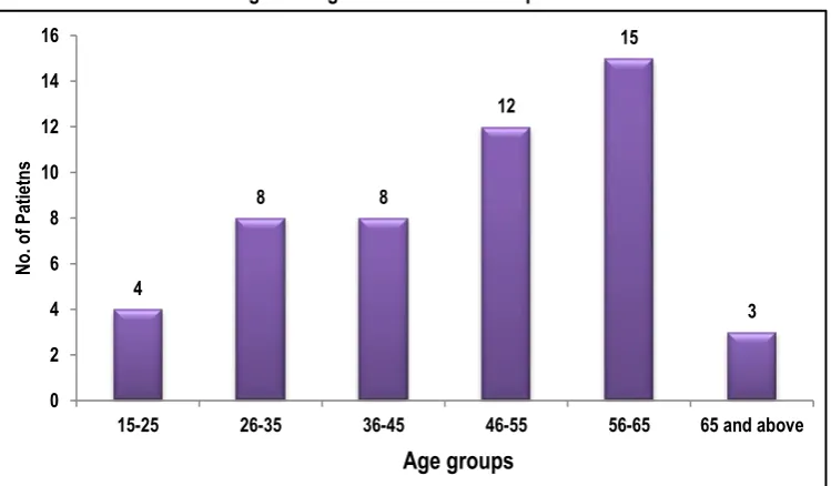
In this study, among the 50 patients included, 31 (62%) were males and 19 (38%) were females. (Figure 1) The distribution of cases in relation to age group is depicted in figure 2.
Figure 1: Sex wise distribution of the patients attended ENT OPD with the symptoms of acute and chronic sinusitis
31 19
Male
Figure 2: Age wise distribution of patients
Table 1: Symptomology of the patients included in the study
Symptom No. of patients Percentage
Nasal discharge 27 54
Nasal obstruction 38 76
Headache 39 78
Disturbance of smell 11 22
Epistaxis 3 6
Sneezing 17 34
Sore throat 1 2
Hawking 1 2
Table 2: Microbiological analysis (Subjects – 50 and isolates – 60)
Microorganisms identified No. of isolates verses specimens
Aerobic organisms
(Both seen on smear and isolated)
31/ 50 (62)
Aerobic organism
(Seen on smear but not isolated)
25/ 50 (50)
Anaerobic organisms 0/ 50 (0)
Fungus 4/ 50 (8)
[Figures in parenthesis denoted percentages]
Table 3: Aerobic microorganisms (bacteria) and its frequency (n=56)
Aerobic microorganisms (bacteria) Frequency
Staphylococcus aureus 21 (37.5)
Klebsiella spp. 4 (7.1)
MRSA 1 (1.7)
Streptococcus 1 (1.7)
Enterobacter spp. 1 (1.7)
Acinetobacter spp. 1 (1.7)
Pseudomonas spp. 1 (1.7)
Citrobacter spp. 1 (1.7)
Gram positive bacilli 5 (8.9)
Gram negative bacilli 11 (19.6)
Gram positive cocci 8 (14.2)
Gram neg coccobacilli 1 (1.7)
[Figures in parenthesis denoted percentages]
4
8 8
12
15
3
0 2 4 6 8 10 12 14 16
15-25 26-35 36-45 46-55 56-65 65 and above
N
o. of
Pa
tie
tn
s
Clinical symptoms of the patients
The clinical diagnosis of acute and chronic sinusitis was confirmed in all 50 patients. History of head ache was present in 39 (78%) of 50 patients. The nasal obstruction and nasal discharge was found among 38 (76%) and 27 (54%) respectively. Other clinical symptoms observed in this study subjects are given in table 1.
Microbiology results
Among 50 patients, bacterial and fungal pathogenic organisms were isolated from 31 samples. Out of 50 patients, 31 samples were positive for aerobic organisms, none were positive for
anaerobes, fungal growth was reported in 4 samples. (Table 2) Further the study was extended to determine the type of microorganism by standard microscopic and biochemical characterization, thereby the maximum isolates was
Staphylococcus aureus (21) followed by gram negative bacilli (11) and gram positive cocci (8). The detailed description of aerobic microorganisms and its frequency was interpreted in table 3. The fungal isolation was Aspergillus flavus and Candida species. The detailed description of fungal isolates and its frequency were depicted in table 4 and Figure 3,4.
Table 4: Fungal isolation and its frequency (n=4)
Fungal isolates Frequency
Aspergillus flavus 2 (50)
Candida albicans 1 (25)
C. tropicalis 1 (25)
[Figures in parenthesis denoted percentages]
Figure 3: KOH mount of the sinonasal polyp tissue showing slender septate fungal hyphae
Figure 4: LPCB mount of Aspergillus flavus
DISCUSSION AND CONCLUSION
The microbiology of infections of the acute and chronic sinus diseases can be anticipated according to the patient's age, clinical presentation, and immunocompetence. In acute sinus disease, viral upper respiratory infections frequently precede bacterial
superinfection by Streptococcus pneumoniae, Haemophilus
influenzae and Moraxella catarrhalis. Staphylococci and respiratory anaerobes are common in chronic sinus infection, which may also be caused by exacerbations of infection with the bacterial species that cause acute disease. Enterobacteriaceae may be found in patients with nosocomial sinusitis who are predisposed to the development of sinusitis by prolonged nasogastric and nasotracheal intubation. Immunosuppressed patients have episodes of sinusitis caused by the usual agents associated with acute sinusitis in immunocompetent patients, and they may also become infected with a broad array of unusual agents, including mycobacterial species, fungi, and protozoa.12
In most of the studies, in order to determine the bacteriologic cause of acute sinusitis, a sample of sinus secretions must be obtained from one of the paranasal sinuses without contamination by normal respiratory or oral flora that colonize mucosal surfaces. When maxillary sinus aspiration is performed on children who have signs and symptoms of acute sinusitis, bacteria are recovered in high density from 70%. In patients with acute, subacute, or chronic sinusitis who are generally well except for
persistent respiratory symptoms, of nasal discharge or cough or both;13 the same technique also included in this study also.
In many studies, the biofilm of bacterial infections may account for many cases of antibiotic refractory chronic sinusitis4 and of 75% of
patients undergoing surgery for chronic sinusitis.14 Biofilms are
complex aggregates of extracellular matrix and interdependent microorganisms from multiple species, many of which may be difficult or impossible to isolate using standard clinical laboratory techniques. These bacteria have their antibiotic resistant increased up to 1000 times when compared to free living bacteria of same species. We attempted to follow a consistent and reproducible technique for isolating bacterial pathogens from the specimens. The non-isolation of bacterial pathogens may be due to technical factors, improper processing, non – infective/ allergic pathology or patients on past infections, now getting treated. The microbiology of chronic sinusitis with polyposis considered as the major isolates of polymicrobial aerobic-anaerobic flora. Fungal colonization of the nose and paranasal sinuses is common in the normal and inflamed sinuses.15 The role of fungi in the
Even though acute and chronic rhinosinusitis have similar symptoms, but severity is more among acute sinusitis. The signs and symptoms of cases were correlated with radiographic results. The most frequent microorganisms found in CRS patients were:
Staphylococcus aureus, (37.5%); followed by Klebsiella pneumoniae (7.1%) etc. There was no growth of bacteria in 19 patients (38%). A short course of antibiotics was effective and patients improved well. The major strength of the study is rigid criteria adopted to select subjects and controls and good laboratory practice adopted to process the samples. Further, this study has certain limitations including
1. This study is made from single centre and limited to aerobic bacterial and fungal organisms.
2. The transversal studies tend to overestimate the long duration of infections and under estimate the short duration infection. Nevertheless this type of transversal analysis has fundamental importance for the knowledge.
3. The study has several important limitations which require discussion. Patient population at the study area may not be a representative of the community at large.
REFERENCES
1. Slavin RG. The diagnosis and management of sinusitis: A practice parameter update. J Allergy Clin Immunol. 2005; 116: 13-47.
2. Spiegel JH. Sinusitis. Otolaryngol Clin North Am. 2004, 37: 221-506.
3. Mustafa M, Patawari P, Iftikhar HM, Shimmi SC, Hussain SS, Sien MM. Acute and chronic rhinosinusitis, pathophysiology and treatment. Int J Pharm Sci Invention. 2015; 4: 30-36
4. Palmer JN. Bacterial biofilms: do they play a role in chronic sinusitis? Otolaryngol Clin North Am. 2005; 38: 1193-1201. 5. Aral M, Keles E, Kaygusuz I. The microbiology of ethmoid and maxillary sinuses in patients with chronic sinusitis. Am J Otolaryngol. 2003; 24: 163-68.
6. Kamath PM, Shenoy VS. Nithin M, Nitish S. Microbiological analysis of paranasal sinuses in chronic sinusitis – a south Indian coastal study. Egypt J ENT All Sci. 2013; 14: 185-189.
7. Niederfuhr A, Kirsche H, Riechelmann H. The bacteriology of chronic rhinosinusitis with or without nasal polyps. Arch Otolaryngol Head Neck Surg. 2009; 135: 234-239.
8. Usha KK, Agatha D, Selvi R. Fungal rhinosinusitis: a clinicomycological perspective. Ind J Med Microbiol. 2015; 33: 120-124.
9. Busaba NY, Siegel NS, Salman SD. Microbiology of chronic ethmoid sinusitis. J Otolaryngol. 2004; 25: 379-384.
10. Buchanan, R.E. and E.N. Gibbons. Bergey’s Manual of Determinative Bacteriology. 8 edn. Williams and Wilken. Co., Baltimore., 1978. pp 1300-1312.
11. Gerhardt P, Murray RGE, Wood WM, Krieg NR. Methods for General and Molecular Bacteriology. ASM Press, Washington DC., 1994; pp 791-800.
12. Wald ER. Microbiology of acute and chronic sinusitis in children and adults. Am J Med Sci. 1998; 316: 13-20.
13. Wald ER. Microbiology of acute and chronic sinusitis in children. J Allerg Clin Immunol. 1992; 90: 452-456.
14. Sanclement JA, Webster P, Thomas J. Bacterial biofilms in surgical specimens of patient with chronic rhinosinusitis. Laryngscope. 2005; 115: 578-582.
15. Ebbens FA, Georgalas C, Fokkens WJ. Fungus as the cause of chronic rhinosinusitis: the case remains unproven. Curr Opin Otolaryngol Head Neck Surg. 2009; 17: 43-49.
16. Ferguson BJ. Definitions of fungal rhinosinusitis. Otolaryngol Clin North Am. 2000; 33: 227-235.
[
Source of Support: Nil.
Conflict of Interest: None Declared.
Copyright: © the author(s) and publisher. IJMRP is an official publication of Ibn Sina Academy of Medieval Medicine & Sciences, registered in 2001 under Indian Trusts Act, 1882. This is an open access article distributed under the terms of the Creative Commons Attribution Non-commercial License, which permits unrestricted non-commercial use, distribution, and reproduction in any medium, provided the original work is properly cited.
Cite this article as: Rupeshkumar TS, Rajesh K, Prabhusaran N, Jesudoss A. Study of Nasal Mucosal Flora in Acute and Chronic Sinusitis. Int J Med Res Prof. 2017; 3(2):144-48.