Bosn J Basic Med Sci 2013; 13 (2): 114-118Abstract
Th e objective of our work was to evaluate the eff ect of interferon-γ (IFN-γ) on cytokine expression in rat acute pancreatitis (AP).
AP was introduced to rats which were divided into Control, AP and IFN-γ group. Rats in the AP and IFN-γ group were sacrifi ced as , and h after IFN-γ treatment. Th e serum amylase (AMA), endotoxin and cytokines were detected. Th e pathological examination and immuno-fl uorescence staining of pancreas for TNF-α, NF-κB and IL- were performed.
Th e serum AMA increased signifi cantly at h and reduced at h after AP. Th e increase in IFN-γ was higher than that in AMA. IL- in-creased in the AP and IFN group, and IFN inin-creased markedly at h after AP. IL- reduced at h after AP compared with AP group. In the AP group, the immunostaining of cytokines increased. In the IFN group, the edema in the pancreas was more severe, and NF-κB and IL- expression was higher than that in the other two groups. IFN-γ can increase serum IL- and reduce IL- in AP.
IFN-γ can increase serum IL- and reduce serum IL- in AP. Th e increase in NF-κB and IL- may exert infl uence on pro-infl ammatory cytokines to deteriorate infl ammation in the pancreas. Th us, to control the IFN-γ might has promise to attenuate pancreatitis.
© Association of Basic Medical Sciences of FB&H. All rights reserved
KEY WORDS: pancreatitis, interferon-γ, nuclear factor-κB, tumor necrosis factor-α, interleukin-
IL-18 and IL-27 in acute pancreatitis
Hongbo Meng1, Jian Gong1, Lin Fang1*, Zhenshun Song1*, Fan Wu1, Bo Zhou1, Mingping Qian1
1Department of General Surgery, Tenth People’s Hospital, Tongji University School of Medicine, 301 Middle Yanchang Road, Shanghai
200072, China. 2Department of Science and Education, Tenth People’s Hospital, Tongji University School of Medicine, 301 Middle
Yanchang Road, Shanghai 200072, China.
INTRODUCTION
Acute pancreatitis is a clinical entity that is believed to have intracellular activation of digestive enzymes and autodiges-tion of the pancreas as its central pathophysiologic cause [, ]. As early as s and s, investigators felt that some mediators released during AP entered the systemic circulation, thereby causing dysfunction of distant organs []. Eff orts to unravel the events in the evolution of tissue damage in AP have shown a number of infl ammatory me-diators to be involved [, ]. Th e proinfl ammatory response is countered by an anti-infl ammatory response, and an im-balance between these two systems leads to localized tis-sue destruction and distant organ damage. Cytokines lie at the heart of the problem and are involved in all aspects
of the cascade leading to systemic infl ammatory response syndrome and multiple organ dysfunction syndrome []. Acute pancreatitis (AP) is a common disease in clinical prac-tice, and its pathogenesis is closely related to the abnormal activation of pancreatin, microcirculatory disturbance, reper-fusion induced injury and release of infl ammatory mediators and cytokines. Recently, increasing attention has been paid to the role of cytokines in the pathogenesis of AP. In the pres-ent study, AP was introduced to rats which were then treated with interferon-γ (IFN-γ) and the cytokines were measured before and after treatment. Th is study aimed to investigate the potential eff ect of IFN-γ on the production of cytokines in AP.
MATERIALS AND METHODS
Preparation of AP animal model
The study was conducted in the Animal Center of Shang-hai Tenth People’s Hospital. Male SD rats (n=) weighing - g were randomly assigned into control group, AP group and IFN-γ group. Rats in the AP group and IFN-γ were sacrifi ced at h, h, h and h after IFN-γ treat-ment (n= per subgroup). In the AP group, rats were fasted
Hongbo Meng and Jian Gong contributed equally * Corresponding authors: Lin Fang and Zhenshun Song,
Department of General Surgery, Tenth People’s Hospital, Tongji University School of Medicine, 301 Middle Yanchang Road, Shanghai 200072, China Tel: +8621-66301057; Fax: +8621-66301051
E-mail: [email protected], [email protected]
Bosn J Basic Med Sci 2013; 13 (2): 115-118
for h. Following anesthesia with diethyl ether and routine sterilization, ligation of duodenum was done to induce AP during which a catheter was inserted in the intestine aiming to avoid intestinal obstruction []. In the IFN-γ group, IFN-γ was injected via the tail vein. In the control group, duodenum was not ligated. After surgery, animals were fasted but given
ad libitum access to water. At diff erent time points, animals were sacrifi ced and the heart, mesenteric lymph nodes, pan-creas, iliac lymph nodes and ilium were collected for use.
Detections
Automatic biochemical analyzer, limulus synthetic ma-trix azo-dye method and ELISA (Wuhan Boster Biotech Co., Ltd) were employed to detect the serum amylase (AMA), endotoxin and cytokines (TNF-α, and IL-), respectively. Under an aseptic condition, the mesen-teric lymph nodes were collected and added to LB medium. One day later, these lymph nodes were seeded into culture plate for bacterial culture. The number of bacterial colo-nies was counted and the type of bacteria also determined.
Pathological examination and immunofl uorescence staining
Th e pancreas was collected and processed for pathological examination with HE staining. Immunofl uorescence staining was done to detect the expression of cytokines in the pancre-as. Th e paraffi n embedded sections were deparaffi nized and dehydrated. After antigen retrieval, sections were treated with . trypsin at oC for min. Following treatment with BSA for blocking, sections were incubated with primary anti-body (:) at oC overnight and then with FITC conjugated secondary antibody. CD was used to label macrophages during which the secondary antibody was labeled with Rho-damine. Incubation was done at oC for h. Mounting was performed with glycerophosphate and sections were observed under a fl uorescence microscope. Th e expression of TNF-α, IL- and NF-κB was detected in these sections.
Statistical analysis
Data were expressed as mean ± standard deviation ( ). Statis-tical analysis was performed with SPSS version . for Win-dows and one way analysis of variance employed for compari-sons. A value of p<. was considered statistically signifi cant.
RESULTS
Changes in serum AMA, endotoxin and IL-
As shown in Table , the serum AMA increased at h after AP and reduced at h, and the increase in AMA was the most obvious in the IFN group. Th e endotoxin increased in diff erent groups to diff erent extents, and the increase was the most evident in the IFN group at h after AP (p<.). Th e amount of bacteria increased in the AP group and IFN group, and was the highest in the IFN group (p<.). TNF-α in-creased in the AP group, but remained unchanged in the IFN group. IL- increased in both AP group and IFN group, and was the highest in the IFN group at h after AP (p<.). IL- increased in both AP group and IFN group, and reached a maximal level at h after AP. At h after AP, the IL- was lower in the IFN group than in the AP group (p <.).
Pathological examination of pancreas
As shown in Figure , the pancreatic cells were intact and swelling of interstitial space was absent (Figure -A). At h after AP, the pancreatic cells were swelling, vascular con-gestion was noted, and swelling of interstitial space was pres-ent accompanied by infi ltration of infl ammatory cells (Figure -B) in the AP group. At h after AP, the above pathologi-cal changes were also found but more severe (Figure -C).
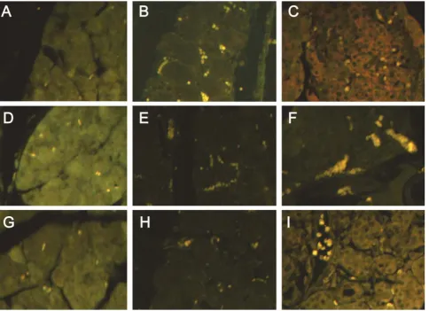
Immunofl uorescence staining of TNF-α, NF-κB and IL- in pancreas
The expression of TNF-α, NF-κB and IL- was detected in the pancreas (Figure ). As shown in the Figure , the
amylase
(Soxhlet unit) Endotoxin (EU)
Migrated colonies (/cm2)
TNF-α (pg/ml)
IL-18 (pg/ml)
IL-27 (pg/ml)
Control 2528.8±630.4 0.19±0.55 0.07±0.14 20.87±2.01 6.58±1.07 30.79±5.65
AP (6 h) 4922.7±1324.2* 0.71±0.34 3.33±2.30* 34.88±7.05* 15.87±14.73 46.13±5.25*
AP (12 h) 5336.9±1591.3* 0.73±0.52* 25.40±12.24* 35.84±14.57 13.73±11.03 68.46±48.82*
AP (24 h) 4159.7±1489.9 1.08±0.03* 63.10±13.39* 34.71±6.98* 30.23±15.59* 46.39±10.76*
AP (48 h) 1710.2±453.1* 0.43±0.05* 50.03±14.35* 23.18±6.69 9.34±2.20* 50.92±13.45*
IFN (6 h) 6880.4±1715.6* 0.68±0.24 78.33±14.37*# 20.26±5.09# 14.72±10.75 47.94±11.90
IFN (12 h) 9603.3±3136.4* 1.21±1.40 130.27±9.70*△
22.35±3.72 15.10±13.21 50.90±12.18*△
IFN (24 h) 10469.5±2099.6*& 1.16±0.97 186.13±23.88*& 20.79±3.23& 27.64±6.44* 47.34±8.27*
IFN (48 h) 2379.8±1708.0 2.14±1.24*☆ 156.83±21.13*☆ 26.28±5.00* 44.01±22.35☆ 54.43±5.02*
F 31.691 5.291 218.276 8.904 8.944 2.715
TABLE 1. Biochemical parameters in rats of diff erent groups (n=10 per group)
Correlation coeffi cient between IL-27 and TNF-α: 0.499, p<0.01; Correlation coeffi cient between IL-27 and IL-18: 0.401, p<0.01; * p<0.05 vs control group;
Bosn J Basic Med Sci 2013; 13 (2): 116-118expression of TNF-α, NF-κB and IL- at a very low level in the control group. The expression of Tα and NF-κB at h after AP and IL- expression at h after AP were markedly increased in the AP group. The TNF-α expression in the IFN group at h was higher than that in the control group but lower than that in the AP at cor-responding time point. The NF-κB expression in the IFN group at h increased significantly. The IL- in the IFN group at h was dramatically increased when com-pared with control group and AP group at after AP.
DISCUSSION
The pathogenesis of AP is complex. Studies have shown that some cytokines play important roles in the pathogen-esis of AP. As an initiator in the occurrence of AP, TNF-α is one of important cytokines aff ecting AP and plays a pro-infl ammatory role in AP at early stage. TNF is the primary member of the inflammatory cytokine family, primary in-ducers of IL- and IL- production, and known to initiate and propagate nearly all of the detrimental consequences of severe sepsis [] nearly all experimental models of
pan-FIGURE 1. Pathological examination of pancreas (HE staining, ×20). A: control group; B: AP group (12 h); C: IFN group (12 h).
Bosn J Basic Med Sci 2013; 13 (2): 117-118
creatitis have implicated TNF as a major pathologic cyto-kine associated with local and systemic tissue destruction. TNF production was closely associated with induction of other genes within their same gene-family []. Likewise, as TNF is produced during AP, so too are its receptors []. IL- is a cytokine that belongs to the IL- superfamily []. IL- levels reflect the severity of acute pancreatitis and display a significant negative correlation with the concen-trations of antioxidative damage factors, serum selenium and glutathione peroxidases (GPx). IL- is one of the key mediators of inflammation in the pathogenesis of acute pancreatitis []. Elevation of serum IL- levels may medi-ate acute pancreatitis associmedi-ated liver injury [, ]. IL- may serve as an additional marker to monitor the severity of infl ammation during pancreatitis since its tissue produc-tion is delayed and appears after that of more commonly investigated cytokines []. IL- appears to protect the pancreas during early induced-induced AP in mice, prob-ably through induction of NO release from an iNOS source. IL- may be a target for new AP therapeutics[]. IL- is mainly produced by monocytes / macrophages and intestinal epithelial cells and endothelial cells also have the ability to secret it. Generally, IL- binds to its receptor ex-erting biological eff ect. In vitro studies reveal that IL- can promote the proliferation of T cells and increase the activity of T cells and NK cells; may elevate the production of some cytokines including TNF-α, IFN-γ, IL- and IL - but reduce the IL- production. In AP patients, the IL- increases sig-nifi cantly and is closely related to the severity of AP. NF-κB is a multifunctional transcriptional factor. Under physiologi-cal conditions, NF-κB binds to IκB leading to the inactivation of NF-κB. Once activated, NF-κB separates from IκB and enter the nucleus initiating the transcription of target genes. There is evidence showing that the NF-κB is significantly activated in AP patients and closely associated with the se-verity of AP. TNF-α is an activator of NF-κB. After binding to receptors on cells, TNF-α may phosphorylate IκB lead-ing to the degradation of IκB which results in activation of NF-κB. Th e activated NF-κB may further increase the syn-thesis and release of TNF-α. In this manner, a large amount of cytokines is released, which further complicates the AP. Interleukin- (IL-) is a heterodimeric cytokine belong-ing to the IL- family and composed of EBI and p pro-tein []. IL- and its receptor WSX- have originally been reported as critical factors for initial commitment of Th differentiation and The Th-promoting effect of IL- has been reported both in in vivo and in vitro experi-ments [, ]. Yoshimura et al found that IL- via IL-R complex (WSX- plus gp) had a suppressive function on cytokine production by fully activated CD+ T cells and significant contribution of STAT to the IL--mediated
cytokine suppression in fully activated CD+ T cells []. Th us, IL-/WSX- have two-sided roles; one is for Th dif-ferentiation and the other for cytokine suppression []. Our results showed IL- increased in AP. Th e changes in IL- might exert effect on AP. Lucas et al. [] reported that IL- could stimulate the generation of IFN-γ by NK cells, promote the proliferation of CD+ T and coordinate the IL- induced diff erentiation of naïve CD+ T cells into Th cells. IL- can up-regulate the ICAM- expression in naïve CD+ T cells, and the interaction between ICAM- and LFA- may promote the diff erentiation of naïve CD+ T cells into Th cells []. In addition, studies also revealed that IL- may exert immunosuppressive eff ect to regulate the immune system, avoid the over-expression of inflam-matory cytokines and inhibit the autoimmune response []. In experiments on mice, IL- was found to attenu-ate the collagen induced arthritis and to inhibit the delayed hypersensitivity. Moreover, Il- could reduce the produc-tion of pro-inflammatory cytokines including IFN-γ []. IFN-γ is mainly produced by T lymphocytes, B lymphocytes, NK cells and monocytes/macrophages, and also known as an immune interferon and plays an important role in the regulation of immune response. In addition, IFN-γ can also promote the proliferation of Th cells and strengthen the im-mune response via Th cells. IFN-γ can also inhibit the activ-ity of Th cells and further suppress the humoral immunity. Th us, IFN-γ is a key factor in the immune response of humans
and possesses multiple bioeff ects. IFN-γ can regulate the ex-pression of CD/, two costimulatory molecules of MHC-I and MHC-MHC-IMHC-I, promote the production of inducible nitric oxide synthesase (iNOS) and activate the oxygen dependent and independent bactericidal system of macrophages leading to the macrophage activation. IFN-γ is a potent activator of macrophages, has the ability to activate macrophages and NK cells and regulate the functions of dendritic cells, T cells and B cells and involves in innate immunity and acquired immunity. IFN-γ can exert synergistic eff ect with LPS to induce the pro-duction of pro-infl ammatory cytokines including IL-, TNF-α and IL- []. In normal intestinal epithelial cells, the TLR expression is at a low level, and LPS fails to induce the NF-κB activation. After incubation with IFN-γ, the TLR expression in the intestinal epithelial cells increases markedly, and sub-sequent stimulation with LPS may induce the potent activa-tion of NF-κB. Th is suggests that IFN-γ can up-regulate the TLR expression in intestinal epithelial cells, facilitate the re-sponse of these cells to LPS and indirectly aff ects NF-κB [].
CONCLUSIONS
Bosn J Basic Med Sci 2013; 13 (2): 118-118are characterized by reduction in IL and increase in IL-. Th is suggests that IFN-γ may inhibit the IL- produc-tion to a certain extent. Currently, the pathway involving in the IFN-γ induced suppression of IL- is still unclear, and might be associated with JAK-STAT mediated tran-scription activation [, ]. Thus, we speculate that to control the IFN-γ may attenuate AP to a certain extent, which provides evidence for the clinical treatment of AP.
ACKNOWLEDGEMENTS
Th is study was supported by the National Natural Science Fund ()
DECLARATION OF INTEREST
Th e authors declare no confl ict of interest.
REFERENCES
[] Leach SD, Gorelick FS, Modlin IM. New perspectives on acute pancreatitis. Scand J Gastroenterol Suppl ;-.
[] Steinberg W, Tenner S. Acute pancreatitis. N Engl J Med. ; ():-.
[] Carey LC. Extra-abdominal manifestations of acute pancreatitis. Surgery ;():-.
[] Mayer J, Rau B, Gansauge F, Beger HG. Infl ammatory mediators in human acute pancreatitis: clinical and pathophysiological implica-tions. Gut ;():-.
[] Norman J. Th e role of cytokines in the pathogenesis of acute pan-creatitis. Am J Surg ;():-.
[] Makhija R, Kingsnorth AN. Cytokine storm in acute pancreatitis. J Hepatobiliary Pancreat Surg ;():-.
[] Su KH, Cuthbertson C, Christophi C. Review of experimental ani-mal models of acute pancreatitis. HPB (Oxford) ;():-. [] Lowry SF. Cytokine mediators of immunity and inflammation.
Arch Surg ;():-.
[] Fink G, Norman J. Pancreatitis induces specific changes in the intrapancreatic expression of the TNF family of genes. Pancreas ;-.
[] de Beaux AC, Goldie AS, Ross JA, Carter DC, Fearon KC. Serum concentrations of infl ammatory mediators related to organ failure in patients with acute pancreatitis. Br J Surg ;():-. [] Lebel-Binay S, Berger A, Zinzindohoue F, Cugnenc P, Th iounn N,
Fridman WH, et al. Interleukin-: biological properties and clini-cal implications. Eur Cytokine Netw ;():-.
[] Perejaslov A, Chooklin S, Bihalskyy I. Implication of interleukin and intercellular adhesion molecule (ICAM)- in acute pancreati-tis. Hepatogastroenterology ;(-):-.
[] Yuan BS, Zhu RM, Braddock M, Zhang XH, Shi W, Zheng MH. Interleukin-: a pro-inflammatory cytokine that plays an im-portant role in acute pancreatitis. Expert Opin Ther Targets ;():-.
[] Ueda T, Takeyama Y, Yasuda T, Matsumura N, Sawa H, Nakajima T, et al. Signifi cant elevation of serum interleukin- levels in pa-tients with acute pancreatitis. J Gastroenterol ;():-. [] Pastor CM, Morel DR, Vonlaufen A, Schiffer E, Lescuyer P,
Frossard JL. Delayed production of IL- in lungs and pancreas of rats with acute pancreatitis. Pancreatology ;():-. [] Ueno N, Kashiwamura S, Ueda H, Okamura H, Tsuji NM,
Hosoha-ra K, et al. Role of interleukin in nitric oxide production and pan-creatic damage during acute pancreatitis. Shock ;():-.
[] Pfl anz S, Timans JC, Cheung J, Rosales R, Kanzler H, Gilbert J, et al. IL-, a heterodimeric cytokine composed of EBI and p protein, induces proliferation of naive CD(+) T cells. Immunity ;():-.
[] Yoshida H, Hamano S, Senaldi G, Covey T, Faggioni R, Mu S., et al. WSX- is required for the initiation of Th responses and resis-tance to L. major infection. Immunity. ;():-. [] Chen Q, Ghilardi N, Wang H, Baker T, Xie MH, Gurney A, et al.
Development of Th -type immune responses requires the type I cytokine receptor TCCR. Nature ;():-. [] Yoshimura T, Takeda A, Hamano S, Miyazaki Y, Kinjyo I, Ishibashi
T, et al. Two-sided roles of IL-: induction of Th diff erentiation on naive CD+ T cells versus suppression of proinfl ammatory
cyto-kine production including IL--induced IL- on activated CD+ T
cells partially through STAT-dependent mechanism. J Immunol ;():-.
[] Yoshida H, Hamano S, Miyazaki Y. Th e double identity of WSX- (IL-R) as an initiator and attenuator of immune responses. Curr Immunol Rev ;-.
[] Lucas S, Ghilardi N, Li J, de Sauvage FJ. IL- regulates IL- re-sponsiveness of naive CD+ T cells through Stat-dependent
and -independent mechanisms. Proc Natl Acad Sci U S A ;():-.
[] Owaki T, Asakawa M, Morishima N, Hata K, Fukai F, Matsui M, et al. A role for IL- in early regulation of Th diff erentiation. J Im-munol ;():-.
[] Yoshida H, Miyazaki Y. Interleukin signaling pathways in regu-lation of immune and autoimmune responses. Int J Biochem Cell Biol ;():-.
[] Miyazaki Y, Shimanoe Y, Wang S, Yoshida H. Amelioration of delayed-type hypersensitivity responses by IL- administration. Biochem Biophys Res Commun ;():-.
[] Zhao J, Kong HJ, Li H, Huang B, Yang M, Zhu C, et al. IRF-/in-terferon (IFN) consensus sequence-binding protein is involved in Toll-like receptor (TLR) signaling and contributes to the cross-talk between TLR and IFN-gamma signaling pathways. J Biol Chem ;():-.
[] Tamai R, Sugawara S, Takeuchi O, Akira S, Takada H. Synergistic eff ects of lipopolysaccharide and interferon-gamma in inducing interleukin- production in human monocytic THP- cells is ac-companied by up-regulation of CD, Toll-like receptor , MD- and MyD expression. J Endotoxin Res ;():-. [] Lighthouse J, Naito Y, Helmy A, Hotten P, Fuji H, Min CH, et al.
Endotoxinemia and benzodiazepine-like substances in compen-sated cirrhotic patients: a randomized study comparing the eff ect of rifaximine alone and in association with a symbiotic preparation. Hepatol Res ;():-.