1535-9778/06/$08.00⫹0 doi:10.1128/EC.5.2.359–367.2006
Copyright © 2006, American Society for Microbiology. All Rights Reserved.
Candida albicans VAC8
Is Required for Vacuolar Inheritance
and Normal Hyphal Branching
Caroline J. Barelle,
1Mathias L. Richard,
2Claude Gaillardin,
2Neil A. R. Gow,
1and Alistair J. P. Brown
1*
School of Medical Sciences, University of Aberdeen, Institute of Medical Sciences, Foresterhill, Aberdeen AB25 2ZD, United Kingdom,1
and Laboratoire de Microbiologie et Ge´ne´tique Mole´culaire, INA P-G UMR-INRA1238 UMR-CNRS2585,
78850 Thiverval-Grignon, France2
Received 5 August 2005/Accepted 11 November 2005
Hyphal growth is prevalent during mostCandida albicansinfections. Current cell division models, which are based on cytological analyses ofC. albicans, predict that hyphal branching is intimately linked with vacuolar inheritance in this fungus. Here we report the molecular validation of this model, showing that a specific mutation that disrupts vacuolar inheritance also affects hyphal division. The armadillo repeat-containing protein Vac8p plays an important role in vacuolar inheritance inSaccharomyces cerevisiae. TheVAC8gene was identified in theC. albicansgenome sequence and was resequenced. HomozygousC. albicans vac8⌬deletion mutants were generated, and their phenotypes were examined. Mutant vac8⌬ cells contained fragmented vacuoles, and minimal vacuolar material was inherited by daughter cells in hyphal or budding forms. Normal rates of growth and hyphal extension were observed for the mutant hyphae on solid serum-containing medium. However, branching frequencies were significantly increased in the mutant hyphae. These observations are consistent with a causal relationship between vacuolar inheritance and the cell division cycle in the subapical compartments ofC. albicanshyphae. The data support the hypothesis that cytoplasmic volume, rather than cell size, is critical for progression through G1.
Vacuolar inheritance is thought to play an important role in cell cycle control and cellular division in the pathogenic fungus
Candida albicans(1). C. albicansis polymorphic and diploid
and has no exploitable sexual cycle (9, 21, 25). In nutrient-rich conditions this fungus exhibits bipolar budding, but under star-vation conditions it undergoes morphological transitions to divide as pseudohyphae or hyphae (27). The degree of nutrient limitation regulates cell division in the growing hypha: branch-ing frequencies decrease as the severity of the nutrient limita-tion increases (1). This is thought to represent a foraging response that allows the fungus to move efficiently to areas of nutrient sufficiency (18), and it has been suggested that this ability promotes fungal invasion of host tissue (19).
During hyphal development inC. albicans, the germ tube grows linearly throughout the first cell cycle, at which point a septum is laid down, thereby dividing the hypha into two dis-tinct compartments. The two compartments are equal in length, and each contains a single nucleus, but the two com-partments differ in total cytosolic volume. The apical compart-ment inherits the majority of the cytoplasm, leaving the sub-apical compartment highly vacuolated and with little cytoplasmic volume (17). This asymmetrical cell division has a profound impact on subsequent cell divisions. The apical com-partment continues to grow at a constant rate and to divide as before. However, the subapical compartment exhibits a growth delay and does not enter a second cell cycle to form a branch until the vacuole volume decreases and the cytosolic volume increases. We have shown previously that these subapical
com-partments are held in G1 until critical cytosolic volume is achieved and they can progress through Start (1). However, a causal relationship between vacuolar inheritance and hyphal branching has not been confirmed.
Vacuolar inheritance in Saccharomyces cerevisiaeis a spa-tially and temporally ordered event (48, 49, 52). In late G1the vacuole orients itself toward the bud site. The onset of S phase results in the formation of a so called “segregation structure” that consists of a stream of small vesicles flowing from the now fragmented maternal vacuole into the bud (50). Reversible dynamic flow of vacuolar material occurs between mother and daughter. Translocation of vacuolar vesicles is dependent upon the actin cytoskeleton and myosin (Myo2p), which acts as the motor for this dynamic process (20, 22). The process of vacu-olar division lasts for approximately 20 min, culminating in M phase, and it results in the accumulation of small vesicles in the daughter cell (13). These subsequently fuse to form a mature vacuole. This phenomenon of vacuole fusion has been studied extensively in vitro and can be divided into three stages: prim-ing, dockprim-ing, and fusion (53, 54).
S. cerevisiaemutants defective in vacuole inheritance or
seg-regation have been isolated and characterized (14, 15, 38, 45, 51). Mutants defective in vacuole partitioning, known asvac
mutants, have been categorized into three groups according to their vacuole morphology (3, 7, 52) and further subdivided into vacuole protein-sorting (vps) mutants (14, 32, 33, 38, 45). Class I mutants have normal vacuole morphology but do not exhibit polarization toward the bud site. Hence, the resultant daughter cells contain little or no maternal vacuolar material. Class II mutants have fewer vacuolar lobes and contain a node on the vacuolar membrane. Large single vacuoles that sometimes dis-play a single contiguous open “figure eight” structure between mother and daughter cells are found in class III mutants (52).
* Corresponding author. Mailing address: School of Medical Sciences, University of Aberdeen, Institute of Medical Sciences, Foresterhill, Aberdeen AB25 2ZD, United Kingdom. Phone: 44-1224-555883. Fax: 44-1224-555844. E-mail: [email protected].
359
on September 8, 2020 by guest
http://ec.asm.org/
The close relationship between vacuole segregation and pro-tein transport pathways has inevitably resulted in mutants de-fective in both processes. The class Dvpsmutants, which mis-sort vacuolar proteins from the Golgi to the cell surface and also display aberrant vacuole segregation, are examples of this. These are categorized as class II mutants according to their vacuole morphology. This overlap makes it difficult to separate vacuole segregation from protein transport and to distinguish between cause and effect. Mutant screens, however, have led to the isolation of a series of non-vpsmutants. One of these mutants isvac8(10, 29, 42, 46).
InS. cerevisiae,vac8mutants exhibit a multilobed or
frag-mented vacuole phenotype and are defective in vacuolar
in-heritance and in cytoplasm-to-vacuole protein targeting (Cvt pathway) (37). However, proteins are transported normally from the Golgi to the vacuole (Vps pathway). As such, they are defined as class I, non-vpsmutants. The Vac8p protein con-tains 11 armadillo (Arm) repeats and is most closely related to
theDrosophilaarmadillo protein (34),-catenin (24), and
pla-koglobin (12). Tandem arrays of Arm repeats, each about 42 amino acids in length, are proposed to form scaffold-like struc-tures that act to support multiprotein complexes. Vac8p is localized to the vacuolar membrane and is required for multi-ple vacuole-related processes. Both myristoylation and pal-mitoylation of Vac8p are required for complete localization. Palmitoylation plays a role in vacuolar inheritance (10, 29, 46)
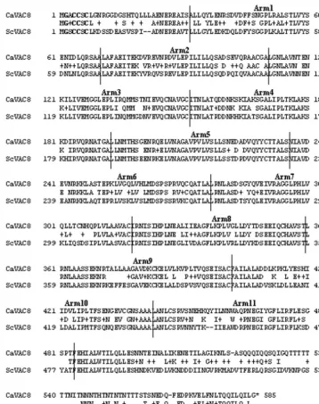
FIG. 1. Comparison of the predictedC. albicansand S. cerevisiae Vac8 proteins. Arm repeats are indicated between vertical lines. The conserved amino-proximal cysteine residues important for palmitoylation (42, 47) and the conserved amino-terminal glycine residue involved in myristoylation (46) are indicated by boldface type.
on September 8, 2020 by guest
http://ec.asm.org/
and is critical for subsequent vacuole fusion in the daughter cell (42, 46). Vacuolar inheritance is dependent upon the for-mation of a complex between the molecular motor Myo2p, the Myo2p-specific binding protein Vac17p, and Vac8p (41). Vac17p provides the specific link between the motor protein and the vacuole. Its expression correlates with the cell cycle, and hence Vac17p plays a critical role in the temporal regula-tion of vacuole inheritance.
In this study, we tested whether a causal relationship exists between vacuolar inheritance and hyphal branching inC. albi-cans. Our approach was to disturb vacuolar inheritance by disrupting theVAC8 locus and then to examine vacuolar in-heritance and hyphal growth in these mutants. Our data con-firm the importance of vacuolar segregation in the growth and division of the hyphal form of this human pathogen.
MATERIALS AND METHODS
Strains and media.Standard yeast media and microbiological techniques were used (39). For liquid growth assays, cells were pregrown in yeast extract-peptone-dextrose (YPD) (39) overnight at 30°C and then diluted to an optical density at 600 nm (OD600) of 0.1 in YPD, YPD containing 1.5 M NaCl, or YPD containing 2.5 M glycerol. Equivalent medium containing 2% (wt/vol) agar was used for the
plate assays, and cell cultures were diluted as described by Palmer et al. (28).C. albicansstrains are listed in Table 1.
DNA manipulation.Based on partial genome sequence data available at the time, the primers 5⬘-GGATCCTCAAATTGACTTCATTACTTG and 5⬘-ATA GAGTTCAAAGAAATGCTACTGGGG were used to PCR amplify 1.1 kb of theVAC8open reading frame (ORF) from theC. albicansgenome. This product was cloned into pGEMT to create pGEMT-VAC8and sequenced using ABI Prism Dye Terminator cycle sequencing kits (Perkin-Elmer) and an ABI 377 automated DNA sequencer. Subsequently, this sequence was used to identify the
VAC8locus in contig4-2360 from the StanfordC. albicansgenomic database. The completeC. albicans VAC8ORF, including 1 kb of 5⬘sequence, was then PCR amplified using the primers 5⬘-CAAGAAGCTTTCAAAATGGG (HindIII site underlined) and 5⬘-CCCAGCTAGCACTGTTGGTCC (NheI site underlined), cloned into pGEMT, and resequenced. The HindIII-NheIVAC8fragment was then cloned into a version of CIp10 (26) that contains theS. cerevisiae CYC1
terminator sequence (2) to create CIp-URA3-VAC8(CAVAC8-5).
Strain construction.To generate thevac8⌬::hisG-URA3-hisGdisruption cas-sette, a short linker region (28 bp) containing unique HindIII and BglII sites was cloned between the Pf1mI and EconI sites of pGEMT-VAC8. Then the hisG-URA3-hisGcassette was released from pMB-7 (11) by digestion with HindIII and BglII and cloned between the HindIII and BglII sites in pGEMT-VAC8. This
vac8⌬::hisG-URA3-hisGcassette was released from pGEMT with ApaI and SacI and transformed intoC. albicansstrain CAI-4 with selection for Ura⫹colonies (11), thereby deleting codons 339 to 459 of the 585-codonVAC8ORF. This generated theVAC8/vac8::hisG-URA3-hisGheterozygote CAVAC8-1 (Table 1). Ura3⫺ VAC8/vac8::hisG segregants were then selected on YPD containing 5-fluoroorotic acid (11), thereby generating strain CAVAC8-2. The second
VAC8allele was disrupted with thevac8::hisG-URA3-hisGcassette to generate thevac8::hisG/vac8::hisG-URA3-hisGhomozygote CAVAC8-3. Ura3⫺segregants were then selected as described before, generating the null mutant CAVAC8-4. TheVAC8gene was reintroduced into CAVAC8-4 on the plasmid CIp-VAC8. This plasmid was linearized with StuI and transformed into CAVAC8-4 to create CAVAC8-5. At each stage of the process, the genotype of strains was confirmed by diagnostic PCR and Southern blotting (36). Diagnostic PCR was carried out with a three-primer mix—VAC8 ATG (5⬘ATGGGTGCCTGCTGTAGTTGT TTAGG 3⬘), VAC8 DEL (5⬘AAGCGGAAATTTCCAGTTGAACTG 3⬘), and HisG (5⬘ACGCGCAGGATATCAATCGGCATGTTTTC 3⬘)—to differentiate between wild-type and disrupted alleles.
Western blotting.Protein extracts were prepared from cell cultures growing exponentially (OD600⫽0.5 to 0.7) in YPD at 30°C. Cells were washed twice in ice-cold sterile water, and protein extracts were prepared as described previously (46). Equal protein loadings from each sample were run on NuPAGE 4 to 12% Tris–N,N-methylenebisacrylamide gels in MOPS (morpholinepropanesulfonic acid) buffer and transferred onto polyvinylidene difluoride membranes according to the manufacturer’s instructions (Invitrogen, Carlsbad, CA). Membranes were incubated with antibodies againstS. cerevisiaeVac8p (46) and then with sheep anti-rabbit antibody conjugated to HRPO (Sigma) and developed using en-hanced chemiluminescence (Amersham Pharmacia Biotech UK Ltd., Bucking-hamshire, England).
Vacuole staining.C. albicansyeast cells were stained with FM4-64 (Molecular Probes Europe BV), as described previously (44). Briefly, yeast cultures were grown in YPD to an OD600of about 0.1, harvested, and resuspended in 0.1 ml of medium containing 40M FM4-64. Samples were incubated at 30°C for 40 min and then washed in YPD. Cells to be grown in the yeast form were then resuspended in 1 ml of fresh YPD and incubated for 2.5 h at 30°C before analysis of the inheritance of vacuoles. To visualize newly synthesized as well as inherited vacuoles, the chase period was reduced to 90 min. To visualize hyphal cells, samples were incubated with FM4-64 as described and then resuspended in 1%
FIG. 2. Confirmation of VAC8 gene deletion. (A) Transformants from each round of disruption were screened by PCR using a mix of three primers: VAC8 ATG, VAC8 DEL, and HisG. VAC8 ATG and VAC8 DEL produce a band of 1.2 kb in the wild-type locus, and the product of VAC8 ATG and HisG is 1.3 kb in a disrupted locus. (B) Western blot of protein extracts from CAF-2, CAVAC8-1, CAVAC8-3, and CAVAC8-5. Equal protein extracts from each strain were loaded into a gel and trans-ferred to a polyvinylidene difluoride membrane, as described in Materials and Methods. The blot was probed with ScVac8p antiserum.
TABLE 1. C. albicansstrains used in this study
Strain Genotype Source or
reference
CAF2-1 URA3/⌬ura3::imm434 14 CAI-4 ⌬ura3::imm434/⌬ura3::imm434 14 CAVAC8-1 ⌬ura3::imm434/⌬ura3::imm434,
⌬vac8::hisG-URA3-hisG/VAC8
This study
CAVAC8-2 ⌬ura3::imm434/⌬ura3::imm434,
⌬vac8::hisG/VAC8
This study
CAVAC8-3 ⌬ura3::imm434/⌬ura3::imm434,
⌬vac8::hisG/⌬vac8::hisG-URA3-hisG
This study
CAVAC8-4 ⌬ura3::imm434/⌬ura3::imm434,
⌬vac8::hisG/⌬vac8::hisG
This study
CAVAC8-5 ⌬ura3::imm434/⌬ura3::imm434,
⌬vac8::hisG/⌬vac8::hisG, CIp-URA3-VAC8
This study
CAVAC8-6 ⌬ura3::imm434/⌬ura3::imm434,
⌬vac8::hisG/⌬vac8::hisG, CIp10-URA3
This study
TABLE 2. Effect ofvac8⌬on vacuolar inheritance in budding cells
Cell characteristic % of cells
a
Wild type vac8⌬
Buds with vacuoles 100 53
Mother cell with 1–3 vacuoles 56 19
Mother cell withⱖ4 vacuoles 44 81
aWild-type and CAVAC8-3 cells were stained with CDCFDA. The absence or
presence of a vacuole(s) was recorded as a percentage of the total number of cells (n⫽50 [each]).
on September 8, 2020 by guest
http://ec.asm.org/
serum at 37°C for 2.5 h (or 1.5 h, where specified). Vacuoles were stained with CDCFDA [5- (and 6-) carboxy-2⬘,7⬘-dichlorofluorescein diacetate] (Molecular Probes Europe BV), as described previously (30). Cells were grown to early log phase, harvested, and resuspended in 1 ml of YCM (1% yeast extract, 1% bactopeptone, 2% dextrose) (35) containing 50M CDCFDA. Cells were incu-bated at 30°C for 30 min, collected by centrifugation, and viewed.
Growth analyses.To analyze the growth of budding cells,C. albicansstrains were grown overnight at 30°C in YPD, and these cells were used to inoculate YPD, YPD containing 2.5 M glycerol, and YPD containing 1.5 M NaCl at a starting OD of 0.1. Growth was monitored at 30°C and 37°C using theA600. For each condition, triplicate cultures were analyzed and the whole experiment was repeated three times. Only reproducible differences in doubling time are re-ported. To examine hyphal growth and branching, approximately 104yeast cells from an overnightC. albicans culture were inoculated onto a poly-L-lysine (Sigma) slide. The slides were incubated overnight in 1% serum at 37°C, washed in H2O, and stained with calcofluor white, as described previously (16). Hyphal growth and branching were measured (1).
Microscopy.Phase-contrast and fluorescence microscopy were performed us-ing an Axioplan 2 microscope (Carl Zeiss Ltd., United Kus-ingdom) with filter sets XF 66, XF 67, and XF 77 (Omega Optical Inc., Brattleboro, VT). Images were generated using a Hamamatsu charge-coupled-device camera.
RESULTS
Isolation and analysis ofC. albicans VAC8.The overall aim of this study was to test whether there is a causal relationship between vacuolar inheritance and hyphal branching inC. albi-cans. To achieve this we needed to generate a C. albicans
mutant in which vacuole inheritance was disrupted. We tar-geted the VAC8 locus, because of its key role in vacuolar inheritance inS. cerevisiae(10, 29, 46).
Initially we identified the homologue ofS. cerevisiae VAC8
on the basis of partialC. albicansgenome sequence informa-tion. Subsequently, the complete VAC8 sequence became available within the Stanford C. albicans genome sequence database. The VAC8 locus was PCR amplified and rese-quenced, revealing a 100% match with the genome sequence
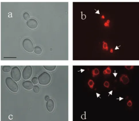
FIG. 3. Effect ofvac8⌬upon vacuolar inheritance in budding cells. Cells were stained with FM4-64 and then visualized after a 2.5-h chase period. Microscopy of budding cells is as follows: phase-contrast image of CAF2-1 cells (a), FM4-64-stained CAF2-1 cells (b), phase-contrast image of CAVAC8-3 cells (c), and FM4-64-stained CAVAC8-3 cells (d). Arrows indicate vacuolar material inherited by daughter cells. Bar, 10m.
FIG. 4. Effect ofvac8deletion upon vacuolar inheritance in hyphal cells. Hyphal cells were stained with FM4-64 and then visualized after a chase period. (A to H) To visualize inherited vacuoles, a chase period of 2.5 h was used. Shown are a phase-contrast image (A) and FM4-64-stained (B) CAF2-1 hyphal cells. (C, E, and G) Phase-contrast image of CAVAC8-3 cells. (D, F, and H) FM4-64-stained CAVAC8-3 cells. Vacuolar inheritance was restored in CAVAC8-5 cells (not shown). (I to L) A shorter chase period (90 min) was used to visualize newly synthesized as well as inherited vacuoles. Shown are a phase-contrast image (I) and FM4-64-stained (J) CAVAC8-3 cells and a phase-contrast image (K) and FM4-64-stained (L) CAVAC8-5 cells. Bar, 10m.
on September 8, 2020 by guest
http://ec.asm.org/
(orf19.745; IPF9747.1). The predicted amino acid sequence of
C. albicansVac8p is 69% identical and 81% similar to that of
S. cerevisiaeVac8p (Fig. 1). The most conserved regions are
the Arm repeats, which are believed to be important for Vac8p function.
C. albicans VAC8 gene disruption. Both alleles of the C.
albicans VAC8 locus were disrupted sequentially using a
vac8::hisG-URA3-hisGcassette that deleted amino acids 339 to
459 and blocked the expression of the 585-amino-acid protein. The disruption of the locus was confirmed by Southern blotting (not shown) and diagnostic PCR (Fig. 2A). Furthermore, Western blotting with antibodies raised against S. cerevisiae
Vac8p (46) indicated that no Vac8p was detectable in the deletion mutant (Fig. 2B).C. albicans vac8⌬cells were viable, suggesting thatVAC8, like its homologue inS. cerevisiae, is not an essential gene (46).
FIG. 5. Branching phenotype of hyphalVAC8 andvac8⌬cells. Individual cells were examined microscopically following calcofluor white staining. Phase-contrast images are shown on the left; calcofluor white-stained cells are shown on the right. (A and B) CAF2-1 hyphal cells. (C and D) CAVAC8-3 cells. (E and F) CAVAC8-3 cells. (G and H) CAVAC8-5 cells. Bar, 20m.
on September 8, 2020 by guest
http://ec.asm.org/
Effect of disruption ofVAC8upon vacuolar inheritance ofC.
albicans.Our rationale for targeting Vac8p was that, based on
the function of its homologue inS. cerevisiae(10, 29, 46), it was likely to play an important role in the inheritance, rather than the synthesis, of vacuoles inC. albicans. To test this we com-pared the vacuoles in wild-type andvac8⌬cells.
Vacuoles were visualized in budding cells by using a stable derivative of fluorescein diacetate (CDCFDA). The number of vacuoles per cell was determined for all cells within a field of view (Table 2). Wild-type (CAF2-1) and vac8⌬ cells (CAVAC8-3) were compared. All wild-type buds contained visible vacuoles, but only 50% of the buds on vac8⌬ cells contained visible vacuoles. Furthermore, more than four vacuoles were visible in most vac8⌬ mother cells, and these were more fragmented than the vacuoles of wild-type mother cells. To gain greater insight into the morphology of the vacuoles invac8⌬cells, we
stained them with the lipophilic styryl dye FM4-64. The de-tectable vacuolar material was greatly reduced if not absent in
vac8⌬ daughter cells, compared with mother cells (Fig. 3). Furthermore, these vacuoles appeared to be more fragmented or multilobed than wild-type vacuoles. Hence, the inactivation ofVAC8caused defects in the vacuolar inheritance of budding
C. albicanscells similar to those seen in S. cerevisiae vac8⌬
mutants.
Vacuolar inheritance was also examined in hyphal cells (Fig. 4).
VAC8 and vac8⌬ cells were stained with FM4-64 and then induced to form hyphae in 1% serum at 37°C for 2.5 h. Vac-uolar staining was evident throughout the length of the wild-type hyphae. The vacuolar staining invac8⌬hyphae was sig-nificantly reduced in comparison with the wild-type hyphae. Furthermore, forvac8⌬cells, most staining was observed in the mother cell, and minimal staining was observed in subapical compartments. Normal vacuole staining was visualized in both yeast and hyphal forms in the reintegrant strain CAVAC8-5 (Fig. 4L). We conclude that, as predicted, the inactivation of
VAC8caused defects in vacuolar inheritance in both the yeast and hyphal growth forms ofC. albicans.
Effects of disruption ofVAC8upon growth ofC. albicansin the yeast form.Our next objective was to determine whetherC.
albicanscell division was affected by the vac8deletion. This
was analyzed in both yeast and hyphal cells using Ura3⫹cells, because ura3 mutants are known to display subtle hyphal growth defects (4, 8, 40).
No significant differences in the growth rates of wild-type andvac8⌬cells were detecttheed on YPD plates at 30°C (not shown). However, vacuolar dysfunction is known to cause sen-sitivity to osmotic stress and/or increased temperature (5, 28, 46). Therefore, we tested the sensitivity ofvac8⌬cells to os-motic stress and elevated temperatures during growth in the yeast form. There were no detectable differences in the growth of wild-type (CAF2-1) andvac8⌬(CAVAC8-3) cells on YPD plates containing 1.5 M NaCl or 2.5 M glycerol at 30°C or 37°C (not shown). However, subtle differences were reproducibly observed in liquid cultures. The doubling time ofvac8⌬cells was 1.25-fold longer than that of wild-type cells in YPD con-taining 1.5 M NaCl at 30°C. A twofold difference in growth rate was observed in this medium at 37°C. Similar differences in growth rate were observed when osmostress was applied with 2.5 M glycerol. At 30°C the doubling time ofvac8⌬cells was 1.25-times longer than that of wild-type cells, and at 37°C this difference was increased to 1.5-fold. Therefore, the aberrant vacuolar inheritance of buddingvac8⌬cells correlated with a slight increase in sensitivity to osmotic stress and temperature.
Effects of theVAC8deletion upon hyphal growth.To quan-tify the effects of theVAC8deletion upon hyphal development, wild-type (CAF2-1), heterozygousVAC8/vac8⌬(CAVAC8-1), homozygous vac8⌬/vac8⌬ (CAVAC8-3), and reintegrant
vac8⌬/vac8⌬/VAC8 (CAVAC8-5) cells were immobilized on
poly-L-lysine slides and incubated in 1% serum at 37°C for 14 h. Hyphal development was stimulated with 1% serum be-cause we had observed the strongest correlation between vac-uolation and hyphal division under these conditions (1). The cells were stained with calcofluor white, which interchelates with chitin, to visualize cell walls and septa (Fig. 5). All hyphal cells within the field of view were counted. There was no significant difference in the number of compartments inVAC8
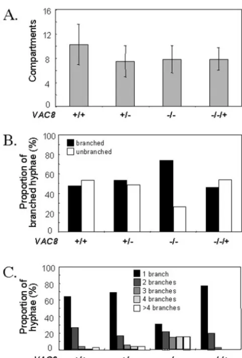
FIG. 6. Quantification of hyphal growth and division ofVAC8and vac8cells. The phenotypes of wild-type (CAF-2),VAC8/vac8 (CAVAC8-1),vac8/vac8(CAVAC8-5), andvac8/vac8/VAC8(CAVAC8-5) cells were quantified using the image types illustrated in Fig. 5. (A) Total length of hyphae determined by total number of compartments. Data are for length of the primary hypha only (n⫽100). (B) Percent branched and un-branched hyphae. Only true branches were counted (n⫽100). Bud-shaped or pseudohyphally Bud-shaped filaments were discounted. (C) Degree of branching was determined by counting the number of true branches emanating from the primary hypha (n⫽300). Branching from secondary hyphae was not included in this analysis.
on September 8, 2020 by guest
http://ec.asm.org/
andvac8⌬hyphae over the duration of the experiment (Fig. 6A). However, there was a significant increase in the proportion of branched hyphae for vac8⌬ cells compared with wild-type, heterozygous, and reintegrant cells (Fig. 6B). Furthermore, there was a significant increase in the number of branches emanating from the primary hyphae of thevac8⌬strain com-pared with the control strains (Fig. 6C). Significantly more branches were evident in CAVAC-3 hyphae than in CAF-2, CAVAC8-1, or CAVAC8-5 hyphae (Fig. 5 and 6). Similar data were generated in three independent experiments. Therefore, the inactivation ofVAC8caused a significant increase in hyphal branching. This was consistent with a role for vacuolar inher-itance in the regulation of hyphal cell division.
DISCUSSION
In true, unconstrictedC. albicanshyphae, there is a corre-lation between cell cycle progression and vacuolar status (1). The more vacuolated the subapical cell, the greater the delay in cell division before a new branch is formed. We have shown previously that, under nutrient-limiting conditions that induce long, sparsely branched hyphae (for example, in media con-taining a low serum concentration), the apical compartment inherits most of the cytoplasmic material while subapical com-partments inherit mostly vacuolar material. Hence, subapical cells are highly vacuolated, and they are also arrested in G1, prior to the point in the cell cycle at which a new branch would emerge from these subapical compartments (1). The addition of nutrients leads to a decrease in vacuole volume and an increase in cytoplasmic content, which correlates with in-creased hyphal branching. Hence, we proposed previously that there is a causal relationship between vacuolar status and cell cycle progression in subapical compartments (1). In this study, we set out to test this hypothesis. Our approach was to disturb vacuolar inheritance by inactivatingVAC8and then to examine the effects upon hyphal growth.
The vacuolar phenotype ofC. albicans vac8⌬cells was sim-ilar to that visualized inS. cerevisiae(10, 29, 46). InS.
cerevi-siae, the vacuoles ofvac8null mutants display a fragmented or multilobed appearance and a reduced degree of vacuolar in-heritance. The C-terminal truncation of S. cerevisiae Vac8p inhibits vacuolar inheritance, and the severity of this pheno-type correlates with the extent of truncation (46). A deletion in
S. cerevisiae VAC8that is analogous to the deletion we created
inC. albicans VAC8causes a dramatic inhibition of vacuolar
inheritance inS. cerevisiae(Fig. 3; Table 2). The Arm repeats within the Vac8p protein (Fig. 1) are believed to be important for the interaction of the vacuole with actin (20). Movement along actin filaments directs the segregation structure toward the daughter cell. The localization of Vac8p on the surface of the vacuole membrane is essential to its interaction with the Myo2p-specific receptor Vac17p. This specific three-way inter-action provides spatial and temporal control over vacuolar inheritance. Although vacuole inheritance is not completely abolished in theC. albicans vac8⌬cells, it is greatly reduced, implying that Vac8p plays a role inC. albicanssimilar to that of itsS. cerevisiaehomologue.
C. albicans vac8⌬mutants still undergo morphogenesis to
form true hyphae (Fig. 4). The fact thatvac8⌬ hyphae grow with normal extension rates suggests that Vac8p inactivation does not inhibit intracellular protein trafficking and vesicular delivery of cell wall synthetic material to the growing tip.
The vac8⌬ hyphae were more branched under low-serum conditions than were wild-type cells (Fig. 5). An increased number of secondary hyphae emerged from the subapical com-partments of thevac8⌬primary hypha (Fig. 5C). In addition, these cells exhibited tertiary and quaternary branching, unlike the controlVAC8 cells (data not shown). We conclude that disturbing vacuolar inheritance causes a defect in hyphal branching patterns inC. albicans(Fig. 6). Aberrant vacuolar inheritance releases subapical cells from the delay in cell cycle progression that is normally observed in hyphae under nutri-ent-limiting conditions.
Bruckmann and coworkers isolated and characterized the
C. albicans VPS34gene, which encodes a phosphatidylinositol
3-kinase (5, 6).C. albicans vps34⌬cells have an enlarged
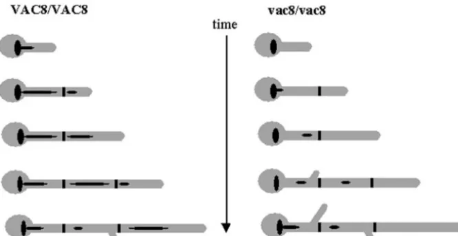
vac-FIG. 7. Model of a growing hypha. The septa are depicted as thick black lines, and the vacuoles are black ellipsoid shapes. As the growing hypha extends and divides, the subapical compartment inherits the majority of the vacuole and the apical cell inherits little. As a direct result of this, the apical cell can continue growing without delay and enter the next stage in the cell cycle. The subapical cell, however, does not have sufficient cell density to pass through Start. Hence, there is a delay in branching that correlates with the degree of vacuolation.
on September 8, 2020 by guest
http://ec.asm.org/
uole (5, 6), but any adverse affects of Vps34p disruption on the recycling of cell surface receptors and signal transduction are currently unknown. On the basis of our model (Fig. 6), which relates cell vacuolation to cellular division, one would expect
vps34⌬cells to display a delay in hyphal branching.
Interest-ingly,C. albicans vps34⌬cells do exhibit delayed hyphal for-mation in liquid serum medium and an inability to switch on solid Spider medium and solid serum-containing medium (5). We note that a mechanistic link between endocytosis and the Rim101 pathway exists through Snf7 (23) and that Rim101 signaling regulates morphogenesis in response to ambient pH (31). However, other endocytosis mutants do not affect Rim101 signaling (23), and therefore it seems likely that, in general, the hyphal defects of vacuole mutants are not medi-ated by effects on the Rim101 pathway.
AnotherC. albicansvacuolar protein-sorting mutant (vps11) has been studied extensively by Palmer et al. (28). This mutant also shows defective hyphal formation in addition to increased sensitivity to temperature, salt, and osmotic stresses. Thevps11
null mutant exhibits diffuse vacuolar staining indicative of a vacuolar biogenesis defect. C. albicans vps11⌬ cells contain small amounts of vacuolar material, and they are unable to form germ tubes as efficiently as wild-type cells. It is not known whether this phenotype is due to the physical lack of a vacuole, to the loss of multiple transport steps in vacuolar protein de-livery, to the loss of Kex2p activity, or simply to a temperature-related growth defect. As this mutant exhibits multiple pheno-types, it is difficult to attribute the defect in hyphal formation to vacuolar biogenesis.
An essential gene that results in altered budding growth (ABG1) has recently been described by Veses and coworkers (43). In hyphal cells, a null mutant displays small, fragmented vacuoles, which is concurrent with an increased branching fre-quency. This observation is entirely consistent with our work-ing model in which decreased vacuolar content causes an in-crease in hyphal cell division.
In this study, we examined the effects of mutations in a gene involved in vacuolar inheritance rather than in vacuolar bio-genesis. Indeed, C. albicans vac8⌬ cells displayed normal growth rates in the absence of osmotic stress. Therefore, we were able to distinguish the effects of disrupting VAC8 on growth rate from those on cell division. Our data strongly support the hypothesis that cytoplasmic volume, rather than total cell volume, is a critical determinant for entry into S phase (1). Highly vacuolated subapical cells do not enter S phase until the cytoplasmic volume increases sufficiently to traverse Start (Fig. 7). This model might be generally applica-ble to the eukaryotic cell division cycle.
ACKNOWLEDGMENTS
We thank Lois Weisman (Department of Biochemistry, University of Iowa, Iowa City, Iowa) for providingS. cerevisiae vac8strains and for communicating data prior to publication. We are also grateful to Norbert Schnell (AstraZeneca) for providing early access to theC. albicans VAC8sequence.
This work was supported by the U.K. Biotechnology and Biological Sciences Research Council (PAC02657), the Wellcome Trust (055015), and the European Commission.
REFERENCES
1.Barelle C. J., E. A. Bohula, S. J. Kron, D. Wessels, D. R. Soll, A. Scha¨fer, A. J. P. Brown, and N. A. R Gow.2003. Asynchronous cell cycle and
asym-metric vacuolar inheritance in true hyphae ofCandida albicans. Eukaryot. Cell2:398–410.
2.Barelle, C. J., C. L. Manson, D. M. MacCallum, F. C. Odds, N. A. R. Gow, and A. J. P. Brown.2004. GFP as a quantitative reporter of gene regulation inCandida albicans. Yeast21:333–340.
3.Bonangelino, C. J., N. L. Catlett, and L. S. Weisman.1997. Vac7p, a novel vacuolar protein, is required for normal vacuolar inheritance and morphol-ogy. Mol. Cell. Biol.17:6847–6858.
4.Brand, A., D. M. MacCallum, A. J. Brown, N. A. Gow, and F. C. Odds.2004. Ectopic expression ofURA3 can influence the virulence phenotypes and proteome ofCandida albicansbut can be overcome by targeted reintegration of URA3 at the RPS10 locus. Eukaryot. Cell3:900–909.
5.Bruckmann, A., W. Kunkel, A. Hartl, R. Wetzker, and R. Eck.2000. A phosphatidylinositol 3-kinase ofCandida albicansinfluences adhesion, fila-mentous growth and virulence. Microbiology146:2755–2764.
6.Bruckmann, A., W. Kunkel, K. Augsten, R. Wetzker, and R. Eck.2001. The deletion ofCAVPS34in the human pathogenic yeastCandida albicanscauses defects in vesicle-mediated protein sorting and nuclear segregation. Yeast
18:343–353.
7.Catlett, N. L., and L. S. Weisman.2000. Divide and multiply: organelle partitioning in yeast. Curr. Opin. Cell Biol.12:509–516.
8.Cheng, S., M. H. Nguyen, Z. Zhang, H. Jia, M. Handfield, and C. J. Clancy.
2003. Evaluation of the roles of fourCandida albicansgenes in virulence by using gene disruption strains that expressURA3from the native locus. Infect. Immun.71:6101–6103.
9.De Backer, M. D., P. T. Magee, and J. Pla.2000. Recent developments in molecular genetics of Candida albicans. Annu. Rev. Microbiol.54:463–498. 10.Flekenstein, D., M. Rhode, D. J. Klionsky, and M. Rudiger.1998. Yel013p (Vac8p), an armadillo repeat protein related to plakoglobin and importin alpha, is associated with the yeast vacuole membrane. J. Cell Sci.111:3109– 3118.
11.Fonzi, W. A., and M. Y. Irwin.1993. Isogenic strain construction and gene mapping inCandida albicans. Genetics134:717–728.
12.Franke, W. W., M. D. Goldschmidt, R. Zimblemann, H. M. Mueller, D. L. Schiller, and P. Cowin.1989. Molecular cloning and amino acid sequence of human plakoglobin, the common junctional plaque protein. Proc. Natl. Acad. Sci. USA86:4027–4031.
13.Gomes de Mesquita, D. S., R. ten Hoopen, and C. L. Woldringh.1991. Vacuolar segregation to the bud ofSaccharomyces cerevisiae: an analysis of morphology and timing in the cell cycle. J. Gen. Microbiol.137:2447–2454. 14.Gomes de Mesquita, D. S., B. van den Haazel, J. Bouwman, and C. L. Woldringh.1996. Characterization of new vacuolar segregation mutants, isolated by screening for loss of proteinase B self-activation. Eur. J. Cell Biol.
71:237–247.
15.Gomes de Mesquita, D. S, J. Shaw, J. A. Grimbergen, M. A. Buys, L. Dewi, and C. L. Woldringh. 1997. Vacuole segregation in the Saccharomyces cerevisiae vac2-1mutant: structural and biochemical quantification of the segregation defect and formation of new vacuoles. Yeast13:999–1008. 16.Gow, N. A. R., and G. W. Gooday.1982. Growth kinetics and morphology of
colonies of the filamentous form ofCandida albicans. J. Gen. Microbiol.
128:2187–2198.
17.Gow, N. A. R., and G. W. Gooday.1982. Vacuolation, branch production and linear growth of germ tubes ofCandida albicans. J. Gen. Microbiol.128:
2195–2198.
18.Gow, N. A. R.1997. Germ tube growth ofCandida albicans. Curr. Top. Med. Mycol.8:43–55.
19.Gow, N. A. R., A. J. P. Brown, and F. C. Odds.2002. Fungal morphogenesis and host invasion. Curr. Opin. Microbiol.5:366–371.
20.Hill, K. L., N. L. Catlett, and L. S. Weisman.1996. Actin and myosin function in directed vacuole movement during cell division inSaccharomyces cerevisiae. J. Cell Biol.135:1535–1549.
21.Hull, C. M., R. M. Raisner, and A. D. Johnson.2000. Evidence for mating of the “asexual” yeastCandida albicans. Science289:307–310.
22.Johnston, G. G., J. A. Pendergast, and R. A. Singer.1991. The Saccharomy-ces cerevisiae MYO2gene encodes an essential myosin for vectorial transport of vesicles. J. Cell Biol.3:539–551.
23.Kullas, A. L., L. Mingchun, and D. A. Davis.2004. Snf7, a component of the ESCRT-III protein complex, is an upstream member of theRIM101pathway inCandida albicans. Eukaryot. Cell3:1609–1618.
24.Macrea, P., C. Turck, and B. Gumbiner.1991. A homologue of the armadillo protein in Drosophila (plakoglobin) associated with E-cadherin. Science
254:1359–1361.
25.Magee, B. B., and P. T. Magee.2000. Induction of mating inCandida albicans
by construction ofMTLaandMTL␣strains. Science289:310–313. 26.Murad, A. M. A., P. R. Lee, I. D. Broadbent, C. J. Barelle, and A. J. P. Brown.
2000. CIp10, an efficient and convenient integrating vector forCandida albicans. Yeast16:325–327.
27.Odds, F. C. 1988. Candida and candidosis. Ballie`re Tindall, London, England.
28.Palmer, G. E., A. Cashmore, and J. Sturtevant.2003.Candida albicans VPS11is required for vacuole biogenesis and germ tube formation. Eu-karyot. Cell2:411–421.
on September 8, 2020 by guest
http://ec.asm.org/
29.Pan, X., and D. D. Goldfarb.1998.YEB3/VAC8encodes a myristylated armadillo protein of theSaccharomyces cerevisiaevacuolar membrane that functions in vacuolar fusion and inheritance. J. Cell Sci.111:2137–2147. 30.Pringle, J. R., R. A. Preston, A. E. M. Adams, T. Stearns, D. G. Drubin, B. K.
Haarer, and E. W. Jones.1989. Fluorescence microscopy methods for yeast. Methods Cell Biol.31:357–435.
31.Ramon, A. M., A. Porta, and W. A. Fonzi.1999. Effect of environmental pH on morphological development ofCandida albicans is mediated via the PacC-regulated transcription factor encoded byPRR2. J. Bacteriol.181:
7524–7530.
32.Raymond, C. K., P. J. O’Hara, G. Eichinger, J. H. Rothman, and T. H. Stevens.1990. Molecular analysis of the yeastVPS3gene and the role of its product in vacuolar protein sorting and vacuolar segregation during the cell cycle. J. Cell Biol.111:877–892.
33.Raymond, C. K., I. Howald-Stevenson, C. A. Vater, and T. H. Stevens.1992. Morphological classification of the yeast vacuolar protein sorting mutants: evidence for a prevacuolar compartment in class Evpsmutants. Mol. Biol. Cell3:1389–1402.
34.Riggleman, B. E., E. Wieschaus, and P. Schedl.1989. Molecular analysis of the Armadillo locus: uniformly distributed transcripts and a protein with novel internal repeats are associated with the Drosophila segment polarity gene. Genes Dev.3:96–113.
35.Rogers, D., and H. Bussey.1978. Fidelity of conjugation inSaccharomyces cerevisiae. Mol. Gen. Genet.162:173–182.
36.Sambrook, J., E. F. Fritsch, and T. Maniatis.1989. Molecular cloning: a laboratory manual, 2nd ed. Cold Spring Harbor Laboratory Press, Cold Spring Harbor, N.Y.
37.Scott, S. V., D. C. Nice III, J. J. Nau, L. S. Weisman, Y. Kamada, I. Keizer-Gunnick, T. Funakoshi, M. Veenhuis, Y. Ohsumi, and D. J. Klionsky.
2000. Apg13p and Vac8p are part of a complex of phophosproteins that are required for cytoplasm to vacuole targeting. J. Biol. Chem.275:25840–25849. 38.Shaw, J. M., and W. Wickner.1991.vac-2: a yeast mutant which distinguishes vacuole segregation from Golgi-to-vacuole protein targeting. EMBO J.10:
1741–1748.
39.Sherman, F.1991. Getting started with yeast. Methods Enzymol.194:3–21. 40.Sundstrom, P., J. E. Cutler, and J. F. Staab.2002. Reevaluation of the role ofHWP1in systemic candidiasis by use ofCandida albicansstrains with selectable markerURA3targeted to theENO1locus. Infect. Immun.70:
3281–3283.
41.Tang, F., E. J. Kauffman, J. L. Novak, J. J. Nau, N. L. Catlett, and L. S. Weisman.2003. Regulated degradation of a class V myosin receptor directs movement of the yeast vacuole. Nature422:87–92.
42.Veit, M., R. Laage, L. Dietrich, L. Wang, and C. Ungermann.2001. Vac8p release from the SNARE complex and its palmitylation are coupled and essential for vacuole fusion. EMBO J.20:3145–3155.
43.Veses, V., M. Casanova, A. Murgui, A. Dominguez, N. A. R. Gow, and J. P. Martinez.2005.ABG1, a novel and essentialCandida albicansgene encoding a vacuolar protein involved in cytokinesis and hyphal branching. Eukaryot. Cell4:1088–1101.
44.Vida, T. A., and S. D. Emr.1995. A new vital stain for visualising vacuolar membrane dynamics and endocytosis in yeast. J. Cell Biol.128:779–792. 45.Wang, Y.-X., H. Zhao, T. M. Harding, D. S. Gomes de Mesquita, C. L.
Woldringh, D. J. Klionsky, A. L. Munn, and L. S. Weisman.1996. Multiple classes of yeast mutants are defective in vacuole partitioning yet target vacuole proteins correctly. Mol. Biol. Cell7:1375–1389.
46.Wang, Y.-X., N. L. Catlett, and L. S. Weisman.1998. Vac8p, a vacuolar protein with armadillo repeats, functions in both vacuole inheritance and protein targeting from cytoplasm to vacuole. J. Cell Biol.140:1063–1074. 47.Wang, Y.-X., E. J. Kauffman, J. E. Duex, and L. S. Weisman.2001. Fusion of
docked membranes requires the armadillo repeat protein Vac8p. J. Biol. Chem.276:35133–35140.
48.Warren, G., and W. Wickner.1996. Organelle inheritance. Cell84:395–400. 49.Weisman, L. S., R. Bacallao, and W. Wickner.1987. Multiple methods of visualizing the yeast vacuole permit evaluation of its morphology and inher-itance during the cell cycle. J. Cell Biol.105:1539–1547.
50.Weisman, L. S., and W. Wickner.1988. Intervacuolar exchange in the yeast zygote: a new pathway in organelle communication. Science241:589–591. 51.Weisman, L. S., S. D. Emr, and W. Wickner.1990. Mutants ofSaccharomyces
cerevisiaethat block intervacuole vesicular traffic and vacuole division and segregation. Proc. Natl. Acad. Sci. USA87:1076–1080.
52.Weisman, L. S.2003. Yeast vacuole dynamics and inheritance. Annu. Rev. Genet.37:435–460.
53.Wickner, W., and A. Haas.2000. Yeast homotypic vacuole fusion: a window on organelle trafficking mechanisms. Annu. Rev. Biochem.69:247–275. 54.Wickner, W.2002. Yeast vacuoles and membrane fusion pathways. EMBO J.
21:1241–1247.