Please cite this article asSoltani T, Salarmoini M, Afsharmanesh M& Tasharrofi S.2019.The Effects of InovoInjection of Ascorbic Acid on Hatchability, Growth Performance, Intestinal Morphology, and Tibia Breaking Strength in 36h PostHatch Fasted Broiler Chickens. Poult. Sci. J. 7(1):43-49.
ISSN: 2345-6604 (Print), 2345-6566 (Online)http://psj.gau.ac.ir DOI:10.22069/psj.2019.15929.1382
The
Effects of
In ovo
Injection of
Ascorbic
Acid on
Hatchability,
Growth
Performance,
Intestinal
Morphology,
and
Tibia
Breaking
Strength
in
36h
Post
Hatch
Fasted
Broiler
Chickens
1 1 1 2
Soltani T, Salarmoini M, Afsharmanesh M & Tasharrofi S
1
Department of Animal Science, College of Agriculture, Shahid Bahonar University of Kerman, Kerman, Iran 2Kerman Agricultural and NaturalResources Research and Education Center, Kerman, Iran
Poultry Science Journal 2019, 7(1):43-49
Introduction
In ovo feeding may help late-term embryos and newly
hatchlings to overcome the limitation of egg nutrients
(Foye et al., 2006). Effects of in ovo injection of
various biologically important compounds (e.g. amino acids, carbohydrates, vitamins, inulin, mannan oligosaccharide, daidzein, minerals and hormones) on hatchability and growth and development of chicks
after hatching have been studied (Tako et al., 2004;
Keralapurath et al., 2010; Cheled-Shoval et al., 2011;
Zhai et al., 2011; Chamani et al., 2012; Tako and
Glahn, 2012; Eslami et al., 2014; Amiri et al., 2015;
Hartono et al., 2015; Yair et al., 2015). Ascorbic acid
(AA) does not exist in eggs and its production in the developing embryos may not be sufficient in particular toward the end of the incubation period when the embryo is most exposed to overheating
(Nowaczewski et al., 2012). So, AA synthesis in the
neonatal chicks is apparently limited. Also, pathological and environmental stressors are known to alter AA synthesis in birds. AA plays an important role in the biosynthesis of corticosterone, a glucocorticoid hormone involved in gluconeogenesis to increase energy supply during stress (Frandson, 1986). Sophisticated results have been reported on the
effect of in ovo injection of AA on hatchability (Ipek
et al., 2003; Nowaczewski et al., 2012; Selim et al.,
2012; Hajati et al., 2014) and growth performance of
the hatchlings (Zakaria, 2001; Ghonim et al., 2009;
Selim et al., 2012; Hajati et al., 2014).
Delay in food and/or water access after hatch due to hatchery treatments such as vaccination, sexing, and transportation and also farm fasting may result in
Abstract
An experiment was conducted to evaluate the efficacy of intra-amnion administration of different doses of ascorbic acid (AA) on hatchability, growth performance, blood metabolites, jejunal morphology, and tibia breaking strength in 36h post hatch fasted broiler chickens. Two hundred eighty-eight
Ross-308 fertile eggs at the 15th day of incubation were divided into four
treatment groups, each containing four replicates of 18 eggs. The treatments included non-injected (control), injected into the amnion with 0.7 mL diluent (distilled water; sham treatment), and injected with 0.7 mL diluent containing 3 or 6 mg AA. The neonatal chicks were deprived of feed and water for 36 h. Hatched chicks were raised till 10 d of age. Hatch percentage was increased
due to in ovo injection of 6 mg/egg AA. In ovo injection of different levels of
AA had no significant effect on body weight gain, feed intake, and feed
conversion ratio. In ovo injection of AA, especially at the 6 mg level,
significantly increased villus height, villus width, and villus height: crypt depth
ratio at 3 d of age. Tibia resistance and breaking strength were improved by in
ovo injection of 6 mg AA when compared to the control at 10 d of age. In
conclusion, it seems in ovo injection of 6 mg AA/egg on 15th d of incubation
could have a positive impact on hatchability, intestinal morphology, and bone characteristics in broiler chickens.
Keywords
Ascorbic acid
In ovoInjection
Intestinal morphology Tibia breaking strength
Corresponding author
Mohammad Salarmoini [email protected]
Article history
additional stress in newly hatched birds (Karadas et
al., 2011). There are very few reports in the literature
related to the effect of in ovo injection of AA on
hatch percentage, intestinal morphology, bone breaking strength and meat quality warrant further research into this area. Thus, the objective of the
present study was to investigate the effects of in ovo
injection of AA at 15th d of incubation and 36 h post-hatch fasting on the above-mentioned traits.
Material and Methods
Two hundred eighty-eight Ross-308 fertile eggs were obtained from a commercial broiler breeder flock (Mahan Company, Kerman, Iran) at 49 weeks of age. The eggs were selected according to quality and
weight (57 ± 2 gr) as specified by Zhai et al. (2011).
In this experiment four treatment groups, each containing four replicates of 18 eggs was used. The treatments included non-injected (control), injected with 0.7 mL diluent (distilled water; sham), and injected with 0.7 mL diluent containing 3 or 6 mg
AA. The AA (L- ascorbic acid, 99%) was purchased from Sigma-Aldrich Company.
Immediately after hatching and drying, 12 chicks (6 male and 6 female) from each replicate were transferred to the floor pens (1×1.2 meters). Chicks are commonly fasted for the first 36 to 72 h post-hatch because of the logistics of commercial
production (Decuypere et al., 2001). Therefore, for
simulation of the commercial conditions, the neonatal chicks were deprived of feed and water for 36 h. Afterward, all birds had free access to feed and water. The ingredients and composition of the starter basal diet (1 to 10 days of age) are shown in Table 1. Diets were fed in mash form and formulated to meet the nutrient requirements of Ross-308 broiler chickens. The initial 32 (±2) ºC room temperature was gradually reduced to 24 (±2) ºC by the third week and remained constant until the end of the experiment. The room’s relative humidity was set at 50±5%. During the study, the birds received a lighting regimen of 23L:1D.
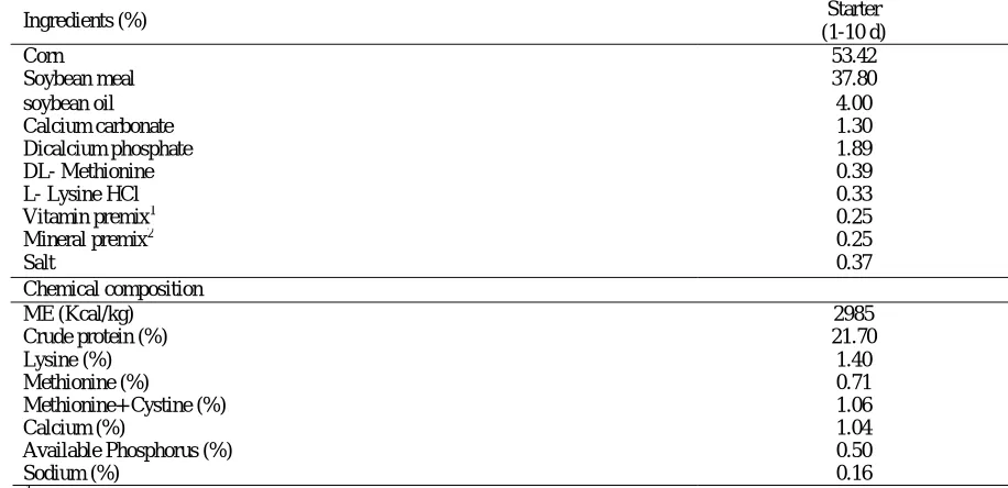
Table 1. Ingredient and nutrient composition of the starter diet
Starter (1-10 d) Ingredients (%)
53.42 Corn
37.80 Soybean meal
4.00 soybean oil
1.30 Calcium carbonate
1.89 Dicalcium phosphate
0.39 DL- Methionine
0.33 L- Lysine HCl
0.25
Vitamin premix1
0.25
Mineral premix2
0.37 Salt
Chemical composition
2985 ME (Kcal/kg)
21.70 Crude protein (%)
1.40 Lysine (%)
0.71 Methionine (%)
1.06 Methionine+ Cystine (%)
1.04 Calcium (%)
0.50 Available Phosphorus (%)
0.16 Sodium (%)
1
Vitamin premix provided the following per kilogram of diet: vitamin A, 9000 IU; vitamin E, 36 IU; cholecalciferol, 2000
IU; vitamin K3, 2 mg; thiamine, 1.8 mg; riboflavin, 6.6 mg; pantothenic acid, 10 mg; niacin, 30 mg; choline chloride, 250
mg; biotin, 0.1 mg; folic acid, 1 mg; pyridoxine 3.0 mg; vitamin B12, 0.015 mg; BHT, 1 mg.
2
Trace mineral premix provided the following in milligrams per kilogram of diet: iron, 50 mg; zinc, 85 mg; manganese, 100 mg; iodine, 1 mg; copper, 10 mg; selenium, 0.2 mg.
In ovo injection procedure
At 15th d of incubation, the injection site (broad end
of the egg) was disinfected with alcohol and then 0.7 mL of each solution was injected into the amnion, using a 23-gauge needle with the depth of 25 mm. The holes were then sealed using commercial glue. All eggs, including those belonging to the non-injected control group remained outside the setter for 20 minutes during the injection procedure. An incubator with automatic temperature and relative
humidity control was used. All eggs were kept at 37.5ºC and 60% relative humidity until they were
transferred to the hatcher (19th d of incubation), after
which 70% relative humidity was applied and egg
rotation was stopped. On the 19th d of incubation, the
eggs were transferred to the hatcher.
Hatchability and growth performance
eggs that hatch. Feed intake (FI) and body weight gain (BWG) of broilers were recorded at 10 days of
age and feed conversion ratio (FCR)was calculated.
Blood metabolites
Blood samples were taken randomly from two male chicks per pen at 3 and 10 d after hatch. The serum was separated and then centrifuged at 900 × g for 10 min. Serum glucose and cholesterol levels were
measured using Pars-Azmoon kits and an
autoanalyzer (Roche Cobas-Mira analyzer).
Intestinal morphology
Two male chicks per pen were sacrificed by cervical dislocation at 3 and 10 d post hatch, then 2 cm tissue sample from the middle of jejunum were cut off, washed in physiological saline solution, and fixed in
10% buffered formalin (100 mL of 40%
formaldehyde, 4 g phosphate, 6.5 g dibasic sodium phosphate and 900 mL of distilled water) for 24 h. Tissues were dehydrated by transferring through a series of alcohols with increasing concentrations, placed into xylol and embedded in paraffin. A microtome was used to make 5 cuts that were 5 µm. The cuts were stained with hematoxylin-eosin. Measurements for villi length and width were taken from the tip of the villus to the valley between individual villi, and measurements for crypt depth were taken from the valley between individual villi to the basolateral membrane. Ten measurements per slide was made for each parameter and averaged into one value (Thompson and Applegate, 2006). In the morphometric study, images were captured using a light microscope (Leica) and a system that analyzed computerized images (Leica Queen 550).
Bone characteristics
After slaughter at d 10, right and left tibias were taken from 2 birds per pen; soft tissues were removed
and stored at −20°C. Right tibia of each bird was
collected to determine bone resistance and breaking strength using an Instron Materials Tester (model 5500, Instron Corp, Canton, MA). Left tibia of each bird was collected to determine the percentage of tibia ash on a fat-free dry weight basis, according to AOAC (2005; method 932.16).
Statistical analysis
Data were statistically analyzed using the GLM procedure of SAS software (SAS Institute, 2005). The statistical model was completely randomized
design. Differences among treatments were
determined by Duncan’s multiple range tests. The
level of significance was reported at P < 0.05.
Results
The hatch percentage of the fertile eggs was increased
(P < 0.01) by in ovo injection of 6 mg AA/egg (Table
2). Although, in ovo injection of either level of AA
caused higher (P > 0.05) hatching weight, but the
effect was not statistically significant (Table 2). BWG, FI, and FCR of chickens are presented in
Table 2. According to the results, in ovo injection of
AA had no significant effect (P > 0.05) on these traits
during 1 to 10 d of age.
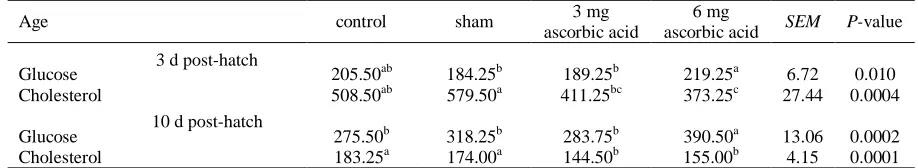
The amount of glucose and cholesterol in blood serum at 3 and 10 d post hatch are shown in Table 3. In ovo injection of 6 mg AA resulted in significantly
higher (P < 0.01) glucose (just at 10 d post-hatch) and
lower (P < 0.01) cholesterol (at either age) levels
when compared to the control and sham groups.
Table 2. Effects of in ovo injection of ascorbic acid on hatchability, hatchlings weight, and also body weight gain, feed intake, and feed conversion ratio of the broiler chicks during 1-10 d of age
control sham 3 mg
ascorbic acid
6 mg
ascorbic acid SEM P-value
Hatchability (%) 70.83b 69.44 b 69.44 b 83.33 a 1.74 0.0002
Hatchlings weight (g) 46.56 46.25 47.60 47.91 0.60 0.103
BWG (g/bird/day) 12.23 11.84 11.17 12.07 0.25 0.063
FI (g/bird/day) 20.99 20.16 19.97 20.28 0.35 0.233
FCR 1.71 1.70 1.78 1.68 0.03 0.088
SEM= standard error of the mean.
Means with no common superscript within each row are significantly (P < 0.05) different.
Table 3. Effects of in ovo injection of ascorbic acid on blood metabolites of chicks at 3 and 10 d post-hatch (mg/dL)
Age control sham 3 mg
ascorbic acid
6 mg
ascorbic acid SEM P-value
Glucose 205.50ab 184.25b 189.25b 219.25a 6.72 0.010
Cholesterol 508.50ab 579.50a 411.25bc 373.25c 27.44 0.0004
Glucose 275.50b 318.25b 283.75b 390.50a 13.06 0.0002
Cholesterol 183.25a 174.00a 144.50b 155.00b 4.15 0.0001
Means with no common superscript within each row are significantly (p < 0.05) different.
SEM= standard error of the mean.
3 d post-hatch
In ovo injection of AA, especially at the 6 mg level, significantly increased villus height (P < 0.01), villus
width (P < 0.05), and villus height: crypt depth ratio (P < 0.01) at 3 d of age (Table 4). The same results were
observed at 10 d of age, except villus width, which did not differ (P > 0.05) between the treatments.
Table 4. Effects of in ovo injection of ascorbic acid on jejunal morphology at 3 and 10 d post-hatch (µm)
villus height : crypt depth crypt depth
villus height villus width
Treatments
10 d 3 d
10 d 3 d
10 d 3 d
10 d 3 d
9.22b
8.1b
107.5ab
43.5ab
991.5c
352.5c
116.0
47.5b
control
9.41b
8.6ab
105.0b
41.0b
988.4c
351.0c
118.0
49.5ab
sham
9.78a
9.4a
104.5b
44.0ab
1022.5b
417.0b
122.5
53.0a
3 mg ascorbic acid
9.87a
9.5a
109.4a
48.5a
1080.5a
463.0a
118.5
54.0a
6 mg ascorbic acid
0.98 0.27
1.50 1.64
2.21 6.89
2.60 1.30
SEM
0.001 0.0078
0.019 0.046
0.0001 0.0001
0.39 0.014
P-value
Means with no common superscript within each column are significantly (P < 0.05) different.
SEM= standard error of the mean.
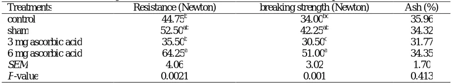
Effects of in ovo feeding of AA on tibia ash,
resistance and breaking strength at 10 d post-hatch are shown in Table 5. These traits were improved by
in ovo injection of 6 mg AA (P < 0.05) compared to the control at 10 d of age. No differences were detected for bone ash.
Table 5. Effects of in ovo injection of ascorbic acid on tibia characteristics at 10 d post- hatch
Treatments Resistance (Newton) breaking strength (Newton) Ash (%)
control 44.75b 34.00bc 35.96
sham 52.50ab 42.25ab 34.32
3 mg ascorbic acid 35.50b 30.50c 31.77
6 mg ascorbic acid 64.25a 51.00a 34.35
SEM 4.06 3.02 1.70
P-value 0.0021 0.001 0.413
Means with no common superscript within each column are significantly (P < 0.05) different.
SEM= standard error of the mean.
Discussion
The results of the present study suggested that in ovo
injection of 6 mg AA could decrease hatching stress and improve hatchability percentage. The results are in agreement with some previous studies (Zakaria and
Al-Anezi, 1996; Ipek et al., 2003; Nowaczewski et
al., 2012; Hajati et al., 2014). Also, Ghonim et al.
(2009) indicated that embryonic mortality and hatchability percentages were significantly improved by AA dipping and spraying methods in ducklings.
In contrast to our finding, Bhanja et al. (2007) and
Selim et al. (2012) reported that in ovo injection of
AA had no effect on hatchability. Improved hatchability could be attributed to the fact that AA is an antioxidant that could reduce synthesis and secretion of corticosteroids and consequently alleviate stress in the last phase of incubation. Newly laid eggs do not have AA, but it will appear by endogenous biosynthesis of 4-day-old embryos which is not an adequate amount of AA in artificial
incubation (Nowaczewski et al., 2012).
In ovo injection of AA had no significant effect on BWG, FI, and FCR during the starter phase. The BWG results are in agreement with the findings of
Ghonim et al. (2009). But, Zakaria (2001), Selim et
al. (2012), and Hajati et al. (2014) reported that in
ovo injection AA resulted in greater BW. The results
for FCR are in agreement with those of Ghonim et al.
(2009) and Hajati et al. (2014), but Selim et al.
(2012) reported better FCR due to in ovo injection of
AA. The results on FI are in agreement with the
findings of Ghonim et al. (2009) and are not in
agreement with those reported by Hajati et al. (2014),
and Selim et al. (2012), who reported higher FI due to
in ovo injection of AA. Improvement in growth
performance due to in ovo supplementation of AA
should be explained by its antioxidant role and its interference with the synthesis of anti-stress hormones of the adrenal glands. Biosynthesis of AA is limited in very young birds and several stressors (for instance: hatch process, transport, and fasting) may alter the biosynthesis or use of AA or both (Pardue and Thaxton, 1986). Anyway, the results on broiler growth performance after hatch are very contradictory.
According to the results, in ovo injection of AA
resulted in significantly higher glucose and lower cholesterol levels in serum. Our findings are in
agreement with Hajati et al. (2015), who reported that
dietary supplementation of vitamin C leads to higher serum glucose. Also, some other studies reported that cholesterol level in serum (Dorr and Nockels, 1971;
EL-gendi et al., 1999) and egg yolk (El-Gendi et al.,
AA. In ovo injection of AA in our study probably reduced synthesis and secretion of corticosteroids (Frandson, 1986) and consequently decreased muscle wasting because of corticosteroids catabolic effects on protein metabolism (McKay and Cidlowski, 2003) and lessened the need for yolk as a protein source which was followed by depression of serum cholesterol derived from yolk use.
In ovo injection of AA significantly improved jejunal morphometric parameters at 3 d of age. no
report is available on the effect of in ovo injection of
AA on intestinal morphology, but similar results
reported for in ovo injection of different nutrients
such as: dextrose (Amiri et al., 2015);
25-hydroxycholecalciferol (Chou et al., 2009), and
carbohydrates (Mousavi et al., 2009). It is well
known that fasting decreases villus maturation and
epithelial layer improvement (Geyra et al., 2001) but,
in ovo feeding of different nutrients (including carbohydrates, proteins, minerals, and vitamins) improves secretion of intestinal mucus and speeds up
maturation of villus epithelial (Bohorquez et al.,
2011).
Tibia resistance and breaking strength were
improved by in ovo injection of AA. There appear to
be no reports on the effect of in ovo injection of AA
on tibia ash, resistance and breaking strength.
Similarly, Weiser et al. (1990) reported that dietary
AA supplementation positively affected bone properties in broilers. Newman and Leeson (1997) revealed that dietary AA can affect bone breaking
strength in laying hens. In contrast, Keshavarz (1996) showed that dietary AA addition did not affect the mineralization of poultry bones. The main role of AA in bone metabolism is in the formation of osteoid (Bhattacharya, 2010) and it is necessary for bone development as a cofactor for the conversion of
vitamin D3 to its active form of 1, 25 (OH)2 D3
(McDowell, 2000; Kutlu, 2001) and also required for the hydroxylation of proline which is necessary for the synthesis of procollagen (Leeson and Summers, 2001).
In conclusion, in ovo injection (into the amnion)
of 6 mg AA/egg on 15th d of incubation, improved hatchability, jejunal morphology, and tibia resistance and breaking strength and had no adverse effect on growth performance of the birds. This may indicate an insufficient endogenous synthesis of AA by chick embryo. The discrepancy of our findings in some parts with the findings of other researchers could be
due to the differences in methodology of in ovo
injection (for example, age of embryo, injection site inside the egg, and injection volume). Therefore, further investigations are needed to highlight the
effect of in ovo injection of AA on embryo growth
and post-hatch growth performance in broiler chicks.
Acknowledgments
The authors are grateful to the office of the vice president in research at Shahid Bahonar University of Kerman, Iran, for providing the facilities and financial support of the experiment.
References
Amiri N, Salarmoini M, Tasharofi S & Eslami M. 2015. Investigation the effects of injection of different nutrients into yolk sac of fertile eggs, with 36 h post-hatch starvation, on hatchability, performance and blood metabolites of broilers. Iranian Journal of Animal Science, 46: 17-27 (in Persian).
AOAC. 2005. Official Methods of Analysis. AOAC
International, Arlington, VA.
Bhanja AB, Mandal AB, Agarwal SK, Majumdar S & Bhattacharyya A. 2007. Effect of in ovo injection of vitamins on the chick weight and post-hatch growth performance in broiler chickens. Proc of
the 16th European Symposium on Poultry
Nutrition, Strasbourg, France. pp. 143-146. Bhattacharya L. 2010. Biochemistry of Nutrition.
First ed. Discovery Publishing Pvt. New Delhi, India.
Bohorquez DV, Bohorquez NE & Ferket PR. 2011. Ultrastructural development of the small intestinal mucosa in the embryo and turkey poult: A light and electron microscopy study. Poultry Science, 90: 842-855. DOI: 10.3382/ps.2010-00939 Chamani M, Tasharofi S, Forudi F, Sadeghi AA &
Aminafshar M. 2012. Evaluation the effects of in
ovo injection of different nutrients on hatch percentage, performance and carcass parameters of broilers. Annals of Biological Research, 3: 3771-3776.
Cheled-Shoval SL, Amit-Romach E, Barbakov M & Uni Z. 2011. The effect of in ovo administration of mannan oligosaccharide on small intestine development during the pre- and posthatch periods in chickens. Poultry Science, 90: 2301-2310. DOI: 10.3382/ps.2011-01488
Chou SH, Chung TK & Yu B. 2009. Effects of
supplemental 25-hydroxycholecalciferol on
growth performance, small intestinal morphology, and immune response of broiler chickens. Poultry Science, 88: 2333-2341. DOI: 10.3382/ps.2009-00283
Decuypere E, Tona K, Bruggeman V & Bamelis F. 2001. The day-old chick, a crucial hinge between breeders and broilers. World’s Poultry Science
Journal, 57: 127-138. DOI:
10.1079/WPS20010010
El-Gendi GM, Iraqi MM & Abdelrahman AA. 1999. Effect of vitamin C supplementation on some productive and physiological parameters in laying hens. Egyptian Journal of Nutrition and Feed, 2: 649-664.
Eslami M, Salarmoini M & Tasharrofi S. 2014. Effects of in-ovo injection of different nutrients on the hatchability and growth performance in broilers. Journal of Livestock Science and Technologies, 2: 1-7.
Foye OT, Uni Z & Ferket PR. 2006. Effect of in ovo
feeding egg white protein, β-hydroxy-β-methyl
butyrate, and carbohydrates on glycogen status and neonatal growth of turkeys. Poultry Science, 85: 1185-1192. DOI: 10.1093/ps/85.7.1185 Frandson RD. 1986. Anatomy and Physiology of
Farm Animals. 7th ed. Lea and Febiger Publisher, Phila, USA.
Geyra A, Uni Z & Sklan D. 2001. Enterocyte dynamics and mucosal development in the posthatch chick. Poultry Science, 80: 776-782. DOI: 10.1093/ps/80.6.776
Ghonim AI, Awad AL, Fattouh MH & El-Shhat AM. 2009. Comparative study of ascorbic acid treatment methods on hatchability traits and growth performance of ducklings. Egyptian Poultry Science Journal, 29: 1085-1099.
Hajati H, Hassanabadi A, golian A, Nassiri Moghaddam H & Nassiri MR. 2014. The effect of in ovo injection of grape seed extract and vitamin C on hatchability, antioxidant activity, yolk sac weight ,performance and ileal microflora of broiler chickens. Research Opinions in Animal and Veterinary Sciences, 4: 633-638.
Hajati H, Hassanabadi A, golian A, Nassiri Moghaddam H & Nassiri MR. 2015. The Effect of grape seed extract and vitamin C feed supplementation on some blood parameters and HSP70 gene expression of broiler chickens suffering from chronic heat stress. Italian Journal
of Animal Science, 14: 1-9. DOI:
10.4081/ijas.2014.3273
Hartono K, Reed S, Ankrah N, Glahn R & Tako E. 2015. Alterations in gut microflora populations and brush border functionality following intra-amniotic daidzein administration. RSC Advances, 5: 6407-6412. DOI: 10.1039/C4RA10962G Ipek A, Sahin U & Yilmaz B. 2003. The effect of in
ovo ascorbic acid and glucose injection in broiler breeder eggs on hatchability and chick weight. Arch Geflügelk, 68: 132- 135.
Karadas F, Surai PF & Sparks NHC. 2011. Changes in broiler chick tissue concentrations of lipid-soluble antioxidants immediately post-hatch. Comparative Biochemistry and Physiology, 160: 68-71. DOI: 10.1016/j.cbpa.2011.05.006
Keralapurath MM, Corzo A, Pulikanti R, Zhai W & Peebles ED. 2010. Effects of in ovo injection of
L-carnitine on hatchability and subsequent broiler performance and slaughter yield. Poultry Science, 89: 1497-1501. DOI: 10.3382/ps.2009-00551 Keshavarz K. 1996. The effect of different levels of
vitamin C and cholecalciferol with adequate or marginal levels of dietary calcium on performance and eggshell quality of laying hens. Poultry
Science, 75: 1227-1235. DOI:
10.3382/ps.0751227
Kutlu HR. 2001. Influences of wet feeding and
supplementation with ascorbic acid on
performance and carcass composition of broiler chicks exposed to a high ambient temperature. Archives of Animal Nutrition, 54: 127-139. DOI: 10.1080/17450390109381972
Leeson S & Summers JD. 2001. Scott's Nutrition of the Chicken. 4th ed. Nottingham University Press, England.
McKay LI & Cidlowski JA. 2003. Physiologic and pharmacologic effects of corticosteroids. Holland-Frei cancer medicine. 6th ed. Hamilton: BC Decker.
McDowell LR. 2000. Vitamins in Animal and Human Nutrition. 2nd ed. Iowa State University Press, Iowa, USA.
Mousavi S, Shivazad M, Chamani M, Sadeghi A & Lotfolahian H. 2009. Study of in ovo feeding as an early nutrition method in broiler chicken. Dynamic Agriculture, 5: 417-425.
Newman S & Leeson S. 1997. Skeletal integrity in layers at the completion of egg production. World’s Poultry Science Journal, 53: 265-277. DOI: 10.1079/WPS19970021
Nowaczewski S, Kontecka H & Krystianiak S. 2012. Effect of in ovo injection of vitamin C during incubation on hatchability of chickens and ducks. Folia Biologica, 60: 93-97.
Pardue SL & Thaxton JP. 1986. Ascorbic acid in poultry. A review. World’s Poultry Science
Journal, 42: 107-123. DOI:
10.1079/WPS19860009
SAS Institute. 2005. SAS User’s Guide. Version 9.1 ed. SAS Institute, Cary, NC.
Selim SA, Gaafar KM & El-Ballal SS. 2012. Influence of in ovo administration with vitamin E and ascorbic acid on the performance of Muscovy ducks. Emirates Journal of Food and Agriculture, 24: 264-271.
Tako E, Ferket PR & Uni Z. 2004. Effects of in ovo feeding of carbohydrates and beta-hydroxy beta- methyl butyrate on the development of chicken intestine. World's Poultry Science Journal, 57: 2023-2028. DOI: 10.1093/ps/83.12.2023
Thompson KL & Applegate TJ. 2006. Feed withdrawal alters small-intestinal morphology and mucus of broilers. Poultry Science, 85: 1535-1540. DOI: 10.1093/ps/85.9.1535
Weiser H, Schlachter M, Probst HP & Kormann AW. 1990. The relevance of ascorbic acid for bone metabolism. Proc of the 2nd Symposium. Kartause, Ittingen, Switzerland.
Yair R, Shahar R & Uni Z. 2015. In ovo feeding with minerals and vitamin D3 improves bone properties in hatchlings and mature broilers.
Poultry Science, 94: 2695-707. DOI:
10.3382/ps/pev252
Zakaria AH & Al-Anez MA. 1996. Effects of ascorbic acid and cooling during egg incubation on hatchability culling, mortality, and body weights of broilers chickens. Poultry Science, 75: 1204-1209. DOI: 10.3382/ps.0751204
Zakaria AH. 2001. Effect of ascorbic acid treatment during egg incubation on the pre-and post-hatching development of broiler chickens. Damascus University Journal for the Agricultural Sciences, 17: 118-130.