Function and Contributes to Secretion and Filamentation
Hallie S. Rane,aStella M. Bernardo,a,bSummer M. Raines,cJessica L. Binder,cKarlett J. Parra,cSamuel A. Leea,b
Section of Infectious Diseases, New Mexico Veterans Healthcare System, Albuquerque, New Mexico, USAa; Division of Infectious Diseases, University of New Mexico Health Science Center, Albuquerque, New Mexico, USAb; Department of Biochemistry and Molecular Biology, University of New Mexico Health Science Center, Albuquerque, New Mexico, USAc
The vacuolar membrane ATPase (V-ATPase) is a protein complex that utilizes ATP hydrolysis to drive protons from the cytosol
into the vacuolar lumen, acidifying the vacuole and modulating several key cellular response systems in
Saccharomyces
cerevi-siae
. To study the contribution of V-ATPase to the biology and virulence attributes of the opportunistic fungal pathogen
Can-dida albicans
, we created a conditional mutant in which
VMA3
was placed under the control of a tetracycline-regulated
pro-moter (tetR-
VMA3
strain). Repression of
VMA3
in the tetR-
VMA3
strain prevents V-ATPase assembly at the vacuolar membrane
and reduces concanamycin A-sensitive ATPase-specific activity and proton transport by more than 90%. Loss of
C. albicans
V-ATPase activity alkalinizes the vacuolar lumen and has pleiotropic effects, including pH-dependent growth, calcium sensitivity,
and cold sensitivity. The tetR-
VMA3
strain also displays abnormal vacuolar morphology, indicative of defective vacuolar
mem-brane fission. The tetR-
VMA3
strain has impaired aspartyl protease and lipase secretion, as well as attenuated virulence in an
in
vitro
macrophage killing model. Repression of
VMA3
suppresses filamentation, and V-ATPase-dependent filamentation defects
are not rescued by overexpression of
RIM8
,
MDS3
,
EFG1
,
CST20
, or
UME6
, which encode positive regulators of filamentation.
Specific chemical inhibition of Vma3p function also results in defective filamentation. These findings suggest either that
V-ATPase functions downstream of these transcriptional regulators or that V-V-ATPase function during filamentation involves
inde-pendent mechanisms and alternative signaling pathways. Taken together, these data indicate that V-ATPase activity is a
funda-mental requirement for several key virulence-associated traits in
C. albicans
.
C
andida albicans
is a major opportunistic human fungal
patho-gen and is responsible for 6.8% of hospital-acquired
infec-tions in the United States (
1
). Despite the availability of several
classes of antifungal drugs, attributable mortality, cost of care, and
length of stay due to invasive candidiasis remain unacceptably
high (
2
,
3
). In addition, resistance to currently available antifungal
drugs is emerging (see reference
4
for a review). Therefore,
devel-opment of new antifungal drug targets remains a critical need. A
diverse set of factors contributing to
C. albicans
virulence have
been identified, including the secretion of aspartyl proteases and
lipases, filamentation, and biofilm formation (
5–8
).
Understand-ing the biology and regulation of these processes and pathways
may illuminate new candidates for antifungal therapy.
The vacuole is a dynamic acidic organelle found in yeast and
plants that is analogous to the mammalian lysosome. It functions
in an array of cellular homeostasis processes and thereby plays an
important role in stress response, adaptation to novel
environ-ments, and cell differentiation (
9–13
). Furthermore, in
C.
albi-cans
, intact vacuolar function is important for filamentation and
virulence (
12–15
). Vacuolar function depends on the
mainte-nance of acidic pH by the vacuolar H-ATPase (V-ATPase), an
enzyme complex that functions in organelle acidification across
eukaryotes (
16
,
17
). The V-ATPase utilizes hydrolysis of ATP to
transport protons from the cytosol into a variety of organelles.
V-ATPase-mediated acidification and membrane energization
are necessary for important vacuolar functions, including calcium
and metal homeostasis (
18
), cargo sorting and membrane
traf-ficking in endocytic and secretory pathways (
19
), and drug
resis-tance (
20
). In
Saccharomyces cerevisiae
, the V-ATPase is expressed
at the vacuolar membrane and the membrane of prevacuolar
compartments and the Golgi compartment.
The V-ATPase complex consists of the V
1and V
osubcom-plexes (
16
). The V
1subcomplex is composed of peripherally
asso-ciated subunits that form the sites of ATP binding and hydrolysis
on the cytosolic side of the membrane. The V
osubcomplex spans
the vacuolar membrane and mediates transport of protons from
the cytosol to vacuolar the lumen. Rotation of V
1and V
osubunits
relative to the catalytic sites couples ATP hydrolysis and active
transport of protons across the vacuolar membrane (
16
). By
driv-ing protons into the vacuole, V-ATPase generates an acidic
lumi-nal pH (pH
⬃
6.25) required for proper activity of various
degra-dative vacuolar enzymes (
21
) and a membrane potential that
energizes secondary transport systems (
17
,
22
). Loss of V-ATPase
function in
S. cerevisiae
leads to a set of growth defects referred to
collectively as the
vma
phenotype. The
vma
phenotype is
charac-terized by the inability to grow on media that are alkaline (pH 7.5
to 8.5), contain high concentrations of calcium, or contain
non-fermentable carbon sources as the sole carbon source (
16
),
whereas growth on acidic media (pH 4.0 to 5.0) is similar to that of
the wild type. Thus, interruption of V-ATPase function interferes
with a variety of key cellular processes and stress responses
likely important for fungal virulence; however, little is known
Received15 May 2013Accepted30 July 2013 Published ahead of print2 August 2013
Address correspondence to Samuel A. Lee, [email protected]. Supplemental material for this article may be found athttp://dx.doi.org/10.1128 /EC.00118-13.
Copyright © 2013, American Society for Microbiology. All Rights Reserved.
doi:10.1128/EC.00118-13
on September 8, 2020 by guest
http://ec.asm.org/
regarding the specific functions of the V-ATPase complex in
C.
albicans
(
17
).
In
S. cerevisiae
, the
VMA3
gene encodes the c subunit of the V
osubcomplex. The c subunit forms a hexameric ring with the c
=
and
c
⬙
subunits of the V-ATPase, which are encoded by
VMA11
and
VMA16
, respectively. This hexameric ring is the main site of
pro-ton transport by the V-ATPase complex.
VMA3
,
VMA11
, and
VMA16
each encode hydrophobic proteins with four or five
trans-membrane domains. Proton transport from the cytosol to
nega-tively charged glutamic acid residues in the c ring involves V
osubunit a (V
oa) (
17
,
23
). The V
oa subunit is the only fungal
V-ATPase subunit encoded by two isoforms,
VPH1
and
STV1
. We
have recently shown that V-ATPase pumps containing Vph1p
versus Stv1p contribute differently to
C. albicans
cell biology and
virulence-related traits (
24
). Whereas
C. albicans VMA3
has not
been previously studied, loss-of-function mutations in the
VMA3
gene have been investigated extensively in
S. cerevisiae
(
25–28
)
and result in pleiotropic effects, including vacuolar alkalinization,
impaired disassembly of the V
oand V
1V
ocomplexes (
28
), and the
vma
growth phenotype (
26
).
In this study, we generated a
C. albicans
tetracycline-regulat-able
VMA3
mutant(tetR-
VMA3
strain) in order to analyze the
contribution of
VMA3
to V-ATPase function, vacuolar
morphol-ogy, and virulence-related phenotypes. Importantly, we
demon-strate that the V-ATPase plays a central role in the induction of
C.
albicans
filamentation; Vma3p-dependent filamentation defects
are dominant and independent of several well-characterized
fila-mentation and pH-responsive signaling pathways.
MATERIALS AND METHODS
Identification of Vma3p.A single potential ortholog of S. cerevisiae Vma3p (http://www.yeastgenome.org/) was identified by a BLASTp search of the Candida Genome Database (http://www.candidagenome .org/). The resulting sequence (orf19.5886) was aligned with that ofS. cerevisiaeVma3p using the software program MAFFT (29). The align-ment was used as a query in the program PRALINE to analyze protein conservation, hydrophobicity, and transmembrane structure (30).
Strains and media.Strains used in this study are listed inTable 1. Throughout, unbuffered medium is used to refer to any growth medium where no buffering agents were added to maintain the pH of the medium. For testing of specific pH-dependent phenotypes, medium was buffered to pH 4.0 or 5.0 using 50 mM succinic acid–50 mM Na2PO4or to pH 7.5
or 8.5 using 50 mM morpholineethanesulfonic acid (MES) hydrate–50 mM morpholinepropanesulfonic acid (MOPS). Standard growth was completed at 30°C in unbuffered yeast peptone dextrose (YPD) (1% yeast extract, 2% peptone, and 2% glucose) supplemented with 80g/ml uri-dine where required. Doxycycline was added to a final concentration of 20
g/ml when needed. For all experiments, cells were inoculated overnight in unbuffered YPD and then reset in fresh unbuffered YPD with and without doxycycline and grown for 24 h at 30°C with shaking in order to completely repressVMA3expression prior to the start of the experiment. Attempted disruption ofVMA3.Primers used in this study are indi-cated inTable 2. We first attempted to generate aVMA3null mutant inC. albicansusing a PCR-based gene disruption strategy (31). The primers VMA3-5DRb andVMA3-3DRb were used to amplify the dpl200-URA3-dpl200-containing plasmid pDDB57 (from A. Mitchell, Carnegie Mellon University). Transformation ofC. albicans BWP17 with the vma3⌬:: dpl200-URA3-dpl200PCR amplicon was performed using the lithium ac-etate method. BecauseS. cerevisiae vmamutants exhibit poor growth at alkaline pH, selective medium was buffered to pH 4.0 or 5.0 to facilitate disruption of the secondVMA3allele. Genomic DNA was extracted from transformants as described previously (32). The transformants were
screened for homologous reintegration via PCR using primersVMA3 -5Det andVMA3-3Det. Disruption of the second allele ofVMA3was at-tempted using theVMA3-5DRb andVMA3-3DRb primers to amplify the plasmid pRS-ARG4⌬SpeI (from A. Mitchell, Carnegie Mellon University) (33). Thisvma3⌬::ARG4PCR amplicon was used to transformC. albicans VMA3/vma3⌬::dpl200-URA3-dpl200 mutants via the lithium acetate method and plated to selective medium without arginine buffered to pH 4.0 or 5.0. The genotype of the resulting mutants was assessed by allele-specific PCR using the primersVMA3-5Det andVMA3-3Det.
Construction of a tetracycline-regulatedVMA3gene.OneC. albi-cans VMA3allele was deleted from the THE1 strain background using PCR-based gene disruption (31) and transformation via the lithium ace-tate method. The primersVMA3-5DRb andVMA3-3DRb (Table 2) were used to amplify the plasmid pDDB57. Strain THE1 was transformed with the resulting PCR amplicon to generate strainVMA3⌬/⫹(Table 1). Cor-rect genomic integration of the gene disruption cassette was confirmed via PCR using the primers VMA3-5Det and VMA3-3Det. Then, the VMA3⌬/⫹strain was plated to fluoroorotic acid (FOA) agar medium, and the resultant FOA-resistant colonies were screened via PCR for the VMA3/vma3⌬::dpl200 genotype using the primers VMA3-5Det and VMA3-3Det. The tetracycline-regulatable system described by Nakayama et al. (34), with modifications allowing PCR-directed targeting as de-scribed by Bates et al. (35), was used to place the remainingVMA3allele under a tetracycline-regulatable promoter. The primers tetVMA3-5DR and tetVMA3-3DR were used to amplify plasmid p99CAU1 (H. Na-kayama, Suzuka University) using the primer design strategy described by Bates et al. (35). This PCR amplicon was inserted upstream of the remain-ingVMA3allele of theVMA3⌬/⫹FOA strain via lithium acetate trans-formation. Transformants were screened for correct insertion of the tetR-VMA3allele using the primers tetVMA3-5Det and tetVMA3-3Det. Expression ofVMA3in the THE1-CIp10 control strain and the tetR-VMA3strain after 24 h of growth in unbuffered YPD with or without doxycycline was assayed using reverse transcriptase PCR (PCR). RT-PCR was performed using the Access RT-RT-PCR system (Promega) accord-ing to the manufacturer’s instructions and usaccord-ing the primers RT-VMA3 -5Det and RT-VMA3-3Det and 5g total mRNA as the template. Absence of contaminating DNA was tested in parallel PCR-based reactions. The correct genotype of these strains was confirmed by Southern blotting fol-lowing standard protocols (36). In brief, genomic DNA prepared from candidate strains was digested with XhoI (New England BioLabs) and run on a 0.6% (wt/vol) agarose gel. A digoxigenin-labeled probe (nucleotide [nt]⫺800 to nt 500 of orf19.5886) was prepared from genomic DNA isolated from strain THE1 with the primersVMA3-5Sblt andVMA3-3Sblt (Table 2) and reagents supplied in the PCR DIG probe synthesis kit (Roche).
Growth at various pHs.The ability of strains to grow on medium without a pH buffer was tested on unbuffered YPD and unbuffered com-plete synthetic medium (CSM) (0.67% yeast nitrogen base without amino acids [YNB], 0.079% complete synthetic mixture, 2% glucose, and 2% agar) with or without doxycycline added. The ability of strains to grow over a pH range was tested on CSM buffered to pH 4.0 to 8.5 with or without doxycycline added. Cells from overnight cultures were washed and counted as previously described (37). Phosphate-buffered saline (PBS) was inoculated with cells from overnight cultures to a starting den-sity of 108cells/ml. Then, a total of five 5-fold dilutions were completed in
96-well plates, and cells were stamped onto agar plates using a multiblot replicator (VP 408H; VP Scientific) and incubated at 30°C for 48 h. Growth at pH 4.0 to 8.5 was also tested in liquid medium: cells from overnight cultures were diluted to an optical density at 600 nm (OD600) of 0.05 in CSM buffered to pH 4.0 to 8.5 and with or without doxycycline added. Then, cells were grown at 30°C using a Biotek Synergy H1M in-strument with double orbital shaking at fast speed and 2-mm frequency, with OD600readings taken at 15-min intervals.
Stress response.The ability of the tetR-VMA3strain to grow on media containing various stressors was tested on agar plates with and without
on September 8, 2020 by guest
http://ec.asm.org/
doxycycline. Plates tested for calcium sensitivity were unbuffered YPD with 200 mM CaCl2and YPD with 200 mM CaCl2buffered to pH 7.5.
Plates used to test the ability of strains to grow on medium containing glycerol as the sole carbon source were unbuffered yeast extract and pep-tone (YEP)–2% ethanol–3% glycerol. The ability of strains to respond to challenge with antifungals was tested on YPD (pH 4.0) with 0.025g/ml caspofungin, 5g/ml fluconazole, or 0.0125g/ml amphotericin B. Tem-perature sensitivity was tested by stamping cells onto unbuffered YPD plates and incubating at 25°C, 30°C, or 37°C for 48 h. Other plates tested were CSM (pH 4.0) plates containing either 1 M NaCl, 200g/ml Congo red, or 50g/ml calcofluor white. Cells were stamped onto media using a multiblot replicator as described above.
Vacuolar acidification assays.Quinacrine staining was performed to visualize acidified vacuoles as described previously (38), with some mod-ifications. First, cells were grown in unbuffered YPD with or without doxycycline for 24 h to ensure complete turnover of extant Vma3p. Then, cells were reset in fresh unbuffered YPD, with or without doxycycline, and grown to early log phase. Cells were cooled on ice for 1 min and resus-pended in 200M quinacrine in YPD buffered with 50 mM sodium phosphate (pH 7.6) for 5 min. Cells were washed twice with 100 mM HEPES–50 mM sodium phosphate–2% glucose (pH 7.6) and resus-pended in the same solution. Cells were visualized via differential
inter-ference contrast (DIC) and fluorescence microscopy. In order to quantify the vacuolar pH, cells were stained with 2=-7= -bis-(2-carboxyethyl)-5-(and-6)-carboxyfluorescein (BCECF)-acetoxymethyl ester (AM) (from Invitrogen), as described previously (24).
V-ATPase assembly and activity assays.Starter cultures were grown for 6 to 8 h in unbuffered YPD with and without doxycycline. Vacuoles were prepared by resetting cells in YPD (pH 4.0) with and without doxy-cycline and growing to an OD600of 1.0 to 1.5 (approximately 18 h).
Vac-uolar membranes were purified by Ficoll density gradient centrifugation (39). For Western blots, 80g of vacuolar protein was separated by SDS-PAGE and transferred to nitrocellulose overnight at 150 mA. The V1A
subunit was visualized with a 1:1,000 dilution of anti-human V1A rabbit
polyclonal antibody (40); the human V1A antibody cross-reacts with the
C. albicansV1A protein.
To quantify ATP hydrolysis in purified vacuolar vesicles, vacuolar ves-icles (15g) were added to an enzymatic assay in which the rate of ATP hydrolysis is coupled to the oxidation of NADH, measured as a loss ofA340
over time (41). Proton transport of purified vacuolar vesicles (30g) was measured via quenching of 1M 9-amino-6-chloro-2-methoxyacridine (ACMA) upon the addition of 0.5 mM ATP–1 mM MgSO4(MgATP) as
described previously (42,43). Fluorescence at 410-nm excitation/490-nm emission was monitored for 1 min prior to MgATP addition and for an TABLE 1C. albicansstrains used in this study
Strain name or description
Parent strain or
description Relevant genotypea Source or reference
BWP17 SC5314 ura3⌬/ura3⌬arg4⌬/arg4⌬his⌬/his1⌬VMA3/VMA3 Wilson et al. 1999 (33)
BWP17-VMA3⌬/⫹ BWP17 ura3⌬/ura3⌬arg4⌬/arg4⌬his⌬/his1⌬ VMA3/vma3⌬::dpl200-URA3-dpl200
This study
THE1 CAI8 ade2⌬::hisG/ade2⌬::hisG ura3⌬::imm434/ura3⌬::imm434 Nakayama et al. 2000
(34) ENO1/eno1⌬::ENO1-tetR-ScHAP4AD-3⫻HA-ADE2 VMA3/VMA3 THE1-CIp10 THE1 ade2⌬::hisG/ade2⌬::hisG ura3⌬::imm434/ura3⌬::imm434 ENO1/
eno1⌬::ENO1-tetR-ScHAP4AD-3⫻HA-ADE2 RP10/RP10::URA3 VMA3/VMA3
Bernardo et al. 2008 (14)
vma3⌬/⫹strain THE1 ura3⌬::imm434/ura3⌬::imm434 VMA3/vma3⌬::dpl200-URA3 -dpl200 ade2⌬::hisG/ade2⌬::hisG ura3⌬::imm434/ura3⌬::imm434 ENO1/eno1⌬::ENO1-tetR-ScHAP4AD-3⫻HA-ADE2
This study
vma3⌬/⫹FOA strain VMA3⌬/⫹
strain
ura3⌬::imm434/ura3⌬::imm434 VMA3/vma3⌬::dpl200 ade2⌬::hisG/ade2⌬::hisG ura3⌬::imm434/ura3⌬::imm434 ENO1/eno1⌬::ENO1-tetR-ScHAP4AD-3⫻HA-ADE2
This study
tetR-VMA3strain VMA3⌬/⫹
FOA strain
ura3⌬::imm434/ura3⌬::imm434 vma3⌬::dpl200::99t-VMA3-URA3 ade2⌬::hisG/ade2⌬::hisG ura3⌬::imm434/ura3⌬::imm434 ENO1/eno1⌬::ENO1-tetR-ScHAP4AD-3⫻HA-ADE2
This study
tetR-VMA3⫹NAT1-PENO1-RIM8strain tetR-VMA3 strain
ura3⌬::imm434/ura3⌬::imm434 vma3⌬::dpl200::99t-VMA3-URA3 ade2⌬::hisG/ade2⌬::hisG ura3⌬::imm434/ura3⌬::imm434 ENO1/ eno1⌬::ENO1-tetR-ScHAP4AD-3⫻HA-ADE2
NAT1-PENO1-RIM8/RIM8
This study
tetR-VMA3⫹NAT1-PENO1-MDS3strain tetR-VMA3 strain
ura3⌬::imm434/ura3⌬::imm434 vma3⌬::dpl200::99t-VMA3-URA3 ade2⌬::hisG/ade2⌬::hisG ura3⌬::imm434/ura3⌬::imm434 ENO1/ eno1⌬::ENO1-tetR-ScHAP4AD-3⫻HA-ADE2
NAT1-PENO1-MDS3/MDS3
This study
tetR-VMA3⫹NAT1-PENO1-UME6strain tetR-VMA3 strain
ura3⌬::imm434/ura3⌬::imm434 vma3⌬::dpl200::99t-VMA3-URA3 ade2⌬::hisG/ade2⌬::hisG ura3⌬::imm434/ura3⌬::imm434 ENO1/ eno1⌬::ENO1-tetR-ScHAP4AD-3⫻HA-ADE2
NAT1-PENO1-UME6/UME6
This study
tetR-VMA3⫹NAT1-PENO1-EFG1strain tetR-VMA3 strain
ura3⌬::imm434/ura3⌬::imm434 vma3⌬::dpl200::99t-VMA3-URA3 ade2⌬::hisG/ade2⌬::hisG ura3⌬::imm434/ura3⌬::imm434 ENO1/ eno1⌬::ENO1-tetR-ScHAP4AD-3⫻HA-ADE2
NAT1-PENO1-EFG1/EFG1
This study
tetR-VMA3⫹NAT1-PENO1-CST20strain tetR-VMA3 strain
ura3⌬::imm434/ura3⌬::imm434 vma3⌬::dpl200::99t-VMA3-URA3 ade2⌬::hisG/ade2⌬::hisG ura3⌬::imm434/ura3⌬::imm434 ENO1/ eno1⌬::ENO1-tetR-ScHAP4AD-3⫻HA-ADE2
NAT1-PENO1-CST20/CST20
This study
aHA, hemagglutinin.
on September 8, 2020 by guest
http://ec.asm.org/
additional 40 s after. Proton transport was calculated as the change in fluorescence for the first 15 s following MgATP addition. For both assays, the V-ATPase inhibitor concanamycin A (100 nM) was used to assess V-ATPase-specific activity.
Vacuolar morphology.For all vacuolar staining, doxycycline was added to the appropriate treatments upon each medium change. Cells
were visualized via DIC and fluorescence microscopy. To simultaneously stain vacuoles with FM4-64 [N -(3-triethylammoniumpropyl)-4-(6-(4-(diethylamino)phenyl)hexatrienyl) pyridinium dibromide] and CMAC (7-amino-4-chloromethyl coumarin), cells were grown in unbuffered YPD for 24 h in the presence or absence of doxycycline. Then, cells were reset in fresh unbuffered YPD with or without doxycycline and grown to TABLE 2Primer sequences used in this study
Primer Primer sequence (5=–3=) Source
VMA3-5DRb TATAATAAATTAAATTAATAGGTAACAATTGAGATTCGATTACAATGAACGCCCACTTGG
GAACAACAACGTTTTCCCAGTCACGACGTT
This study
VMA3-3DRb TGAAAATATGAAAGACTATAGTAAGTAGGGCCATATGACCATTGTTGATGGATATCAGCT
CCCACTTTTCTGTGGAATTGTGAGCGGATA
This study
VMA3-5Det AATGCGATGAGACTTTGCAT This study
VMA3-3Det GACTGGCGAAAACTAGTGGG This study
VMA3-5SB CCCACTAGTTTTCGCCAGTC This study
VMA3-3SB TGAGCATTCACAGTACCAGG This study
tetVMA3-5DR TCAAGTTTTGGGATTGTTGCACAACATCTAAGATTTGTTGAGTCAAAGCAGAATCAGC
CAATGGGAAAGCGTAATACGACTCACTATAGGG
This study
tetVMA3-3DR TTCCCTTCTGCAATTGAGTCCATCTGTAATCTAATCAAACAGCAAACATACCATAAATC
AGACATATTTTCTAGTTTTCTGAGATAAAGCTG
This study
tetVMA3-5Det AGCAGCAATAACTGGTCTGG This study
tetVMA3-3Det GGTTCCCTTCTGCAATTGAG This study
tetINS-3Det CTAGTTTTCTGAGATAAAGCTG This study
VMA3-5Sblt TGAGGTGACAGAAGCAGCAA This study
VMA3-3Sblt CAACAGATAAACCAGCACCC This study
RT-VMA3-5Det GCCGGTATTATTGCCATTTA This study
RT-VMA3-3Det ACGTCTTGAGAAGCTCTTG This study
ENO-SF TTGATAATTCAGGAATATTACAAC Milne et al. (48)
uUME6-5DR CCTGTTATTATAATCAAGGTTAGATATATAATTGGCTCATTATTGCTTTGCTTTACATA
ATTGGTGATAGCGTTAGTATCGAATCGACAGC
This study
uUME6-3DR TTGTATCTTCTCCATAAGGCGAATTTGGTGCTGAAGAAGTTGAATCGGGTGTAACCATA
TGGGTAATCATTGTTGTAATATTCCTGAATTATC
This study
UME6-5Det TCAATTAGAAACCAACAGAGG This study
UME6-3Det CAGCAGCACTAACACTGACAC This study
UME6-3Det2 GAGCCAGATGTAAATATGGAG This study
uRIM8-5DR CATCAGGTGAAAGTGTATTGCTTAAAGAGCCCACCTTCCCACACCCATAGTTGTAAGT
AGGGCAAATAATCGTTAGTATCGAATCGACAGC
This study
uRIM8-3DR TTGAATGGAAATGGGCGGAGTCGTTAAATAGTTTGGGGGTGGGTAGTATTTTTGATAC
TGCTCGTCTCATTGTTGTAATATTCCTGAATTATC
This study
RIM8-5Det CTACACTCAACCTGTTCCTAG This study
RIM8-3Det TAGCGCGTGCATCAACATCAG This study
RIM8-3Det2 TTCTCCTGGTAACCAGACCT This study
uCST20-5DR GACAATCCTCACTTTAAGTCTAACGTATATACGCGTACACCATCTTATACTCCACAT
ACATATTGGATTCCGTTAGTATCGAATCGACAGC
This study
uCST20-3DR GTAGATGAGAAGACTCATTTGGATCTGTTATTGATGTTTGTGTAGGATTGTTCTCTGAA
AGTATGCTCATTGTTGTAATATTCCTGAATTATC
This study
CST20-5Det GTAGTTTGTTAGTAGCGGGTCT This study
CST20-3Det AGTGGCTATTCAGACCTGAGT This study
CST20-3Det2 TGATTCTGGATTGCTCTCGT This study
uMDS3-5DR TCTTGAGACTTTTTAACCACTAAGACTTGTGGGTTATATAATAGCAGTTCACATTTGTA
GCAAATCTAGACGTTAGTATCGAATCGACAGC
This study
uMDS3-3DR GATCATCCTTTTCGGTGGGGGGTAATTGTAGACTGTAACAGGCGCTTGCTGTAGGT
ATTAACGTAGACATTGTTGTAATATTCCTGAATTATC
This study
MDS3-5Det GCTTCCTCCTCCCATATCCTC This study
MDS3-3Det TGTCGCTGCTACTGTCTGTCA This study
MDS3-3Det2 CAGGGATGGTGTAGTTGAGT This study
uEFG1-5DR TAACATTTAATTTATATTCCAAGAGTTAATTGATTAAACAACTTGGTCCAAGAATTCAT
TACCAGGCGTGCGTTAGTATCGAATCGACAGC
This study
uEFG1-3DR TTTGCTGGGGCATACCGTTATTGTAATTTCCGTTCATTTGATTGTAATAGGGTATAGAAT
ACGTTGACATTGTTGTAATATTCCTGAATTATC
This study
EFG1-5Det CCTTTGTGTCCCTTGCATAC This study
EFG1-3Det GCAACAGTGCTAGCTGATTG This study
EFG1-3Det2 GTTGTTGCATTGTCGATACA This study
on September 8, 2020 by guest
http://ec.asm.org/
early log phase. Cells were resuspended to an OD600of 2 to 4 in unbuffered
YPD with 40M FM4-64, incubated for 15 min at 30°C, and then reset in fresh unbuffered YPD and incubated for 45 min at 30°C. Next, cells were resuspended to an OD600of 0.1 in 10 mM HEPES–5% glucose (pH 7.4).
CMAC was added to a concentration of 100M, and cells were incubated at room temperature for 15 min and examined via microscopy using Texas Red (FM4-64) and 4=,6-diamidino-2-phenylindole (DAPI) (CMAC) filters. To create a three-dimensional (3D) image of the vacuole, FM4-64 staining was completed as previously described (44), except that prior to staining, cells were grown for 24 h in unbuffered YPD with or without doxycycline to ensure complete turnover of Vma3p. A Zeiss Apo-tome system was used for capturing Z-stack images. 3D image assembly was completed using AxioVision 4.7 software (Zeiss).
Secretion and filamentation assays.Secretion was assessed on solid media: extracellular protease secretion was assayed on unbuffered bovine serum albumin (BSA) plates (45), and lipase secretion was assayed on unbuffered YNB plus 2.5% Tween 80 plates (46). All plates were prepared with and without 20g/ml doxycycline. First, cells were grown in YPD with or without doxycycline for 24 h. Then, 3l cells were spotted onto plates. BSA plates were incubated at 30°C for 48 h, and Tween 80 plates were incubated at 37°C for 5 days.
Filamentation was assessed on solid and in liquid media. Solid media tested were YPD with 10% fetal calf serum (FCS), medium 199 supple-mented withL-glutamine, Spider medium as previously described (47),
and RPMI–L-glutamine. All but Spider medium were prepared with 2% (wt/vol) agar; Spider medium was prepared with 1.35% (wt/vol) agar. Filamentation assays were completed (i) with all media buffered to pH 4, and (ii) on standard filamentation media, unbuffered YPD plus FCS, un-buffered M199 (pH 7.5), and unun-buffered Spider (pH 7.2) agar, and on RPMI agar buffered to pH 7.0 with 165 mM MOPS. All plates were pre-pared with and without 20g/ml doxycycline. Three microliters cells from overnight cultures were spotted to agar plates, and plates were incu-bated at 37°C for 5 days. Filamentation in liquid medium was tested in RPMI–L-glutamine (pH 4.0) in the presence or absence of doxycycline.
Medium was inoculated with cells from overnight cultures to a starting density of 5⫻106cells/ml. Cells were grown at 37°C with shaking at 200
rpm for 2 to 24 h. Cells were visualized via light microscopy at selected time points. We were unable to assess filamentation in liquid fetal calf serum at pH 4.0 due to denaturing of serum proteins at low pH.
We also assessed the effect of chemical inhibition ofVMA3on fila-mentation in the wild-type strain SC5314. Filafila-mentation was tested in RPMI–L-glutamine buffered to pH 7.0 with 165 mM MOPS. Medium was
inoculated with cells from overnight cultures to a starting density of 5⫻ 106cells/ml, and either 10M bafilomycin A1 in dimethyl sulfoxide
(DMSO) or 5M concanamycin A in DMSO was added. The final con-centration of DMSO was 3% for bafilomycin A1 and 1% for concanamy-cin A; therefore, to eliminate the possibility of DMSO effects on filamen-tation, a 1% DMSO-only control was used. Cells were grown at 37°C with shaking at 200 rpm for 2 to 24 h and were visualized via light microscopy at selected time points.
Overexpression of positive regulators of filamentation.A PCR-based transformation method using nourseothricin as a positive selection marker was used to overexpressRIM8,MDS3,UME6,EFG1, orCST20by inserting theENO1promoter directly upstream of each gene (48) in the tetR-VMA3strain. The genotype of the five resulting strains, listed in Table 1, was tetR-VMA3⫹PENO1-RIM8, tetR-VMA3⫹PENO1-MDS3,
tetR-VMA3⫹PENO1-UME6, tetR-VMA3⫹PENO1-EFG1, and
tetR-VMA3⫹PENO1-CST20. Amplicons for transformation were generated via
PCR using the primers shown inTable 2and the plasmid pNAT1-ENO1 (from S. Bates, University of Exeter).C. albicanstetR-VMA3cells were transformed using the lithium acetate method, with a 4-h growth step in YPD added after heat shocking the cells in order to allow integration and translation of theNAT1gene before exposing the cells to nourseothricin, as described previously (48). Correct integration of the PCR amplicons was confirmed by allele-specific PCR using one primer inside the
ampli-con (ENO-SF) (Table 2) and one primer within the open reading frame of the gene targeted for overexpression (Table 2). Integration was further confirmed using an alternative allele-specific PCR with up- and down-stream primers flanking the region targeted for insertion (Table 2). Then, tetR-VMA3⫹PENO1-RIM8, -MDS3, -UME6, -EFG1, or -CST20cells were
spotted to filamentation-inducing media as described above to determine whether overexpression of these positive regulators of filamentation would rescue the filamentation defect observed in the tetR-VMA3strain in the presence of doxycycline. The tetR-VMA3⫹PENO1-UME6strain was
further investigated; first, tetR-VMA3and tetR-VMA3⫹PENO1-UME6
cells were grown in unbuffered YPD with or without doxycycline for 24 h. Then, cells were washed twice in 1⫻PBS and resuspended in PBS. Cells in PBS were visualized via light microscopy at 0 h. Finally, cells were seeded in unbuffered YPD plus 10% FCS to a concentration of 5⫻106cells per
ml and incubated at 37°C, 200 rpm, for 24 h, with visualization via light microscopy at selected time points.
Biofilm formation.Biofilm formation was tested using the XTT-re-duction assay as previously described (37). Biofilms were formed in RPMI–L-glutamine buffered to pH 4.0 to 8.5. Each treatment was
per-formed in quadruplicate, and each experiment was repeated twice. Macrophage killing assays.Thein vitromodel of macrophage infec-tion was performed as previously described (12). The J774A.1 murine macrophage cell line was purchased from ATCC. Macrophage cells were grown in unbuffered high-glucose Dulbecco’s modified Eagle medium (DMEM) supplemented with 10% FCS at 37°C with 5% CO2for 72 h.
Next, fresh unbuffered DMEM plus 10% FCS was seeded with 2⫻105
macrophage cells/ml, and 0.75 ml of this solution was used to seed Lab-Tek chambered slides (Nalge-Nunc). The slides were incubated at 37°C, 5% CO2, overnight. Spent medium was removed, and adherent
macro-phage cells were washed twice with PBS. Overnight cultures ofC. albicans strains were washed three times in PBS, andC. albicanscells were added to unbuffered DMEM plus 10%FCS, with or without doxycycline, to a mul-tiplicity of infection (MOI) of 2.C. albicanscells were coincubated with adherent macrophage cells overnight at 37°C with 5% CO2. Then, cells
were washed twice with PBS, and macrophage viability was assessed using the Invitrogen Live/Dead viability/cytotoxicity kit, following the manu-facturer’s instructions. Live macrophages from 12 separate fields of each chamber were counted, and the results were analyzed for statistical differ-ences using one-way analysis of variance (ANOVA), followed by the Tukey’s multiple-comparison test (GraphPad Prism 5.01). The experi-ment was performed independently three times, and a representative ex-periment is presented.
RESULTS
Genetic analysis and disruption of
VMA3
.
Vma3p is a highly
conserved protein (
49
) largely composed of hydrophobic
resi-dues. A BLASTp search of the
Candida
genome database using the
S. cerevisiae
Vma3p sequence as a query revealed a single
161-amino-acid predicted protein with 87.5% identity and 94.4%
sim-ilarity to
S. cerevisiae
Vma3p. Hydrophobic residues such as
iso-leucine, phenylalanine, valine, and leucine comprise 39% of
C.
albicans
and 40% of
S. cerevisiae
Vma3p. Transmembrane
struc-ture prediction of Vma3p using the software program Phobius
(
50
) revealed four transmembrane domains in the open reading
frame which are present at identical locations in
C. albicans
and
S.
cerevisiae
Vma3p. Given its high degree of structural conservation,
we anticipated that genetic deletion of
VMA3
in
C. albicans
would
mimic findings for
S. cerevisiae
in that it should prevent V
oassem-bly, eliminate all V-ATPase function, and allow us to assess the
contribution of
VMA3
and V-ATPase function to
C. albicans
physiology and virulence-related phenotypes.
Therefore, we attempted to construct a
vma3
⌬
null mutant in
C. albicans
. Because
vma
mutants in
S. cerevisiae
and
C. albicans
grow poorly on alkaline media, we buffered all selective medium
on September 8, 2020 by guest
http://ec.asm.org/
plates to either pH 4.0 or 5.0 to assist in the selection for positive
transformants. One allele of
VMA3
was readily deleted in the
BWP17 background using the PCR-based “mini-Urablaster”
cas-sette (
31
) to generate a
VMA3
/
vma3
⌬
::
dpl200
-
URA3
-
dpl200
strain. However, in our hands, we were unable to recover
second-allele deletion strains at pH 4.0 to pH 5.0 or on unbuffered
me-dium. Therefore, we constructed a conditional
VMA3
mutant, the
tetR-
VMA3
strain, using a tetracycline-repressible system (
34
,
35
)
in which
VMA3
expression is suppressed in the presence of
doxy-cycline. We used THE1-CIp10, a strain from the THE1
back-ground in which the
URA3
gene has been integrated into the
ge-nome, as an additional control (
14
). Strain construction was
confirmed via Southern blot analysis (see Fig. S1 in the
supple-mental material) and RT-PCR (
Fig. 1
). RT-PCR analysis indicated
that in the presence of doxycycline, the
VMA3
transcript was
ab-sent in the tetR-
VMA3
strain after 24 h (
Fig. 1
). The
VMA3
tran-script was present in the tetR-
VMA3
strain in the absence of
doxy-cycline and in the THE1-CIp10 strain both with and without
doxycycline (
Fig. 1
). After 6 and 18 h of treatment with
doxycy-cline, the
VMA3
transcript was still present in the tetR-
VMA3
strain. In all subsequent experiments, all strains were grown for
24 h in the presence or absence of doxycycline prior to the start
of the experiment to ensure complete disruption of
VMA3
.
The tetR-
VMA3
strain exhibits the
vma
phenotype.
Cells
car-rying genetic disruptions of V-ATPase subunits develop the
vma
phenotype in
S. cerevisiae
, characterized by pH-conditional
lethal-ity (
16
). To test for pH-dependent growth, tetR-
VMA3
cells were
spotted on medium adjusted to a broad pH range (pH 4.0 to 8.5).
tetR-
VMA3
strain growth was comparable to that of the
THE1-CIp10 control strain under derepressing conditions (
Fig. 2
). After
addition of doxycycline to repress
VMA3
expression, tetR-
VMA3
strain growth was decreased at an alkaline pH (pH 7.5 and pH 8.5)
(
Fig. 2
) but not at an acidic pH (pH 4.0 and pH 5.0) (
Fig. 2
). The
vma
phenotype was also observed in liquid CSM buffered to pH 4
to 8.5 (data not shown). The tetR-
VMA3
cells grew at nearly
wild-type levels on nonbuffered YPD and at wild-wild-type levels on CSM
under derepressing and repressing conditions (
Fig. 2
), likely due
to acidification of the surrounding medium or of key cellular
components. This phenotype has previously been observed in
S.
cerevisiae vma
mutants (
26
).
VMA3
is involved in stress responses.
The
S. cerevisiae vma3
⌬
mutant grows poorly on media containing high concentrations of
calcium or on nonfermentable carbon sources (
17
). It also
exhib-its increased cold sensitivity (
51
) and reduced resistance to a
vari-ety of stress conditions (
16
). We thus plated
C. albicans
tetR-VMA3
cells after 24 h of growth in unbuffered YPD with and
without doxycycline on the following: (i) CSM plus 200 mM
CaCl
2, both unbuffered and buffered to pH 7.5, (ii) unbuffered
YEP plus 2% glycerol as the sole carbon source, or (iii) CSM, pH 4,
containing the antifungals caspofungin, fluconazole, and
ampho-tericin B. Repression of
C. albicans VMA3
expression significantly
reduced tetR-
VMA3
growth under most conditions (
Fig. 3A
).
No-tably, tetR-
VMA3
cells had increased sensitivity to caspofungin
(0.025
g/ml) but did not have increased sensitivity to fluconazole
(5
g/ml) and had modestly increased sensitivity to amphotericin
B (0.0125
g/ml) (
Fig. 3A
). The dependence of V-ATPase
func-tion on ergosterol has been elucidated previously (
52
). Finally, we
tested the ability of the tetR-
VMA3
strain to grow at various
tem-peratures after 24 h of growth in unbuffered YPD with and
with-out doxycycline. Like
S. cerevisiae vma
⌬
mutants, the tetR-
VMA3
strain showed enhanced sensitivity to lower temperatures under
repressing conditions (
Fig. 3B
). tetR-
VMA3
growth was also
tested on medium containing 1.5 M NaCl, 50
g/ml calcofluor
white, or 200
g/ml Congo red; no difference in growth compared
to that of the wild type was observed (data not shown).
VMA3
is necessary for vacuolar acidification.
V-ATPase
pro-ton transport acidifies the vacuolar lumen, and we anticipated that
the tetR-
VMA3
vacuolar pH would be altered when
VMA3
was
repressed. We stained the tetR-
VMA3
strain with quinacrine, a
basic dye that accumulates inside acidic compartments, such as
the vacuole (
44
), to determine whether vacuolar acidification was
defective. After 24 h of growth in unbuffered YPD, the vacuoles of
tetR-
VMA3
cells accumulated quinacrine comparably to results
for the THE1-CIp10 wild-type control (
Fig. 4A
). After 24 h of
FIG 1RT-PCR analysis ofVMA3expression. RNA was extracted fromTHE1-CIp10 and tetR-VMA3cells after 6, 18, and 24 h of incubation in unbuffered YPD with doxycycline (DOX). For comparison, RNA was also extracted from THE1-CIp10 and tetR-VMA3cells incubated for 6, 18, and 24 h in unbuffered YPD without doxycycline. Reverse transcriptase PCR (RT-PCR) was used to amplify the 300-bpVMA3transcript using the primers RT-VMA3-5Det and RT-VMA3-3Det. After 6 and 18 h of treatment with doxycycline, theVMA3 transcript was still present in the tetR-VMA3strain. After 24 h of treatment with doxycycline, theVMA3gene was completely repressed.
FIG 2Growth on unbuffered YPD, unbuffered complete synthetic media (CSM), and CSM buffered to pH 4 to pH 8.5 at 30°C. The triangle at the bottom of each column indicates decreasing cell densities (1.0⫻108, 2.0⫻
107, 4.0⫻106, 8.0⫻105, 1.6⫻105, and 3.2⫻104cells/ml, from left to right).
In both columns, the top row is the THE1-CIp10 strain, and the bottom row is the tetR-VMA3strain. In the presence of doxycycline, the tetR-VMA3strain exhibits poor growth at pH 7.5 and pH 8.5.
on September 8, 2020 by guest
http://ec.asm.org/
growth in unbuffered YPD with doxycycline, tetR-
VMA3
vacuoles
did not stain with quinacrine. Fluorometric vacuolar pH
mea-surements using BCECF, a pH-sensitive fluorophore that
accu-mulates in the fungal vacuole (
42
,
53
,
54
), validated the results
obtained with quinacrine (
Fig. 4B
). Depletion of Vma3p in the
tetR-
VMA3
strain led to vacuolar alkalinization, as indicated by an
increase in the vacuolar pH from 6.17 to 6.77 upon repression of
VMA3
expression. Together, these results indicate that Vma3p is
necessary for vacuolar acidification in
C. albicans
.
VMA3
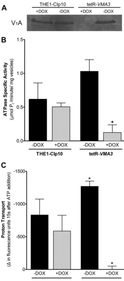
is required for V-ATPase assembly and activity.
The
vacuolar alkalinization measured in tetR-
VMA3
cells upon
deple-tion of Vma3p suggests that V-ATPase activity was drastically
compromised. To directly establish the effect of
VMA3
suppres-sion on V
oV
1complex assembly and catalytic activity, we purified
vacuolar membrane vesicles from cells grown in unbuffered YPD
with and without doxycycline for 24 h by density gradient
centrif-ugation. Western blots using an antibody against the catalytic
sub-unit A of V
1(V
1A) did not detect theV
1A subunit in vacuolar
membrane fractions under repressing conditions (
Fig. 5A
),
indi-cating that V-ATPase complexes are not assembled. This assembly
defect suggests that as in
S. cerevisiae
(
23
), deletion of the V
oc
subunit in
C. albicans
prevents assembly of V
o, and consequently,
V
1cannot assemble at the membrane.
The V
1domain is the site of ATP hydrolysis in the V-ATPase
complex, whereas the V
odomain is the site of proton transport.
The lack of intact V-ATPase complex upon depletion of Vma3p is
thus predicted to result in a significant reduction in both ATP
hydrolysis and proton transport. We measured ATP hydrolysis
spectrophotometrically using a coupled enzymatic assay (
41
), and
proton transport was measured fluorometrically using ACMA
(
42
,
43
). Both assays were performed in the presence and absence
of 100 nM concanamycin A, a specific V-ATPase inhibitor. Under
tetR-
VMA3
repression, concanamycin A-sensitive ATP hydrolysis
decreased by 88% (
Fig. 5B
) and proton transport decreased by
⬎
99% (
Fig. 5C
). These results are in agreement with the
alkalin-ization of the vacuoles in the tetR-
VMA3
strain upon addition of
doxycycline, as evidenced by both quinacrine and BCECF
exper-iments, and confirm that
VMA3
is required for both ATPase
hy-drolysis and proton transport by the V-ATPase in
C. albicans
.
Interruption of
VMA3
leads to abnormal vacuolar
morphol-ogy.
Our results indicate that
VMA3
is a fundamental V-ATPase
component required for V-ATPase activity and maintenance of
vacuolar function in
C. albicans
. We next costained vacuoles with
FM4-64 and CMAC to determine if lack of V-ATPase function
also altered vacuolar morphology. FM4-64 is a lipophilic dye that
is endocytosed and transported to the vacuole, where it stains
vacuolar membranes. CMAC is a dye that is thought to passively
permeate the cell membrane and accumulate in the vacuolar
lu-men via the action of glutathione pumps; CMAC accumulation is
independent of pH (
55
). For tetR-
VMA3
cells grown in
unbuf-fered medium under repressing conditions, excessive FM4-64
vacuolar membrane staining coincided with CMAC staining of
the lumen, indicating intravacuolar accumulation of endocytosed
membranes (
Fig. 6A
). This phenotype was confirmed by
perform-ing Z-stack fluorescence microscopy of FM4-64-stained cells (see
Movie S1 in the supplemental material). When treated with
doxy-cycline, tetR-
VMA3
cells contained a single spherical or obloid
vacuole containing multiple membrane compartments. In
con-trast,
S. cerevisiae vma3
⌬
cells have a single enlarged vacuole (
56
).
The vacuolar morphology of the tetR-
VMA3
strain after 24 h of
growth in unbuffered YPD with and without doxycycline was
fur-ther assessed using thin-section electron microscopy. The
vacuo-lar morphology of the THE1-CIp10 control strain, both with and
without doxycycline, has been studied previously (
14
). Under
re-pressing conditions, the tetR-
VMA3
strain accumulates folds of
vacuolar membrane on the interior of the vacuole (
Fig. 6B
). We
observed single, enlarged vacuoles with interior membrane
struc-tures in the majority of cells examined (data not shown),
indica-tive of a vacuolar fission defect (
56
).
VMA3
contributes to protease and lipase secretion.
The
se-cretion of degradative enzymes is involved in
C. albicans
patho-genesis (
5
,
8
). We assayed
in vitro
secretion of aspartyl proteases
and lipases on unbuffered BSA and unbuffered YNB-Tween 80
media, respectively (
Fig. 7
).
C. albicans
cells, spotted on medium
FIG 3Growth under stress conditions. The triangle at the bottom of eachcolumn indicates decreasing cell densities (1.0⫻108, 2.0⫻107, 4.0⫻106,
8.0⫻105, 1.6⫻105, and 3.2⫻104cells/ml, from left to right). In both
columns, the top row is the THE1-CIp10 strain, and the bottom row is the tetR-VMA3strain. (A) The abilities of the tetR-VMA3strain to respond to high-calcium stress, to use glycerol as a nonfermentable carbon source, and to resist challenge with antifungal agents were tested on agar plates incubated at 30°C. Calcium sensitivity was tested on unbuffered YPD plus 200 mM CaCl2
and on YPD buffered to pH 7.5 plus 200 mM CaCl2. Ability to use glycerol as
a nonfermentable carbon source was tested on unbuffered YEP with 2% glyc-erol and 3% ethanol. Susceptibility to antifungal agents was tested on YPD buffered to pH 4 with 0.0125g/ml amphotericin B (AMB), 0.025g/ml caspofungin (CAS), or 5g/ml fluconazole (FLU) added. Under repressing conditions, the tetR-VMA3strain grows poorly on medium containing high concentrations of calcium, glycerol, or caspofungin. (B) tetR-VMA3strain growth at various temperatures was tested on unbuffered YPD plates incu-bated at 25°C, 30°C, or 37°C. Under repressing conditions, the tetR-VMA3 strain exhibits increased sensitivity to low temperature (25°C).
on September 8, 2020 by guest
http://ec.asm.org/
containing BSA as the sole nitrogen source, secrete secreted
aspar-tyl proteases (SAPs) that digest the BSA, creating a halo of
prote-olysis around the colony (
45
). Like the THE1-CIp10 control
strain, the tetR-
VMA3
strain exhibited normal proteolytic activity
under derepressing conditions. The addition of doxycycline
com-pletely inhibited extracellular proteolytic activity of the
tetR-VMA3
strain (
Fig. 7
). Similarly, wild-type
C. albicans
cells secrete
lipases on YNB-Tween 80 medium, creating a halo of
precipita-tion around the colony. Under repressing condiprecipita-tions, tetR-
VMA3
exhibited decreased lipolytic activity on YNB-Tween 80 agar
(
Fig. 7
).
VMA3
is required for filamentation.
Since repression of
VMA3
impairs secretion of degradative enzymes involved in
pathogenesis, we asked whether other virulence-associated traits
are associated with V-ATPase function in
C. albicans
. We assessed
in vitro
filamentation by the tetR-
VMA3
strain on solid and in
liquid media buffered to pH 4.0, allowing us to discriminate
be-tween filamentation defects and pH-specific growth defects.
Un-der repressing conditions, the tetR-
VMA3
strain did not filament
on solid media that are either weak inducers or strong inducers of
filamentation (
Fig. 8A
). In contrast, the tetR-
VMA3
strain grown
under nonrepressing conditions produced robust hyphal
struc-tures comparable to those of the THE1-CIp10 control strain. We
also assessed
in vitro
filamentation of the tetR-
VMA3
strain on
standard filamentation media: unbuffered YPD plus FCS,
unbuf-fered M199 (pH 7.5), and unbufunbuf-fered Spider pH 7.2 agar, as well as
RPMI agar buffered to pH 7.0 with 165 mM MOPS. The
tetR-VMA3
strain was also unable to filament under these conditions
(data not shown). Under repressing conditions, filamentation of
the tetR-
VMA3
strain was dramatically reduced in liquid RPMI
(pH 4.0) at 37°C even at 24 h of incubation (
Fig. 8B
). To further
validate the importance of V-ATPase in filamentation, we treated
the wild-type
C. albicans
strain SC5314 with specific chemical
in-hibitors of Vma3p. Concanamycin A and bafilomycin A1 are
po-tent V-ATPase inhibitors that bind specifically to V
oc (Vma3p),
blocking rotation of the hydrophobic c ring and preventing
proton transport and ATP hydrolysis (
57–59
). Treatment with
concanamycin A (
Fig. 8C
) and bafilomycin A1 (data not shown)
inhibited filamentation. This lack of filamentation upon
pharma-cological inhibition of V-ATPase further supports the genetic data
indicating that V-ATPase-mediated proton transport is essential
for
C. albicans
filamentation under a variety of
in vitro
conditions.
The tetR-
VMA3
strain exhibits the
vma
phenotype in
bio-films.
The adoption of a biofilm lifestyle by
Candida
species on
both biotic and abiotic surfaces has been identified as a major
factor in their pathogenicity and virulence (
60
,
61
). Therefore, we
tested biofilm formation in RPMI (pH 4.0 to 8.5). The
THE1-CIp10 control strain formed robust biofilms in RPMI buffered to
pH 7.5 or 8.5 and biofilms of lesser metabolic activity in RPMI
buffered to pH 4.0 or 5.0 in either the presence or absence of
doxycycline (data not shown). At alkaline pH, the tetR-
VMA3
strain exhibited decreased metabolic activity relative to that of the
control strain in the presence of doxycycline. However, at acidic
pH, the tetR-
VMA3
strain consistently generated metabolic
activ-ity similar to that of controls when coincubated with doxycycline,
indicating that tetR-
VMA3
exhibits the
vma
growth phenotype in
both the biofilm and planktonic states.
The tetR-
VMA3
strain is attenuated in macrophage killing.
Phagocytes, such as macrophages and neutrophils, constitute the
host’s first line of defense against
Candida
infection. After
phago-cytosis, survival of
C. albicans
within the host is dependent upon
induction of phagocyte death (
62
). We therefore used a murine
macrophage killing assay to analyze the contribution of
VMA3
to
in vitro
virulence (
Fig. 9
). We first tested planktonic growth in
unbuffered DMEM over 30 h; there was no significant difference
in growth between the tetR-
VMA3
strain with and without
doxy-cycline (data not shown). Next, after 24 h of coincubation in
un-buffered DMEM plus 10%FCS, the THE1-CIp10 strain
effi-ciently killed the macrophage cell line, as did the tetR-
VMA3
strain without doxycycline. Upon repression of the
VMA3
gene, the tetR-
VMA3
strain displayed significantly attenuated
macrophage killing.
Overexpression of key positive regulators involved in
fila-mentation regulation does not rescue the tetR-
VMA3
filamen-tation defect.
In order to better understand the role of V-ATPase
activity in filamentation, we sought to ascertain whether
V-FIG 4Vacuolar acidification. (A) Quinacrine staining of cells grown in unbuffered YPD with and without doxycycline. Quinacrine accumulates in the vacuole under acidic conditions. Under derepressing conditions, both the THE1-CIp10 and tetR-VMA3strains accumulate quinacrine in vacuoles. Under repressing conditions, the tetR-VMA3strain does not accumulate quinacrine. (B) BCECF quantification of vacuolar pH in cells grown in unbuffered YPD. After treatment with doxycycline, the tetR-VMA3strain exhibits alkalinized vacuoles. Asterisks (ⴱ) denotes statistical significance,P⬎0.001, compared to all other treatments.on September 8, 2020 by guest
http://ec.asm.org/
ATPase activity is an absolute requirement for filamentation or if
the severe filamentation defect in the tetR-
VMA3
strain could be
overcome by overexpression of positive transcriptional regulators
of filamentation. Thus, we generated strains that overexpressed
FIG 5V-ATPase assembly and activity. (A) Western blot using anti-V1A
an-tibody in vacuolar vesicles purified after 24 h of growth in unbuffered YPD⫾ doxycycline. Eighty micrograms of vacuolar protein was loaded per lane. The V1A subunit (Vma1p) is not detected in vacuolar membrane vesicles from the
tetR-VMA3strain under repressing conditions, indicating that the V-ATPase complex is not properly assembled at the vacuolar membrane. (B) Concana-mycin-A-sensitive ATP hydrolysis in vacuolar vesicles purified after 24 h of growth in unbuffered YPD with or without doxycycline. ATPase-specific ac-tivity was measured in purified vacuolar vesicles using a spectrophotometric enzyme assay in which ATP hydrolysis is coupled to NADH oxidation. Loss of Vma3p leads to an 88% decrease in concanamycin-A-sensitive ATP hydroly-sis. (C) Proton transport in vacuolar vesicles purified after 24 h of growth in unbuffered YPD with or without doxycycline. ATP-dependent proton trans-port across purified vacuolar membranes was measured via fluorescence quenching of ACMA upon the addition of ATP and MgSO4. Repression of
VMA3leads to⬎99% reduction in proton transport activity. Asterisks (ⴱ) denote statistical significance,P⬍0.05, compared to results for all other treat-ments.
FIG 6Vacuolar morphology of the tetR-VMA3strain. (A) FM4-64 and CMAC double staining of cells grown in unbuffered YPD for 24 h with and without doxycycline. FM4-64 (red) stains vacuolar membranes, and CMAC (blue) stains the vacuolar lumen. Under repressing conditions, FM4-64 and CMAC staining reveal the accumulation of membranous structures on the interior of the vacuole in the tetR-VMA3strain. (B) Thin-section electron microscopy of the tetR-VMA3strain after 24 h of growth in unbuffered YPD with and without doxycycline. “N” denotes the nucleus, and “V” denotes the vacuole. Under repressing conditions, the tetR-VMA3strain displays aberrant vacuolar ultrastructure, indicated by enlarged vacuoles with inclusions of vac-uolar membrane on the interior of the vacuole.
FIG 7Secretion on BSA and Tween 80 agar. Cells were grown for 24 h in unbuffered YPD with and without doxycycline. Then, secreted aspartyl pro-tease (Sap) secretion was assayed on unbuffered YNB-BSA agar plates (72 h at 30°C), and lipase secretion was determined on unbuffered Tween 80 agar plates (120 h at 37°C). When grown under derepressing conditions, the tetR-VMA3strain secretes aspartyl proteases and lipases at levels comparable to those for THE1-CIp10, evidenced by halos of clearance surrounding the col-ony. When grown under repressing conditions, the tetR-VMA3strain exhibits dramatically reduced Sap and lipase secretion.
on September 8, 2020 by guest
http://ec.asm.org/
the positive regulators of filamentation
UME6
,
RIM8
,
CST20
,
MDS3
, and
EFG1
in the tetR-
VMA3
background. Next, we
as-sayed filamentation of these strains on solid filamentation agar
with and without doxycycline. Under repressing conditions,
over-expression of
RIM8
,
MDS3
,
CST20
, and
EFG1
did not rescue the
filamentation defect in the tetR-
VMA3
strain on unbuffered YPD
plus 10% FCS agar (
Fig. 10A
) or on unbuffered M199 (pH 7.5)
agar (data not shown).
UME6
is a key regulator of filamentation; overexpression of
UME6
results in constitutive filamentation and can rescue
fila-mentation defects caused by mutations in other genes (
63–65
),
including
RIM8
and
MDS3
, which regulate pH-dependent
signal-ing pathways. In nonrepresssignal-ing conditions, the
P
ENO1-
UME6
col-ony formed robust filaments in a three-dimensional manner,
rather than spreading only along the plate’s surface (
Fig. 10A
). The
colony was embedded into the agar more deeply and protruded
above the surface to a greater extent than the colonies formed by
the other strains studied. Therefore, to further analyze the effect of
overexpression of
UME6
in the tetR-
VMA3
background, we
stud-ied the tetR-
VMA3
⫹
NAT1-P
ENO1-UME6
strain in liquid rich
me-dium (unbuffered YPD) and filamentation meme-dium (unbuffered
YPD plus 10% FCS). As expected, overexpression of
UME6
led to
filamentation in liquid YPD without environmental induction
(
Fig. 10B
). However, upon repression of
VMA3
, the
tetR-FIG 8Filamentation on hypha-inducing media. (A) Filamentation on YPDplus 10% FCS, M199, Spider, and RPMI agar plates buffered to pH 4. Cells were grown for 24 h in unbuffered YPD with and without doxycycline and then spotted to agar plates and incubated for 5 days at 37°C. When grown under derepressing conditions, the tetR-VMA3strain produces filamentous struc-tures comparable to those of the THE1-CIp10 control strain. When grown under repressing conditions, the tetR-VMA3strain exhibits dramatically re-duced filamentation on all media tested. (B) Filamentation in liquid culture after 24 h of incubation. Strains were grown in RPMI–L-glutamine with and without doxycycline buffered to pH 4 at 37°C, 200 rpm. When grown under derepressing conditions, the THE1-CIp10 and tetR-VMA3strains produced hyphae. When grown under repressing conditions, the tetR-VMA3strain ex-hibited substantially decreased hyphal growth. (C) Filamentation byC. albi-cansSC5314 in the presence or absence of 5M concanamycin A1, a V-ATPase inhibitor specific to Vma3p. Strains were grown for 24 h in RPMI–L -glutamine buffered to pH 7 at 37°C, 200 rpm. The addition of concanamycin A inhibited wild-type filamentation.
FIG 9In vitromodel of macrophage infection.C. albicanscells were grown for 24 h in YPD with and without doxycycline before coincubation with macro-phage cells in unbuffered DMEM plus 10% FCS at an MOI of 2. (A) Counts of live macrophage cells from 12 separate fields after 24 h of coincubation withC. albicansstrains. The asterisk denotes statistical significance,P⬍0.01, com-pared to all results for other treatments. Each experiment was performed in triplicate; a representative experiment is shown. (B) Live (green) and dead (red) macrophage cells were costained with calcein AM and ethidium bromide homodimer, respectively, and visualized by fluorescence microscopy. Repre-sentative images from the 24-h time point are shown.