BOSNIAN JOURNAL OF BASIC MEDICAL SCIENCES 2008; 8 (2): 116-120&
Abstract
The study was designed with the main intent to assess and explain the differ-ences between athlete’s heart syndrome and the heart of healthy non-athletes, and to distinguish between physiological and pathological heart condition. Prolonged athletic training causes changes in heart that are termed “athlete’s heart syndrome”. Athlete’s heart diagnosis and related issues are a great chal-lenge due to complementary morphological, functional and electro-physiolog-ical changes that may indicate both physiologelectro-physiolog-ical and pathologelectro-physiolog-ical condition. The study included subjects, of those were active athletes and were in control group. The study protocol included one clinical examina-tion, one electrocardiogram and one echocardiograph for each subject. Aver-age Aver-age was ,±, in the athletes and ,±, in control group. Sig-nificantly higher average left ventricle (LV) mass (,g vs. ,g) and LV mass index (,g/m vs. ,g/m) was found in the athletes (p<,).
Th e study showed increased mass and wall thickness with usual inner dimensions of athlete’s heart. Systolic and diastolic function of athlete’s heart is normal. Ath-lete’s heart with these features is a healthy heart.
KEY WORDS: athlete’s heart, left ventricle hypertrophy, ultrasound.
ATHLETE’S HEART
SYNDROME AND
ECHOCARDIOGRAPHIC
CHANGES
Amir Kreso¹*, Amila Arslanagić²
¹ Public Institution Center for Sports Medicine, Sarajevo Canton,
Patriotske lige , Sarajevo, Bosnia and Herzegovina
² Clinic for Heart Diseases and Rheumatism, University of Sarajevo Clinics Centre,
Bolnička , Sarajevo, Bosnia and Herzegovina
BOSNIAN JOURNAL OF BASIC MEDICAL SCIENCES 2008; 8 (2): 117-120
Introduction
“Athlete’s heart syndrome” is a well known condi-tion that includes structural, electrophysiological and functional adaptation of myocard to an increased physical activity (training), which depends on the intensity, duration and type of the Activity (,,,). Left ventricle hypertrophy in athletes frequently re-sembles pathological conditions (hypertension or hypertrophic cardiomyopathy) and differential diag-nosis is particularly important in active athletes (,,). Different data on the nature (physiological vs. patho-logical) of left ventricle hypertrophy (LVH) in ath-letes and veterans were collected in the past (,,). Pathological left ventricle hypertrophy is a risk fac-tor for disease and death in mature age (,). Early detection of pathological LVH may reduce cardiac complications in athletes during training. Echocardiography is capable of analyzing struc-tural and functional changes in myocard in athlete’s heart and distinguish between physi-ological and pathphysi-ological hypertrophy (,,). The study was designed with the objective of dem-onstrating echocardiographic features in athletes in comparison to those in healthy non-athletes.
Subjects and Methods
The study is designed as a monocentric, open, pro-spective, comparative analysis within groups of active athletes classified according to the type of athletic activity and within the group of healthy in-dividuals engaged in no recreational athletic activity. Subjects were examined and analyzed in Public Institution Center for sports medicine and Pub-lic Institution Center for students’ healthcare. Th e study included subjects, of those were athletes
with at least two years of active training and were con-trol group subjects with no athletic activity whatsoever.
Analyzed echocardiographic parameters: - Ao - aorta width
- LA - left atrium - RVD - right ventricle
- IVSd - intraventricular septum diastolic thickness - LVIDd – left ventricle diastolic internal diameter - LVPWd – posterior wall diastolic thickness - IVSs – intraventricular septum systolic thickness - LVIDs - left ventricle systolic internal diameter - LVPWs – left ventricle posterior wall systolic
thickness
- EF-systolic function of left ventricle - E/A- diastolic function of left ventricle
Other parameters signifi cant in heart ultrasound and function assessment:
- Blood pressure (mmHg) - Pulse (beats per min) - Body mass (kg) - Body mass index - Body height (cm)
- Body Surface Area - BSA (m)
Th e subjects were classifi ed in groups according to the estimation of their athletic activity or lack thereof. Ba-sic analyzed parameters were compared among groups. Th e results were analyzed by the mode of descriptive statistic. In analyzing the significance of the differ-ences between means the threshold was set at p<,. We also analyzed left ventricle mass in-dex. Left ventricle mass and body surface area were used to calculate left ventricle mass index. Body surface area (BSA) was calculated according to Mosteller’s formula:
BSA (m²) = ( [height (cm) x mass (kg) ] / )½
Left ventricle mass is calculated from the measured parameters: LVID - left ventricle diastolic internal diameter, LVPWd posterior wall diastolic thickness and IVSd - intraventricular septum diastolic thickness. Left ventricle mess was calculated according to the formula: LVM(g)= , * [(LVIDd +IVSd + PWTd)- LVIDd] – ,
Left ventricle mass index was calculated according to Penn’s formula:
LVMI (g/m) = LVM (g)/BSA (m)
Deveroux criteria indicate hypertrophy when LVMI> g/m in men and LVMI> g/m in women.
Finally, we calculated average thickness of left ventricle wall according to the formula:
(IVST + PWT)/ LVID.
Results
Average age of the athletes was ,±,(SD) years, while average values of body mass, body surface area
Legend: MIN-minimal value, MAX- maximal value, SD – standard deviation, BMI – body mass index, BSA – body surface area
TABLE 1. Basic characteristics of the athletes (n=100)
MIN MAX Mean ± SD
Age (years) 14 65 20,51 ± 8,51
Body mass (kg) 60 121 81,12 ± 2,53
Height (cm) 158 194 181,26 ± 7,76
BMI 19,59 34,49 24,68 ± 3,38
BSA (m2) 1,38 3,16 2,05 ± 0,37
Blood pressure
BOSNIAN JOURNAL OF BASIC MEDICAL SCIENCES 2008; 8 (2): 118-120(BSA) and blood pressure were , ±, (SD) kg, ,±,(SD)m and (,)/(,) mmHg
re-spectively (Table ).
Average age of the non-athletes was , ±, (SD) years, while body mass, body surface area (BSA) and blood pressure were ,±, (SD) kg, ,±,(SD) m and (,)/(,) mmHg respectively (Table ).
Ultrasound fi ndings
Average left ventricle mass index (LVMI in g/m) (± SD)
is ,±, grams in athletes and ,±, grams in non-athletes. Th e diff erence between groups is statisti-cally signifi cant (p<,). Average posterior wall diastolic thickness (LVPWd in cm) (± SD) is ,±, cm in ath-letes and ,±, cm in non-athath-letes. Th e diff erence be-tween groups is statistically signifi cant (p<,) (Table ).
Th e values are given as mean ± standard deviation (SD)
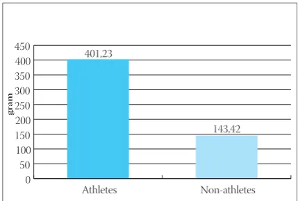
Average left ventricle mass (± SD) is ,±, gram in athletes and ,±, gram in non-athletes. Th e difference between groups is statistically significant (p<,) (Table , Graph ).
Average values of left ventricle mass index in all sub-jects is illustrated in Graph . Average LV mass index (±SD) is ,±, g/m in athletes and ,±, g/m in non-athletes. The difference between groups is statistically significant (p<,) (Table ).
Discussion
The paper presents the results of our study on simi-larities and differences in heart size using echocar-diography as a basic tool. The set of parameters was compared between the groups of active athletes and
Legend: MIN-minimal value, MAX- maximal value, SD – standard deviation, BMI – body mass index, BSA – body surface area
TABLE 2. Basic characteristics of the non-athletes (n=50)
Legend: AV – mean, SD – standard deviation, MAX – maximal value, MIN – minimal value, MED – median, LVM – left ventricle mass
TABLE 4. Average left ventricle mass (g)
Legend: AV – mean, SD – standard deviation, MAX – maximal value, MIN – minimal value, MED – median
TABLE 5. Average left ventricle mass index in g/m2
TABLE 3. Ultrasound heart parameters in all subjects
MIN MAX Mean ±SD
Age (years) 18 33 21,48 ± 2,53
Body mass (kg) 49 102 69,46 ±12,25
Height (cm) 160 200 174 ± 0,09
BMI 29,74 17,36 22,92 ± 3,01
BSA (m2) 2,83 1,10 1,69 ± 0,37
Blood pressure
(mmHg) 115/77 155/92 125(20,5)/85(15,75)
LV mass in athletes LV mass in non-athletes
Min 212,57 116,52
Max 749,00 160,18
Med 390,57 144,31
AV 401,23 143,43
SD 100,55 12,86
Athletes Non-athletes
Min 119,61 46,71
Max 289,62 135,02
Med 193,83 85,81
AV 196,05 83,98
SD 35,42 19,48
(P = <0,001) , S All subjects Athletes Non-athletes
LVM (left ventricle mass in g) 401,23±100,55 143,42±12,86 LVMI (LV mass index in g/m2) 196,05±35,42 83,98±19,48
LVPWd (LV posterior wall
dia-stolic thickness in cm) 1,51±0,17 0,78±0,09 IVSd (IV septum diastolic
thick-ness in cm) 1,55±0,21 0,83±0,09
LVIDd (LV diastolic internal
diameter in cm) 4,93±0,40 4,97±0,29
Average LV wall thickness (IVSd
BOSNIAN JOURNAL OF BASIC MEDICAL SCIENCES 2008; 8 (2): 119-120
the control group of healthy subjects that pursue no athletic activity, even for recreational purposes. Th eir study yielded signifi cant data on clinical examination of changes in electrocardiographs and echocardiogra-phy. Th e results obtained in our study are comparable. Athletes pursuing endurance sports (bicycling, rowing/
canoe and “cross country skiing”) exhibit signifi cantly larger left ventricle (,). This group also exhibits significant changes in echocardiography and electro-cardiography ( ). Our group did not include ath-letes of this profile so we were unable to obtain the data. On the other hand, athletes pursuing technical sports (alpine skiing, judo etc.) most frequently show no changes in electrocardiograph. Furthermore, their electrocardiographs are normal or close to normal. In order to establish clinical importance of abnormal ECG in athletes, Pelliccia et al. compared ECG changes with echocardiographicaly assessed myocard morphol-ogy using different criteria, in athletes engaged in different sports. ECG was distinctly changed in , mildly changed in and normal or with mi-nor changes in subjects. Abmi-normal ECG was as-sociated with male sex, young age, strength sports and large heart dimensions. Structural cardiovascu-lar disorder was rarely responsible for ECG changes in trained athletes. It suggests that the bizarre ECG changes may be a part of “athlete’s heart syndrome”. Pelliccia et al. () published their study of athletes competing in Olympic sports. Athletes participating in
this study suffered from no cardiovascular disorders and maintained blood pressure < / mmHg almost constantly. Average age was years (range -) and of them were male. Our study group included only male subjects and their blood pressure was also main-tained below / mmHg. Echocardiography showed left ventricle posterior wall thickness above mm in athletes. In our study, this record was found in ath-letes, with values ranging between , mm and mm. Th ese values were recorded in athletes pursuing strength sports such as weightlifting. We found normal left ventri-cle internal diameter along with normal systolic and dia-stolic function, which is concordant with the cited study. Considering that “athlete’s heart syndrome” only par-tially develops due to the training itself, two studies demonstrated significant heritability of left ventricle posterior wall thickening, thus myocardial changes in athletes may be genetic in part (). Possible genetic im-plications in athlete’s heart should be better addressed in the future. Modern non-invasive techniques facili-tate examination of myocard metabolism in athletes. Finally, although studies confirm athlete’s heart as a physiological change, there are beliefs that intensive training may cause development of malignant ven-tricular arrhythmia and be associated with sudden death. Also, possible role of ergogenous aids (dop-ing) cannot be completely excluded. In addition, the fact that heart remains enlarged in numerous athletes after cessation of training is increasingly addressed.
Conclusion
Demographic diff erences and heart size between athletes and non-athletes were compared using echocardiography. Signifi cant diff erences (p<,) were found in athletes in: IVSd (, cm), LVPWd (, cm ), LVM (, g), LVMI (, g/m) and average LV thickness (, cm) in comparison with non-athletes: IVSd (, cm), LVPWd (, cm),
LVM (, g), LVMI (, g/m) and average LV thickness (, cm).
BOSNIAN JOURNAL OF BASIC MEDICAL SCIENCES 2008; 8 (2): 120-120References
() Donelly D.K., Howard T.M. Electrocardiography and prepartici-pation physical examination. Curr. Sports Med. Rep. ; (): -
() Th ompson P.D. Historical concepts of the athlete’s heart. Medi-cine & Science in Sports & Exercise ; -.
() Fagard R. Athlete’s heart. Heart ; : -
() Hanne-Paparo N., Wendkos M.H., Bruner D.T. Th e wave ab-normalities in the electrocardiogram of top –ranking athletes without demonstrable organic heart disease. Am. Heart J. ; -
() Pelliccia A., Maron B.J. Th e athlete’s heart: remodeling, electro-cardiogram and preparticipation screening. Cardiol. Rev. ; (): -
() Popp R. et al. Cardiac anatomy viewed systematically with two dimensional echocardiography Chest ; :
() D’Andrea A., D’Andrea L., Caso P., Scherillo M., Zeppilli P., Ca-labrò R. Th e usefulness of Doppler myocardial imaging in the study of the athlete’s heart and in the diff erential diagnosis be-tween physiological and pathological ventricular hypertrophy. Echocardiography ; () : -
() Rich B.S., Havens S.A. Th e athletic heart syndrome. Curr. Sports Med. Rep. ; () : -
() Lang R.M., Bierig M., Devereux R.B., Flachskampf F.A., Foster E., Pellikka P.A., Picard M.H., Roman M.J., Seward J., Shanewise J.S., Solomon S.D., Spencer K.T., Sutton M.S., Stewart W.J. Recom-mendations for chamber quantifi cation: a report from the Ameri-can Society of Echocardiography’s Guidelines and Standards Committee and the Chamber Quantification Writing Group, developed in conjunction with the European Association of Echocardiography, a branch of the European Society of Cardiol-ogy. J. Am. Soc. Echocardiogr. ; () : -
() Spirito P. et al. Noninvasive assessment of left ventricular diastolic function: comparative analysis of Doppler echocardiography and radionucleotid angiografi c techniques. JAAC ; :
() Reindell H. et al. Herz Kleiskraufankheitenund Sport, JochanAm-brosius Barth, Munchen,
() Medved R. Historijat sportskog srca. Športnomedicinske objave, ; :
() Balady G.J., Cadigan J.B., Ryan T.J. Electrocardiogram of the ath-lete; an analysis of professional football players. Am. Cardiol. ; (): -
() Pelliccia A., Spataro A., Maron B.J. Prospective echocardiographic screening for coronary artery anomalies in elite competitive athletes. Am. J. Cardiolog. Am J Cardiol. ; (): -.
() Pelliccia A., Maron B.J, Culasso F, et al. Clinical signifi cance of ab-normal electrocardiographic patterns in trained athletes. Circula-tion ; : –.
() Boman K., Olofsson M., Dahlöf B., Gerdts E., Nieminen M.S., Papademetriou V., Wachtell K., Devereux R.B. Left ventricular structure and function in sedentary and physically active subjects with left ventricular hypertrophy (the LIFE Study). Am. J. Cardiol. ; () : -
() Plum B.M., Zwinderman A.H., van der Laarse A. et al. Th e ath-lete’s heart. A meta analysis of cardiac structure and function . Circulation ; : -
() Gottdiener J.S., Bednarz J., Devereux R., Gardin J., Klein A., Man-ning W.J., Morehead A., Kitzman D., Oh J., Quinones M., Schiller N.B., Stein J.H., Weissman N.J. American Society of Echocardiog-raphy recommendations for use of echocardiogEchocardiog-raphy in clinical trials. J. Am. Soc. Echocardiogr. ; () : -
() Pelliccia A., Maron B.J. Athlete’s heart electrocardiogram mim-icking hypertrophic cardiomyopathy. Curr. Cardiol. Rep. ; ():-