INTRODUCTION
The renin-angiotensin system is an important mecha-nism involved in the regulation of blood pressure in vivo. As an antagonist of angiotensin II receptor type 1 (AT1R), val-sartan (VAL) has antihypertensive effects as well as multiple cardiovascular protective effects, such as anti-inflammatory, anti-platelet aggregation, and anti-atherosclerosis effects. However, these pleiotropic functions of VAL are related not only to the regulation of the renin-angiotensin system, but also to the increased synthesis and biological activity of intra-vascular nitric oxide (NO) [1-3]. NO is a second messenger
molecule with high free-radical activity, and it can maintain the homeostasis of the cardiovascular system by regulating vascular tension, cardiac contractility, platelet aggregation, and immune-inflammatory responses [4-6]. Endothelial NO synthase (eNOS) is one of the key enzymes involved in NO synthesis [7]. Su et al. [8] reported that VAL could activate the phosphatidylinositide 3-kinase (PI3K)/Akt/eNOS path-way, thus increasing NO synthesis in endothelial cells. In addition, Tsai and Lee et al. [9,10] reported that the adenos-ine monophosphate-activated protein kinase (AMPK)/eNOS signaling pathway is also a key mechanism in NO synthesis in endothelial cells. Furthermore, Ha et al. [11] showed that VAL could increase the phosphorylation of AMPK in THP-1 cells (a human acute monocytic leukemia cell line), thus playing an inhibitory role in inflammation. However, it is still not clear whether eNOS activation and NO synthesis in endothelial
Nitric oxide synthesis-promoting effects of valsartan
in human umbilical vein endothelial cells via the
Akt/adenosine monophosphate-activated protein
kinase/endothelial nitric oxide synthase pathway
Yingshuai Zhao1,2, Liuyi Wang2,3*, Shanshan He1, Xiaoyan Wang1, Weili Shi1
1Department of Cardiology, People’s Hospital of Zhengzhou University, Zhengzhou, Henan, China, 2Department of General Medicine, Henan Provincial People’s Hospital, Zhengzhou, Henan, China, 3Department of General Medicine, People’s Hospital of Zhengzhou University, Zhengzhou, Henan, China
ABSTRACT
Valsartan (VAL), an antagonist of angiotensin II receptor type 1, has antihypertensive and multiple cardiovascular protective effects. The pleio-tropic functions of VAL are related to the increased synthesis and biological activity of intravascular nitric oxide (NO). In this study, the role and mechanisms of VAL in the synthesis of NO were examined in human umbilical vein endothelial cells (HUVECs). Ten µmol/L of VAL was used to treat EA.hy926 cells for 30 minutes, 1, 3, 6, 12, and 24 hours, and three concentrations of VAL (i.e., 10, 1, and 0.1 µmol/L) were used to treat EA.hy926 cells for 24 hours. The cells were divided into five groups: Control, VAL, VAL + Compound C (adenosine monophosphate-activated protein kinase [AMPK] inhibitor, 1 µmol/L), VAL + LY294002 (Akt [protein kinase B] inhibitor, 10 µmol/L), and VAL + L-nitro-arginine methyl ester (L-NAME, endothelial NO synthase [eNOS] inhibitor, 500 µmol/L) groups. The NO content in the VAL-treated HUVEC line (EA.hy926) was detected using the nitrate reductase method, and western blot was used to detect the phosphorylation of Akt, AMPK, and eNOS, as well as the changes in total protein levels. VAL increased NO synthesis in EA.hy926 cells in time- and dose-dependent manners (p < 0.05) and the intracellular phosphorylation levels of Akt, AMPK, and eNOS at the corresponding time points. LY294002, Compound C, and L-NAME could inhibit the VAL-promoted NO synthesis. VAL activated Akt, AMPK, and eNOS, thus promoting NO synthesis and playing a protective role in endothelial cells. These results partially explained the mechanisms underlying the cardiovascular protective effects of VAL.
KEY WORDS: Valsartan; nitric oxide; protein kinase B; adenosine monophosphate-activated protein kinase; endothelial nitric oxide synthase
DOI: http://dx.doi.org/10.17305/bjbms.2017.1319 Bosn J Basic Med Sci. 2017;17(2):132-137. © 2017 ABMSFBIH
such as AMPK. In this study, VAL and inhibitors of AMPK, Akt (protein kinase B), and eNOS were used to treat human umbilical vein endothelial cells (HUVECs) [EA.hy926] to fur-ther explore the role and molecular mechanisms of VAL in NO synthesis in endothelial cells.
MATERIALS AND METHODS
Reagents
VAL was purchased from Dalian Melone Biotechnology Co., Ltd. (Dalian, China). The antibodies phospho-AMPKα (Thr172), AMPKα, phospho-Akt (Ser473), Akt, phos-pho-eNOS (Ser1177), and eNOS were purchased from Cell Signaling Technology (Beverly, MA, USA). The β-actin anti-body was purchased from Beijing ZSGB-Bio Origene Co., Ltd. (Beijing, China). Horseradish peroxidase-labeled goat anti-mouse and goat anti-rabbit secondary antibodies were purchased from Santa Cruz (Dallas, TX, USA). Compound C was purchased from Sigma-Aldrich (St. Louis, MO, USA). LY294002 and L-nitro-arginine methyl ester (L-NAME) were purchased from MedChem Express (Monmouth Junction, NJ, USA).
Serial subcultivation of EA.hy926 cells
The HUVEC line (EA.hy926) was purchased from the Cell Bank of Type Culture Collection Committee, Chinese Academy of Sciences (Shanghai, China). Cells were cultured in 10% of fetal bovine serum (Bailing Biotechnology Co., Ltd., Lanzhou, China) containing high-glucose Dulbecco’s modi-fied Eagle’s medium (Beit-Haemek, Israel) at 37°C in 5% CO2. The medium was changed once every 3 days. When the cells grew to 80% confluence, serial subcultivation was performed at a ratio of 1:2. The cells used in the experiments were in loga-rithmic growth phase and in good condition.
Detection of NO
Due to an extremely low NO content in the supernatant of the cell culture (in preliminary experiments, the NO content was not detected in the cell culture supernatant), the nitrate reductase method (Griess Reagent; Beyotime Biotechnology Co., Ltd., Hangzhou, China) was used to determine the intra-cellular NO concentration [12]. Ten µmol/L of VAL was used to treat EA.hy926 cells for 30 minutes, 1, 3, 6, 12, and 24 hours, and three concentrations of VAL, i.e., 10, 1, and 0.1 µmol/L, were used to treat EA.hy926 cells for 24 hours. To clarify the mechanisms underlying the effect of VAL on NO synthesis in EA.hy926 cells, the cells were divided into five groups as follows: Control, VAL, VAL + Compound C (AMPK inhib-itor, 1 µmol/L), VAL+ LY294002 (Akt inhibinhib-itor, 10 µmol/L),
and VAL+ L-NAME (eNOS inhibitor, 500 µmol/L) groups. After treatment with the corresponding drugs, the cells in each group were washed with pre-cooled phosphate-buffered saline (PBS) twice, and then, an appropriate amount of cell lysis buffer for nitric oxide assay (Beyotime Biotechnology Co., Ltd.) was added to the culture dishes for 30 seconds on ice, to induce lysis. After 15 minutes of centrifugation at 12000 r/ min, the supernatant was obtained, and the protein concen-tration was detected using the bicinchoninic acid (BCA) method (Beyotime Biotechnology Co., Ltd.). According to the results, the protein concentration for each group was adjusted to 1 µg/µL using the lysate. Each group included five repetitive wells, and the NO content in each group was detected and cal-culated in strict accordance with the instructions of the NO assay kit (Beyotime Biotechnology Co., Ltd.).
Western blot analysis
After treatment with the corresponding drugs, the cells in each group were washed with pre-cooled PBS twice and then scraped using a cell scraper and placed in a centrifuge tube for 5 minutes of centrifugation at 3000 r/min. After the supernatant was discarded, the cell lysate was added for 30 minutes on ice for lysis. The product was centrifuged at 12000 r/min for 15 minutes, and the supernatant was obtained for total protein determination using the BCA method. The total protein (30 µg) from each sample was then examined by sodium dodecyl sulfate-polyacrylamide gel electrophoresis, followed by transfer to a polyvinylidene difluoride membrane (90V for 2 hours) [Millipore, Billerica, MA,USA] and blocking for 2 hours in 5% skim milk at room temperature. The corre-sponding primary antibodies (dilution, 1:1000) were added for overnight incubation at 4°C, followed by washing with Tris-buffered saline with Tween 20 (TBST) [4×5 minutes], 2-hour incubation with the corresponding secondary antibodies (dilution, 1:2000) at room temperature, and washing with TBST. The membrane was then placed in the ImageQuant LAS4000 (General Electric Company, Marlborough, MA, USA), and hypersensitive ECL chemiluminescence solution (Beyotime Biotechnology Co., Ltd.) was added for exposure.
Statistical analysis
RESULTS
Impacts of VAL on NO content in EA.hy926 cells
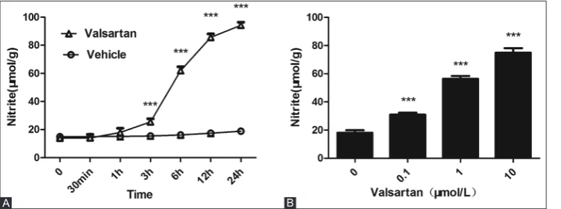
VAL (10 µmol/L) was used to treat EA.hy926 cells for 30 minutes, 1, 3, 6, 12, and 24 hours. Compared with the control group (treated with the solvent), VAL increased the intracellular NO concentration in a time-dependent man-ner, and the intergroup difference became statistically signif-icant after 3 hours of treatment (p < 0.001); after 24 hours, the concentration reached a peak (Figure 1A). Three con-centrations of VAL, i.e., 10, 1, and 0.1 µmol/L, were used to treat EA.hy926 cells for 24 hours. Compared with the control group, VAL increased the intracellular NO concentration in a dose-dependent manner (p < 0.001) (Figure 1B). Therefore, in subsequent experiments, the cells were treated with 10 µmol/L VAL.
Effects of VAL on PI3K/Akt, AMPK, and eNOS
and their phosphorylation levels in EA.hy926 cells
FIGURE 2. Effects of valsartan (10 µmol/L) on Akt, adenosine monophosphate-activated protein kinase (AMPK) and endothelial nitric oxide synthase (eNOS) phosphorylation and expression in human umbilical vein endothelial cells. Ctrl: control; Antibodies: p-AMPKα (Thr172) - phospho-AMPKα (Thr172), AMPKα, p-Akt (Ser473) - phospho-Akt (Ser473), Akt, p-eNOS (Ser1177) - phospho-eNOS (Ser1177),
After EA.hy926 cells were treated with 10 µmol/L VAL for 30 minutes, 1, 3, 6, 12, and 24 hours, the western blot analysis showed that the phosphorylation levels of Akt, AMPK, and eNOS increased significantly. In particular, the phosphoryla-tion levels of Akt and eNOS began to increase after the 1-hour treatment period, and that of AMPK increased significantly after the 6-hour treatment period. However, VAL had no sig-nificant effect on the total expression levels of these three pro-teins (Figure 2).
Effects of the Akt/AMPK signaling pathway on
eNOS activation and NO synthesis induced by
VAL in EA.hy926 cells
To clarify whether VAL promotes NO synthesis in EA.hy926 cells via AMPK/Akt-activated eNOS, the cells were divided into five groups as follows: Control, VAL, VAL + Compound C (AMPK inhibitor, 1 µmol/L), VAL+ LY294002 (Akt inhibitor, 10 µmol/L), and VAL+ L-NAME (eNOS inhibitor, 500 µmol/L) groups. The cells were pretreated with
FIGURE 1. Effects of valsartan (VAL) on the nitric oxide (NO) concentration in human umbilical vein endothelial cell (HUVECs). (A) Time-effect relationship of VAL (10 µmol/L) on NO production in HUVECs. (B) Dose-Time-effect relationship of VAL on NO production in HUVECs. The experiment was performed in five repetitive wells and the mean values (± standard deviation) are reported. ***p<0.001 versus vehicle-treated group.
the corresponding inhibitors for 1 hour. In VAL group, VAL could significantly activate Akt and AMPK after the 3- and 6-hour treatment periods, thus the intracellular eNOS phos-phorylation level, changes in total protein, and the effects on NO synthesis in each group were detected at 3 and 6 hours. The results of western blot analyses showed that at 3 hours, in VAL+ LY294002 and VAL+ L-NAME groups, LY294002 and L-NAME, respectively, inhibited VAL-induced eNOS activation, but in VAL + Compound C group, Compound C showed no significant inhibitory effects (Figure 3A). At 6 hours, in VAL + Compound C and VAL+ L-NAME groups, Compound C and L-NAME, respectively, both inhibited VAL-induced eNOS activation, but in VAL+ LY294002 group, LY294002 did not show significant inhibitory effects (Figure 3C). With respect to NO levels, at 3 hours, in VAL+ LY294002 and VAL+ L-NAME groups, LY294002 and L-NAME, respectively, significantly inhibited VAL-induced NO synthesis (iNOS) (p < 0.001), but in VAL + Compound C group, Compound C showed no significant inhibitory effects (p > 0.05) (Figure 3B). At 6 hours, in VAL+ LY294002, VAL + Compound C and VAL+ L-NAME groups, LY294002, Compound C, and L-NAME, respectively, all significantly inhibited VAL-iNOS (p < 0.001). However, the inhibi-tory effects of LY294002 and Compound C were relatively weaker, while L-NAME could completely inhibit VAL-iNOS (p < 0.001) (Figure 3D).
DISCUSSION
Atherosclerosis is an important pathological change that causes chronic ischemic cardio-cerebral diseases. The pathogenesis of this disease is complex, and several theories have been developed to explain the process, among which many scholars currently support the “response to injury” theory [13]. Hypertension is a common risk factor of this disease, and endothelial dysfunction is a “bridge” connecting the two. Some studies have shown that patients with hyper-tension might exhibit endothelial dysfunction in the early stage [14]. Therefore, controlling hypertension and improving the endothelial function are important for the prevention of atherosclerosis.
A large number of experimental studies [1-3] have shown that VAL, a clinical first-line antihypertensive drug, could increase in vivo synthesis and biological activity of NO, thus exhibiting a variety of cardiovascular protective effects, e.g. blood vessel relaxation, anti-inflammation, and anti-plate-let aggregation. Nevertheless, the specific mechanisms of VAL action require further elaboration. Vascular endothelial cells are an important source of NO synthesis in vivo [15]. In this study, VAL was used to treat HUVECs (EA.hy926); it increased NO synthesis in the EA.hy926 cells in time- and dose-depen-dent manners and this result was related to the activation of the PI3K/Akt/eNOS pathway, consistent with the results of
FIGURE 3. Effects of Akt/adenosine monophosphate-activated protein kinase on valsartan (VAL)-mediated endothelial nitric oxide (NO) synthase (eNOS) phosphorylation and NO production in human umbilical vein endothelial cells (HUVECs). (A) The variation of VAL (10 µmol/L)-induced eNOS activation after HUVECs were incubated with LY294002 [LY] (10 µmol/L), Compound C [CompC] (1 µmol/L), L-nitro-arginine methyl ester (L-NAME) (500 µmol/L) for 3 hours. (B) The variation of VAL (10 µmol/L)-induced nitric oxide (NO) produc-tion after HUVECs were incubated with LY294002 (10 µmol/L), Compound C (1 µmol/L), L-NAME (500 µmol/L) for 3 hours. (C) The vari-ation of VAL (10 µmol/L)-induced eNOS activvari-ation after HUVECs were incubated with LY294002 (10 µmol/L), Compound C (1 µmol/L), L-NAME (500 µmol/L) for 6 hours. (D) The variation of VAL (10 µmol/L)-induced NO production after HUVECs were incubated with LY294002 (10 µmol/L), Compound C (1 µmol/L), L-NAME (500 µmol/L) for 6 hours. ***p<0.001 versus vehicle-treated group. ###p<0.001 versus VAL group. Ctrl: control; Antibodies: p-eNOS (Ser1177) - phospho-eNOS (Ser1177); eNOS; beta-actin.
A B
Su et al. [8]. Furthermore, our results showed that AMPK was also involved in this process, although Compound C (an AMPK inhibitor) used by Su et al. [8] did not affect NO syn-thesis induced by VAL in endothelial cells. This inconsistency could be explained in several ways. First, the AMPKα subunit has two subtypes, α1 and α2. The α1 is the main subtype that promotes eNOS activation and NO synthesis [16], while α2 is mainly associated with endothelial cell differentiation and angiogenesis [17]. Although these subtypes are both expressed in vascular endothelial cells, the expression ratio of AMPKα1 to α2 differs significantly among different donors [18]. In the study of Su et al. [8], the expression of the AMPKα1 subtype may be lower compared to the AMPKα1 expression in our study, possibly explaining the negligible effects on VAL-iNOS in the human aortic endothelial cells observed in their study. However, because VAL did not affect the total protein and phosphorylation levels of AMPK in human aortic endothe-lial cells in their study, additional experiments are necessary to verify this hypothesis. Another explanation is the excessive concentration of Compound C (10 µmol/L); Labuzek et al. [19] reported that Compound C itself could increase intracellular inflammation activating inflammation-induced NO synthase, another important kinase involved in NO synthesis when inflammation occurs in endothelial cells [20,21]. Therefore, we speculate that a high concentration of Compound C might not inhibit VAL-iNOS, and might even increase the NO con-centration in endothelial cells.
In our study, we first applied VAL to endothelial cells for various time periods and found that VAL in turn increased the phosphorylation levels of Akt, eNOS, and AMPK. AMPK is an upstream signaling molecule of eNOS, but AMPK was activated significantly later than eNOS in this study. Accordingly, we speculate that after VAL was applied to the endothelial cells, it first activated Akt, which promoted the increased phosphorylation of its downstream signaling molecule eNOS. When AMPK was activated, eNOS was in an equilibrium state with respect to phosphorylation and dephosphorylation, and the phosphorylation level of eNOS did not further increase. To verify this hypothesis, the endo-thelial cells were divided into five groups: Control, VAL, VAL + Compound C (AMPK inhibitor, 1 µmol/L), VAL+ LY294002 (Akt inhibitor, 10 µmol/L), and VAL+ L-NAME (eNOS inhib-itor, 500 µmol/L), and the effects on the phosphorylation level of eNOS and NO synthesis were observed. As shown in Figure 3A and B, eNOS activation and NO synthesis in the endothelial cells treated with 3-hour VAL treatment were associated with the activation of Akt, while AMPK was not involved in VAL-induced eNOS activation and NO synthe-sis. As shown in Figure 3C and D, eNOS activation and NO synthesis in the endothelial cells treated with 6-hour VAL
treatment were not only associated with the activation of Akt, but also with the activation of AMPK, and, in our opinion, the latter association was much more important. In addition, L-NAME could significantly inhibit VAL-iNOS either at 3 or 6 hours, indicating that VAL-iNOS was closely related to the activation of eNOS. Therefore, these results ultimately indi-cate that VAL could activate Akt, AMPK, and eNOS at differ-ent time points, thus increasing NO synthesis in endothelial cells.
Our study had some shortcomings. First, our results only revealed that VAL could activate AMPK contained in vascular endothelial cells, and upstream signaling was not examined. Recent studies have found at least three upstream kinases of AMPK, including liver kinase B1 (LKB1), Ca2+/calmod-ulin-dependent protein kinase (CaMKK), and transforming growth factor-β-activated protein kinase1(TAK1) [22-24], and the first two are well-supported. Ha et al. [11] reported that in THP-1 cells, VAL could activate AMPK via LKB1, but the precise kinase(s) associated with VAL activation of AMPK in endothelial cells are unknown. Additional studies on the acti-vation of upstream kinase(s) of AMPK by VAL may provide support for the conclusion that VAL increases NO synthesis in endothelial cells via the AMPK/eNOS pathway. Second, our results indicated that VAL could increase NO synthesis in endothelial cells via the PI3K/Akt/AMPK/eNOS pathway as a selective AT1R antagonist; however, some studies observed partial pharmacological effects of VAL that were independent of its role in antagonizing the AT1R [11,25]. We did not deter-mine whether the obstruction of the AT1R was involved in the effects of VAL on NO synthesis.
CONCLUSION
Our study not only validated that VAL could activate the PI3K/Akt/eNOS pathway, and thus increase NO synthesis in endothelial cells, but also revealed for the first time that VAL could increase NO synthesis in endothelial cells by activating the AMPK/eNOS pathway. These results partially explain the mechanisms underlying the antihypertensive effects of VAL as well as its simultaneous inhibitory effects on the occurrence and development of atherosclerosis.
ACKNOWLEDGMENTS
This study was supported by Henan Provincial Scientific and Technological Projects (No. 122102310589).
DECLARATION OF INTERESTS
REFERENCES
[1] Sander GE, Giles TD. Nebivolol and valsartan as a fixed-dose combination for the treatment of hypertension. Expert Opin Pharmacother 2015;16(5):763-70.
http://dx.doi.org/10.1517/14656566.2015.1020790.
[2] Xu Y, Hu X, Wang L, Jiang Z, Liu X, Yu H, et al. Preconditioning via angiotensin Type 2 receptor activation improves therapeutic efficacy of bone marrow mononuclear cells for cardiac repair. PLoS One 2013;8(12):e82997.
http://dx.doi.org/10.1371/journal.pone.0082997.
[3] Kalinowski L, Matys T, Chabielska E, Buczko W, Malinski T. Angiotensin II AT1 receptor antagonists inhibit platelet adhe-sion and aggregation by nitric oxide release. Hypertenadhe-sion 2002;40(4):521-7.
http://dx.doi.org/10.1161/01.HYP.0000034745.98129.EC.
[4] Duan L, Lei H, Zhang Y, Wan B, Chang J, Feng Q, et al. Calcitonin gene-related peptide improves hypoxia-induced inflammation and apoptosis via nitric oxide in H9c2 cardiomyoblast cells. Cardiology 2016;133(1):44-53.
http://dx.doi.org/10.1159/000439123.
[5] Sunshine SB, Dallabrida SM, Durand E, Ismail NS, Bazinet L, Birsner AE, et al. Endostatin lowers blood pressure via nitric oxide and prevents hypertension associated with VEGF inhibition. Proc Natl Acad Sci U S A 2012;109(28):11306-11.
http://dx.doi.org/10.1073/pnas.1203275109.
[6] Campelo AE, Cutini PH, Massheimer VL. Testosterone modulates platelet aggregation and endothelial cell growth through nitric oxide pathway. J Endocrinol 2012;213(1):77-87.
http://dx.doi.org/10.1530/JOE-11-0441.
[7] Lundberg JO, Gladwin MT, Weitzberg E. Strategies to increase nitric oxide signalling in cardiovascular disease. Nat Rev Drug Discov 2015;14(9):623-41.
http://dx.doi.org/10.1038/nrd4623.
[8] Su KH, Tsai JY, Kou YR, Chiang AN, Hsiao SH, Wu YL, et al.
Valsartan regulates the interaction of angiotensin II Type 1 receptor and endothelial nitric oxide synthase via Src/PI3K/Aktsignalling. Cardiovasc Res 2009;82(3):468-75.
http://dx.doi.org/10.1093/cvr/cvp091.
[9] Tsai HY, Lin CP, Huang PH, Li SY, Chen JS, Lin FY, et al. Coenzyme Q10 attenuates high glucose-induced endothelial progenitor cell dysfunction through AMP-activated protein kinase pathways. J Diabetes Res 2016;2016:6384759.
http://dx.doi.org/10.1155/2016/6384759.
[10] Lee CH, Lee SD, Ou HC, Lai SC, Cheng YJ. Eicosapentaenoic acid protects against palmitic acid-induced endothelial dysfunc-tion via activadysfunc-tion of the AMPK/eNOS pathway. Int J Mol Sci 2014;15(6):10334-49.
http://dx.doi.org/10.3390/ijms150610334.
[11] Ha YM, Park EJ, Kang YJ, Park SW, Kim HJ, Chang KC. Valsartan independent of AT1 receptor inhibits tissue factor, TLR-2 and -4 expression by regulation of Egr-1 through activation of AMPK in diabetic conditions. J Cell Mol Med 2014;18(10):2031-43.
http://dx.doi.org/10.1111/jcmm.12354.
[12] Zhong X, Xiu LL, Wei GH, Liu YY, Su L, Cao XP, et al. Bezafibrate enhances proliferation and differentiation of osteoblastic MC3T3-E1 cells via AMPK and eNOS activation. Acta Pharmacol
Sin 2011;32(5):591-600.
http://dx.doi.org/10.1038/aps.2011.15.
[13] Sitia S, Tomasoni L, Atzeni F, Ambrosio G, Cordiano C, Catapano A, et al. From endothelial dysfunction to atherosclerosis. Autoimmun Rev 2010;9(12):830-4.
http://dx.doi.org/10.1016/j.autrev.2010.07.016.
[14] Taddei S, Virdis A, Mattei P, Salvetti A. Vasodilation to acetyl-choline in primary and secondary forms of human hypertension. Hypertension 1993;21(6 Pt 2):929-33.
http://dx.doi.org/10.1161/01.HYP.21.6.929.
[15] Wyatt AW, Steinert JR, Mann GE. Modulation of the L-arginine/ nitric oxide signalling pathway in vascular endothelial cells. Biochem Soc Symp 2004;71:143-56.
http://dx.doi.org/10.1042/bss0710143.
[16] Goirand F, Solar M, Athea Y, Viollet B, Mateo P, Fortin D, et al. Activation of AMP kinase alpha 1 subunit induces aortic vasorelax-ation in mice. J Physiol 2007;581(Pt 3):1163-71.
http://dx.doi.org/10.1113/jphysiol.2007.132589.
[17] Nagata D, Mogi M, Walsh K. AMP-activated protein kinase (AMPK) signaling in endothelial cells is essential for angiogenesis in response to hypoxic stress. J Biol Chem 2003;278(33):31000-6. http://dx.doi.org/10.1074/jbc.M300643200.
[18] Ewart MA, Kennedy S. AMPK and vasculoprotection. Pharmacol Ther 2011;131(2):242-53.
http://dx.doi.org/10.1016/j.pharmthera.2010.11.002.
[19] Labuzek K, Liber S, Gabryel B, Buldak L, Okopien B. Ambivalent effects of compound C (dorsomorphin) on inflammatory response in LPS-stimulated rat primary microglial cultures. Naunyn Schmiedebergs Arch Pharmacol 2010;381(1):41-57.
http://dx.doi.org/10.1007/s00210-009-0472-2.
[20] Yehuda I, Madar Z, Leikin-Frenkel A, Tamir S. Glabridin, an isofla-van from licorice root, downregulates iNOS expression and activity under high-glucose stress and inflammation. Mol Nutr Food Res 2015;59(6):1041-52.
http://dx.doi.org/10.1002/mnfr.201400876.
[21] Cortese-Krott MM, Kulakov L, Opländer C, Kolb-Bachofen V, Kröncke KD, Suschek CV. Zinc regulates iNOS-derived nitric oxide formation in endothelial cells. Redox Biol 2014;2:945-54. http://dx.doi.org/10.1016/j.redox.2014.06.011.
[22] Thibert C, Perret C, Billaud M. AMPK potentiation by LKB1 iso-forms. Oncotarget 2015;6(34):35139-40.
DOI: 10.18632/oncotarget.6127.
[23] Hurley RL, Anderson KA, Franzone JM, Kemp BE, Means AR, Witters LA. The Ca2/calmodulin-dependent protein kinase kinases are AMP-activated protein kinase kinases. J Biol Chem 2005;280(32):29060-6.
http://dx.doi.org/10.1074/jbc.M503824200.
[24] Herrero-Martín G, Høyer-Hansen M, García-García C, Fumarola C, Farkas T, López-Rivas A, et al. TAK1 activates AMPK-dependent cytoprotective autophagy in TRAIL-treated epithelial cells. EMBO J 2009;28(6):677-85.
http://dx.doi.org/10.1038/emboj.2009.8.
[25] Iwashita M, Sakoda H, Kushiyama A, Fujishiro M, Ohno H, Nakatsu Y, et al. Valsartan, independently of AT1 receptor or PPAR gamma, suppresses LPS-induced macrophage activation and improves insulin resistance in cocultured adipocytes. Am J Physiol Endocrinol Metab 2012;302(3):E286-96.