R E V I E W
Open Access
Antibacterial coating of implants in
orthopaedics and trauma: a classification
proposal in an evolving panorama
Carlo Luca Romanò
1*, Sara Scarponi
1, Enrico Gallazzi
1, Delia Romanò
1and Lorenzo Drago
2Abstract
Implanted biomaterials play a key role in current success of orthopedic and trauma surgery. However, implant-related infections remain among the leading reasons for failure with high economical and social associated costs. According to the current knowledge, probably the most critical pathogenic event in the development of implant-related infection is biofilm formation, which starts immediately after bacterial adhesion on an implant and effectively protects the microorganisms from the immune system and systemic antibiotics. A rationale, modern prevention of biomaterial-associated infections should then specifically focus on inhibition of both bacterial adhesion and biofilm formation. Nonetheless, currently available prophylactic measures, although partially effective in reducing surgical site infections, are not based on the pathogenesis of biofilm-related infections and unacceptable high rates of septic complications, especially in high-risk patients and procedures, are still reported.
In the last decade, several studies have investigated the ability of implant surface modifications to minimize bacterial adhesion, inhibit biofilm formation, and provide effective bacterial killing to protect implanted biomaterials, even if there still is a great discrepancy between proposed and clinically implemented strategies and a lack of a common language to evaluate them.
To move a step forward towards a more systematic approach in this promising but complicated field, here we provide a detailed overview and an original classification of the various technologies under study or already in the market. We may distinguish the following: 1. Passive surface finishing/modification (PSM): passive coatings that do not release bactericidal agents to the surrounding tissues, but are aimed at preventing or reducing bacterial adhesion through surface chemistry and/or structure modifications; 2. Active surface finishing/modification (ASM): active coatings that feature pharmacologically active pre-incorporated bactericidal agents; and 3. Local carriers or coatings (LCC): local antibacterial carriers or coatings, biodegradable or not, applied at the time of the surgical procedure, immediately prior or at the same time of the implant and around it. Classifying different technologies may be useful in order to better compare different solutions, to improve the design of validation tests and, hopefully, to improve and speed up the regulatory process in this rapidly evolving field.
Keywords:Orthopaedics, Trauma, Implant, Joint, Prosthesis, Biofilm, Infection, Coating, Antibacterial, Classification
* Correspondence:[email protected] 1
Department of Reconstructive Surgery of Osteo-articular Infections C.R.I.O. Unit, IRCCS Galeazzi Orthopaedic Institute, Via R. Galeazzi 4, 20161 Milan, Italy Full list of author information is available at the end of the article
Introduction
Even if current peri-operative infection prevention methods, like antibiotic prophylaxis, have significantly reduced the incidence of surgical site infections (SSI), up to 2.5 % of primary hip and knee arthroplasties and to 10 % of revision arthroplasties can still be compli-cated by periprosthetic joint infection (PJI) [1]. More-over, according to recent analysis, these figures could even be underestimated and are on the rise [2], while multi-resistant pathogens are often retrieved [3]. The occurrence of PJI is a devastating complication, often requiring implant removal, with high morbidity and possible mortality raise [4] and elevated economical and social costs [5].
Implant-related infections are the results of a complex interaction of various factors, including bacterial load, microorganism’s and host’s type, surgical procedure and technique, and type of implant and of antibacterial prophylaxis.
In fact, even elective surgery may not be performed in a completely sterile environment, and operating rooms have been shown to become contaminated within the first few hours of service [6, 7]. While in the majority of cases the rather low bacterial load eventually present at surgery may be generally overcome by the host’s immunological defense and the systemic antibiotic prophylaxis [8], in some patients a SSI may eventually develop, especially in high-risk patients, in which relevant co-morbidities may increase the relative risk for infection up to 20 times compared to normal population [9–11]. Similarly, the most complex surgical procedures and techniques have been shown to be more prone to septic complications [12]. In this context, implant features, including size, shape, material, and intended use, also play an important role [13], while, for example, local antibacterial prophylaxis, like the use of antibiotic-loaded bone cement or bone grafts has been shown to reduce the incidence of implant-related in-fections [14, 15]. In line with these considerations, a strong recommendation was delivered, in a recent international consensus meeting on PJI, concerning the need for devel-oping effective antibacterial surfaces that prevent bacterial adhesion and colonization of implants and proliferation into the surrounding tissues [16].
In the present review, we provide a detailed descrip-tion and an original classificadescrip-tion of the technologies already available or under investigation, aimed at min-imizing implant-related infections in orthopedics and trauma surgery.
Rationale of antibacterial coating of implants
Prophylactic systemic antibiotics are administered routinely to patients who receive an orthopedic device to prevent peri-operative infection [8]. However, systemic administra-tion of antibiotics has many potential disadvantages includ-ing the need for a correct timinclud-ing of administration, the
relatively low drug concentration at the target site, and the limited ability to kill bacteria eventually present on the implant surface or embedded in biofilms.
When Anthony Gristina first proposed the concept of a
“race for the surface”, more than three decades ago, he described a simplified model of implant-related infection, whereby host and bacterial cells compete in determining the ultimate fate of the implant [17]. According to this model, when the host cells colonize the implant surface first, the probability of attachment of bacterial cells is very low and vice versa. Further studies have now made it clear that the process is much more complex and only partially understood.
Bacteria have a highly successful and diversified strategy to adhere and survive on virtually all natural and synthetic surfaces [18, 19]. Surface characteristics of a biomaterial such as roughness, hydrophobicity, and electrostatic charge play only conditional roles [20], while a number of potential receptors for bacterial adhesive ligands are offered by the protein film that covers an implant immediately after its placement into the host body [21–23]. Complement, albu-min, and several other host proteins and lipids are the main components of this conditional protein film [24–26]. The process of bacterial adhesion can be divided into a reversible phase, based on nonspecific interactions between implant surface and bacterial adhesions, and an irreversible phase, mediated by molecular and cellular interactions and closely associated with expression of biofilm-specific gene clusters in reversibly attached bacteria. All the process from bacterial adhesion to the production of a mature biofilm is extremely efficient and is normally completed within 12 to 18 h [27–30].
On the host side, the detailed process of implant osteo and tissue integration is also incompletely unveiled [31, 32]. According to Gristina’s model, host cells, once attached to implant surfaces, should lead to periprosthetic bone regen-eration and remodeling, protecting the biomaterial against bacterial colonization [33]. However, bacteria may survive to osteointegration or to fibrous tissue encapsulation of an implant, while peri-implant fibrous tissue may even prevent direct contact between host immunity cells and bacterial molecules, while the presence of the implant has been shown to impair innate local host response [34–36].
At the same time, any coating technology should prove to be safe in the short and long term, should not interfere with osteointegration or induce bacterial resistance in the long run, and should be easy to implement in the clinical practice and at an affordable cost. Also, since bacterial colonization, from microbial adhesion to an established mature biofilm layer only takes few hours [39], any anti-bacterial protection should act at the exact time of surgery and for at least some hours or days thereafter.
Classification of antibacterial coating technologies
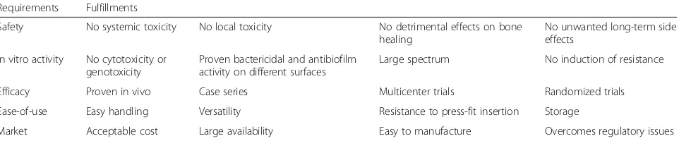
Local antibacterial implant protection can be achieved in different ways. Currently, there is not a single, uni-versally accepted classification of coating technologies, standardized validation methods are lacking and regu-latory aspects appear somewhat inadequate in view of the clinical needs and expectations. Table 1 summa-rizes the basic requirements that an “ideal” coating technology should fulfill to meet the needs of a wide-spread clinical use.
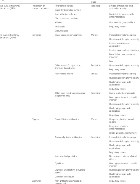
According to their strategy of action, antibacterial coat-ings can be classified in at least three groups (Table 2):
1. Passive surface finishing/modification (PSM). Here all surface chemistry and/or structure modifications aimed at preventing or reducing bacterial adhesion, without releasing bactericidal agents to the
surrounding tissues, are included.
2. Active surface finishing/modification (ASM). Active coatings, included in this class, feature pharmacologically active pre-incorporated antibacterial agents, like antibiotics, antiseptics, metal ions, or other organic and inorganic compounds.
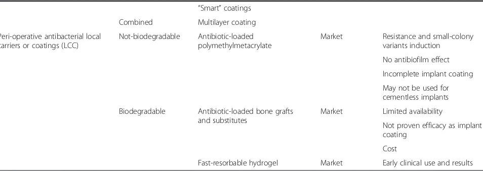
3. Peri-operative antibacterial local carriers or coatings (LCC). According to this strategy, implant
protection is achieved through a biodegradable or not-biodegradable antibacterial carrier or coating, applied during surgery on the implant or around it; the coating may simply act by delivering high local concentrations of one or more pre-loaded antibacterials, or it may have a direct or synergistic antibacterial activity. Antibiotic-loaded
polymethylmethacrylate is probably the very first
example of this coating strategy, used since many years to protect joint implants.
Although necessarily schematic, this classification may be helpful to compare different technologies both in the clinical practice and concerning regulatory aspect, that should probably differ for different classes.
Passive surface finishing/modification
Chemical and/or physical modifications of the surface layer of an existing biomaterial may result in a substantial change of its susceptibility to bacterial colonization.
Surface characteristics of implants, like surface rough-ness and chemistry, hydrophilicity, surface energy or potential and conductivity play in fact crucial roles in bacterial adhesion and subsequent biofilm formation. Surface physio-chemical modification of an implant is also relatively simple and economic to achieve and to industrialize.
Ultraviolet light irradiation, for example, is able to in-crease “spontaneous” wettability on titanium dioxide, which in turn can inhibit bacterial adhesion without compromising osteogenesis on titanium alloy implants [40, 41]. A bacterial anti-adhesive surface can also be achieved by modifying the crystalline structure of the surface oxide layer [42].
Some polymer coatings, like the hydrophilic poly-methacrylic acid, polyethylene oxide, or protein-resistant polyethylene glycol can be applied to the surface of titanium implants and result in significant inhibition of bacterial adhesion [43–45]. Even if some of these coatings may impair local osteoblast function, the use of additional bioactive molecules as sericin and RGD motif could restore and even improve the im-paired cell function [46]. Hydrophobic and super-hydrophobic surface treatment technologies have also shown a great repellent antibacterial effect in preclin-ical studies [47, 48].
Other researchers have focused on controlling the bio-logical response to biomaterials via alterations in surface structure and design [49, 50]. In this regard, changing the implant surface at a nanometric scale, at which bacterial adhesion does not simply follow the roughness of the
Table 1A list of requirements to be fulfilled by the“ideal”antibacterial implant coating strategy
Requirements Fulfillments
Safety No systemic toxicity No local toxicity No detrimental effects on bone healing
No unwanted long-term side effects
In vitro activity No cytotoxicity or genotoxicity
Proven bactericidal and antibiofilm activity on different surfaces
Large spectrum No induction of resistance
Efficacy Proven in vivo Case series Multicenter trials Randomized trials
Ease-of-use Easy handling Versatility Resistance to press-fit insertion Storage
Table 2Classification of antibacterial implant protection strategies
Strategy Features Examples Development
stage
Limits
Passive surface finishing/ modifications (PSM)
Prevention of bacterial adhesion
Hydrophilic surface Preclinical Limited antibacterial and antibiofilm activity Super-hydrophobic surface
Anti-adhesive polymers Possible interference with osteointegration Nano-patterned surface
Albumin Unknown long-term effects
Hydrogels Regulatory issues
Biosurfactants
Active surface finishing/ modifications (ASM)
Inorganic Silver ions and nanoparticles Market Incomplete implant coating
Questionable long-term toxicity
Limited versatility and applicability
Limited large-scale applications
Possible bacterial resistance induction
Costs
Other metals (copper, zinc, titanium dioxide etc.)
Preclinical Questionable long-term toxicity
Regulatory issues
Non-metals: Iodine Clinical Incomplete implant coating
Questionable long-term toxicity
Challenging large-scale application
Regulatory issues
Other non-metal ions (selenium, grapheme, etc.)
Preclinical Poorly studied compounds
Coating resistance to press-fit insertion
Questionable long-term toxicity
Challenging large-scale application
Regulatory issues
Organic Coated/linked antibiotics Market Unique application to nail coating
Long-term effects on osteointegration
Single antibiotic (gentamicin)
Covalently linked antibiotics Preclinical Incomplete implant coating.
Questionable long-term toxicity
Challenging large-scale application
Regulatory issues
Antimicrobial peptides No data on in vivo or clinical effects
Cytokines Coating resistance to press-fit
insertion
Enzymes and biofilm disrupting agents
Questionable long-term toxicity
Challenging large-scale application
Chitosan derivatives
Synthetic Non-antibiotic antimicrobial compounds
surface but also is dependent on other variables like the quantity of adsorbed proteins, can in fact suppress bacteria adhesion [51].
Treating more specifically protein surfaces and/or pro-tein–bacteria interactions may also be a successful strategy of inhibiting bacterial adhesion to a specific biomaterial [52, 53]. Friedman et al., using a rabbit model, demon-strated reduced bacterial adherence on pure titanium samples and decreased infection rates of implants coated with cross-linked albumin [54].
More recently, novel strategies include production of self-assembled mono- or multilayers, surface grafting or hydrogels, or the use of biosurfactants and microbial amphyphilic compounds with excellent anti-adhesive properties [55, 56].
In summary, a number of anti-adhesive surface modi-fications have been proposed for different purposes, but only a few will probably be suitable for clinical use. In particular, a strong anti-adhesive layer cannot be used for coating of fixation surfaces of joint arthro-plasty since it could also interfere with implant osteoin-tegration, leading to early mechanical failure [48, 49]. Another challenge of designing anti-adhesive tech-nologies relates to the current inability to find a uni-versal treatment that can be applied to all surfaces and biomaterials, all bacterial species and under all (in-growth and nonin(in-growth) implants. Moreover, passive coating methods should be preferred as long as their antibacterial ability is strong enough to prevent bio-film formation, but the ability of passive coatings to resist bacterial adhesion is generally limited and varies greatly depending on the bacterial species and loads [57]. In vivo efficacy and long-term effects of these new technologies both on host’s cells and on bacterial resistance are also poorly understood and need to be further investigated before clinical applications and market introduction.
Active surface finishing/modification
Surface modifications may include pharmacologically active pre-incorporated antibacterial agents or compounds, like antibiotics, antiseptics, metal ions, or organic molecules. Such pharmacologically activated coatings may change the implant from a passive, pharmacologically inert medical de-vice, to something more and more similar to a drug agent, with difficult to predict long-term effects and challenging regulatory issues.
Historically, two main strategies have been proposed for effective antibacterial surface treatment either“contact kill-ing” or drug eluting, while in terms of durability, we can distinguish between degradable and non-degradable coat-ings. Killing of bacteria can be achieved by interfering with cell respiration or division, cell wall formation or bacterial signaling network as well as inhibition of the transition of planktonic phenotype of bacteria into a sessile type [58].
Antibacterial surface technologies can employ metals (silver, zinc, copper, etc.), non-metal elements (e.g., iodine, selenium), organic substances (antibiotics, anti-infective peptides, chitosan, other substances), and their combinations.
Antibacterial activity of the majority of metal coatings is closely linked to the ionic or nano form rather than to the bulk material [59]. Silver is the most prevalent metal used in biomedical applications. Dissolved silver cations are biochemically active agents that interfere with bacterial cell membrane permeability and cellular metabolism. Silver also contributes to formation of reactive oxygen species and other mechanisms that potentially influence prokaryotic cells [60]. There has been concern, however, about the toxicity of silver ions [61]. Research efforts have focused on the development of silver coating technologies that reduce or even eliminate toxicity while maintaining antibacterial effects [62, 63]. Despite demonstrated clinical efficacy and safety in recent comparative studies [64, 65], routine use of silver-coated implants remains rather
Table 2Classification of antibacterial implant protection strategies(Continued) “Smart”coatings
Combined Multilayer coating
Peri-operative antibacterial local carriers or coatings (LCC)
Not-biodegradable Antibiotic-loaded polymethylmetacrylate
Market Resistance and small-colony variants induction
No antibiofilm effect
Incomplete implant coating
May not be used for cementless implants
Biodegradable Antibiotic-loaded bone grafts and substitutes
Market Limited availability
Not proven efficacy as implant coating
Cost
limited. The main obstacle to a broader usage of such technology is cytotoxicity on bone cells, that prevented until now coating of the intra-medullary part of the pros-thesis. In addition, cost issues and the inability to apply the technology to a variety of prosthetic implants and de-vices further reduces its application outside oncological or highly selected cases.
Copper and zinc also have potent antibacterial effects on a wide spectrum of bacterial species [66, 67]; however, potential toxic side effects of these metals remain a strong concern [68]. Proposed solutions include copper- and zinc-based nanomaterials or, alternatively, controlled release [69]. The risk of bacterial resistance to metallic coatings remains a potential limitation for their widespread use [70]. Concern also exists about the mechanical properties of im-plant nanocoatings since damage may occur during surgical implantation especially in cementless implants inserted via press-fit methods [71].
Another interesting technology is related to modification of commonly used alloys, like titanium. The anti-infective potential of titanium dioxide layers has been widely investi-gated and proven effective in vitro both alone [72] or in combination with other substances [73].
Non-metal elements like hydrogen, chlorine, iodine, or oxygen are commonly used in biomedicine for their anti-infective properties. Selenium bound covalently onto the surface of titanium or titanium alloy implant discs has been shown to preventStaphylococcus aureus
and Staphylococcus epidermidis attachment without affecting osteoblast viability [74]. Selenium catalyzes the formation of superoxide radicals and subsequently inhibits bacterial adhesion and viability. In addition, selenium nanoparticles can inhibit bacterial growth and biofilm formation [75].
Ongoing research is also directed to determine the clinical applicability of carbon substances like graphene or carbon nanotubes, that can be synthesized in multi-functional layers [76]; however, the most interesting technology today under study, related to non-metal elements, is probably iodine coating of titanium alloys, that has recently demonstrated clinical efficacy in a continuous series of 222 patients with excellent results [77].
Several organic compounds with antibacterial properties have the potential to be linked to the surface of implants conferring them anti-infective properties. A large number of studies have investigated the efficacy of surfaces coated with covalently linked antibiotics [78–82]. Clinical effective-ness of such implants is most likely limited to infections caused by bacteria that are sensitive to the specific anti-biotic that has been coupled. In addition, strong forces such as covalent binding are insufficiently sensitive to react to weak external stimuli [83]. In fact, despite the theoretical advantages for non-eluting systems, this concept is lim-ited by the fragility of the coatings and killing activity
potential of bacteria which might not be directly adja-cent to the implant. To overcome these issues, combi-nations of antibiotics with other compounds have been proposed either alone or in association with a particu-lar mechanism of controlled release [84]. Antibiotics such as gentamicin, vancomycin, and others have been loaded into porous hydroxyapatite (HA) coatings on titanium implants. The antibiotic-HA coatings exhibit significant improvement in preventing infection com-pared with standard HA coatings in vivo, but there are still many unresolved issues regarding the methodology of antibiotic incorporation into the HA coating and the optimal release kinetics and possible detachment of the coating at the time of press-fit insertion.
Biodegradable polymers and sol–gel coatings are also utilized to form controlled release antibiotic-laden coat-ings on titanium implants [85, 86]. Clinical applications of antibiotic-loaded D-poly-lactate acid/gentamycin intra-medullary coated nail have been recently reported with early positive results [87].
Some antiseptic agent such as chlorhexidine, chloroxyle-nol, or poly-hexamethylenebiguanide have demonstrated efficacy and might be an alternative to avoid the risk of drug resistance. Chlorhexidine can be adsorbed to the TiO2 layer on titanium surfaces and is released gradually over several days [88]. Its release pattern is similar to that of antibiotic-laden coatings with an initial rapid release rate followed by slower but sustained release [89].
Another promising approach involves coating implants with antimicrobial peptides, cytokines, or other molecules critical for host response to bacteria invasion. This hetero-geneous group of substances has proven experimentally their efficacy against a wide range of pathogens [90]. Anti-microbial peptides, like antibiotics, function via damage of the cell wall and inhibition of key bacterial protein synthe-sis. In addition, they exert influence upon inflammation, tissue healing, and apoptotic events [91]; resistance to anti-microbial peptides has been reported less frequently than to antibiotics [92]. Initial experiments demonstrated that a thin layer of antimicrobial peptides affixed onto the surfaces of metal alloys exhibit excellent antibacterial effects against typical pathogens related to PJI [93].
Long-term impact of permanently coated implants with antibiotics and other organic compounds, never used before either for local or general administration, does raise concerns regarding possible induction of bacterial resist-ance, local, and general toxicity and possible detrimental effects on implant osteointegration, ultimately preventing clinical applications until now.
Still more complex approaches involve the development of multifunctional surface layers, like functional polymer brush coating, that combine anti-adhesive and antimicro-bial substances and other compounds able to enhance tissue integration [97], while “smart coatings”, sensitive and responsive to a variety of stimuli, including the pres-ence of bacteria [98], are another fascinating but futuristic research pathway that poses a number of open questions, like feasible coating manufacturing process, non-adverse reactions in vivo, mechanical resistance, or preservation of intended functionalities throughout the life of the device.
Peri-operative antibacterial carriers or coatings
Instead of pre-manufactured surface modifications, either with or without pharmacologically active agents, a differ-ent approach to implant protection may be to provide a traditional implant with an antibacterial carrier or coating at the time of surgery. The separation of the protective solution from the implant until surgery may reduce the regulatory requirements and increase the applicability of a universal antibacterial coating to many different already existing implants and biomaterials.
Local administration of antibiotics historically attracted much attention in orthopaedics. Buchholz et at. first popularized the incorporation of antibiotics into poly-methylmethacrylate (PMMA) bone cement for local anti-biotic prophylaxis in cemented total joint arthroplasty [99]. Clinical studies have shown that antibiotic-loaded bone cement can decrease deep infection rates of cemen-ted total hip arthroplasties and revision rates due to sup-posed “aseptic” loosening when combined with systemic antibiotic administration [100] and this solution has been found both effective and economically sound, especially in high-risk patients [101, 102]. However, PMMA was not designed as a local delivery carrier of antibiotics and may have some limits. Antibiotic-loaded PMMA may not over-come biofilm formation and may be associated with the development of antibiotic-resistant“small-colony variants” [103, 104], while the increasing use of cementless implants worldwide, especially at the hip site, make this a possible option only for a restricted number of patients.
Other porous materials for local antibiotic delivery like collagen sponges [105], cancellous bone [106], and calcium phosphate cements [107, 108] were not specifically designed to protect implanted biomaterials, and their use for routine infection prevention in joint prosthesis is limited by their insufficient in vitro, in vivo, and
clinical evidence of efficacy in this specific application, their inability to be applied as a coating to all implants’ surfaces, and their relatively high costs and possible interference with primary implant fixation and long-term osteointegration.
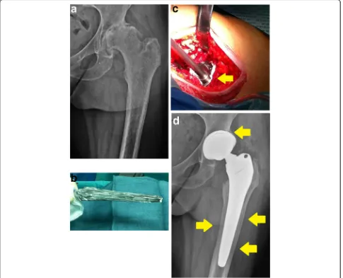
Biocompatible hydrogels do represent a possible alternative solution, as they have demonstrated to be able to deliver local pharmacological agents and may be designed to meet the desired elution pattern [109]. Recently, a fast-resorbable hydrogel coating, that can be loaded intra-operatively with various antibacterials, has been introduced in the European market [110].
Fig. 1“Defensive Antibacterial Coating”, DAC® (Novagenit Srl, Mezzolombardo, Italy): a fast-resorbable hydrogel coating, composed of covalently linked hyaluronan and poly-D,L-lactide, is spread onto a cementless hip prosthesis. The hydrogel is loaded intra-operatively with one or more antibiotics that are released within 48 to 72 h, providing antibacterial and antibiofilm protection to the implant
Table 3Tested antibacterials to be loaded with DAC hydrogel coating at concentrations ranging from 2 to 10 % ([113] and Novagenit Srl data on file)
Antibacterial family Tested antibiotics
Aminoglycosides Gentamicin
Tobramycin
Amikacin
Carbapenems Meropenem
Glicopeptides Vancomycin
Teicoplanin
Quinolones Ciprofloxacin
Cyclic lipopeptides Daptomycin
Rifamycins Rifampicin
Glycylcyclines (Tetracyclines) Tigecyclin
Oxazolidinones Linezolid
Antifungals Amphotericin B
Fluconazole
Based on the facts that bacterial colonization occurs within the very first hours after implant and that short-term systemic prophylaxis is equally effective as the long-term one to prevent PJIs [111], this novel coating technology introduced the “short-term local protection” of the implant. A short-term local delivery system may in fact meet the requirements needed to win the“run to the surface”, while limiting possible long-term unwanted side effects [112]. This novel fast-resorbable hydrogel coating,“Defensive Antibacterial Coating”, DAC® (Novagenit Srl, Mezzolombardo, Italy), composed of cova-lently linked hyaluronan and poly-D,L-lactide, (Fig. 1), is designed to undergo complete hydrolytic degradation in vivo within 48 to 72 h, being able to completely release a variety of different antibacterials, including glycopep-tides, amynoglycosides, and fluoquinolones (Table 3), at
concentrations ranging from 2 % to 10 %. The hydrogel showed a synergistic antibacterial activity with various an-tibiotics and antibiofilm agents in vitro [113], while in vivo it has been proven effective a rabbit model of highly contaminated implant both with [114] and without sys-temic prophylaxis [115]. An ongoing European multicen-ter clinical trial (Fig. 2) is currently investigating the safety and the efficacy of the device [116].
Conclusions
A tremendous amount of research has been done in the last two decades in order to better protect implanted biomate-rials from bacterial colonization, but we are still probably at the beginning of the journey. In fact, in spite of the highly successful advances in preclinical studies, a remarkable dis-crepancy still exists between the proposed strategies and
their clinical applications [117, 118]. While most of the coatings under study will not probably be suitable for orthopedic implants, due to cytotoxicity, immunoreactivity, or genotoxicity problems, clinical application of those successfully tested in vitro and in vivo can still be limited by biotechnological, regulatory, economic, and medico-legal issues.
Improving collaborative efforts amongst governments, regulatory agencies, industry leaders, and health care payers will probably allow more and more our patients to benefit from these very promising technologies.
A comprehensive classification and a common language are needed to standardize appropriate tests and realistic regulatory requirements, in order to favor the transition from preclinical studies to effective patient’s protection.
Consent
Written informed consent was obtained from the patient for the publication of this report and any accompanying images.
Competing interests
CLR is a co-inventor of the DAC hydrogel technology and has received reimbursements, fees, and funding from the following companies, involved in the development of some of the technologies cited in the text: DePuySynthes Inc., Zimmer-Biomet Inc., Link Italia Srl, Heraeus GmbH, Tecres SpA, AdlerOrtho Srl. LD has received reimbursements, fees, and funding from the following companies, involved in the development of some of the technologies cited in the text: Zimmer-Biomet Inc., Heraeus GmbH. DR, EG, and SS have no competing interests.
Authors’contributions
CLR and LD conceived, drafted, and finalized the paper. EG, DR, and SS collected and analyze relevant bibliography and contributed to writing and finalizing the manuscript. All authors read and approved the final manuscript.
Author details
1Department of Reconstructive Surgery of Osteo-articular Infections C.R.I.O.
Unit, IRCCS Galeazzi Orthopaedic Institute, Via R. Galeazzi 4, 20161 Milan, Italy.
2
Laboratory of Clinical Chemistry and Microbiology, I.R.C.C.S. Galeazzi Orthopaedic Institute, Milan, Italy.
Received: 4 August 2015 Accepted: 16 September 2015
References
1. Lentino JR. Prosthetic joint infections: Bane of orthopedists, challenge for infectious disease specialists. Clin Infect Dis. 2003;36:1157–61.
2. Dale H, Hallan G, Hallan G, Espehaug B, Havelin LI, Engesaeter LB. Increasing risk of revision due to deep infection after hip arthroplasty. Acta Orthop. 2009;80:639–45.
3. Aggarwal VK, Bakhshi H, Ecker NU, Parvizi J, Gehrke T, Kendoff D. Organism profile in periprosthetic joint infection: Pathogens differ at two arthroplasty infection referral centers in Europe and in the United States. J Knee Surg. 2014, 10, doi:10.1055/s-0033-1364102.
4. Zmistowski B, Karam JA, Durinka JB, Casper DS, Parvizi J. Periprosthetic joint infection increases the risk of one-year mortality. J Bone Joint Surg Am. 2013;95:2177–84.
5. Kurtz SM, Lau E, Watson H, Schmier JK, Parvizi J. Economic burden of periprosthetic joint infection in the United States. J Arthroplast. 2012;27:61–5. 6. An YH, Friedman RJ. Prevention of sepsis in total joint arthroplasty. J Hosp
Infect. 1996;33:93–108.
7. Humphreys H. Surgical site infection, ultraclean ventilated operating theatres and prosthetic joint surgery: Where now? J Hosp Infect. 2012;81:71–2.
8. Bratzler DW, Houck PM. Surgical Infection Prevention Guidelines Writers Workgroup; American Academy of Orthopedic Surgeons. Antimicrobial prophylaxis for surgery: an advisory statement from the National Surgical Infection Prevention Project. Clin Infect Dis. 2004;38:1706–15.
9. Illingworth KD, Mihalko WM, Parvizi J, Sculco T, McArthur B, el Bitar Y, et al. How to minimize infection and thereby maximize patient outcomes in total joint arthroplasty: a multicenter approach: AAOS exhibit selection. J Bone Joint Surg Am. 2013;95:e50.
10. Pruzansky JS, Bronson MJ, Grelsamer RP, Strauss E, Moucha CS. Prevalence of modifiable surgical site infection risk factors in hip and knee joint arthroplasty patients at an urban academic hospital. J Arthroplast. 2014;29:272–6.
11. Aggarwal VK, Tischler EH, Lautenbach C, Williams Jr GR, Abboud JA, Altena M, et al. Mitigation and education. J Arthroplast. 2014;29:19–25. 12. Namba RS, Inacio MC, Paxton EW. Risk factors associated with surgical site
infection in 30,491 primary total hip replacements. J Bone Joint Surg Br. 2012;94:1330e8.
13. Moriarty TF, Schlegel U, Perren S, Richards RG. Infection in fracture fixation: can we influence infection rates through implant design? J Mater Sci Mater Med. 2010;21:1031e5.
14. Jamsen E, Furnes O, Engesaeter LB, Konttinen YT, Odgaard A, Stefansdottir A, et al. Prevention of deep infection in joint replacement surgery. Acta Orthop. 2010;81:660e6.
15. van de Belt H, Neut D, Schenk W, van Horn JR, van der Mei HC, Busscher HJ. Infection of orthopedic implants and the use of antibiotic-loaded bone cements. A review. Acta Orthop Scand. 2001;72:557e71.
16. Cats-Baril W, Gehrke T, Huff K, Kendoff D, Maltenfort M, Parvizi J. International consensus on periprosthetic joint infection: description of the consensus process. Clin Orthop Relat Res. 2013;471:4065–75.
17. Gristina AG, Naylor P, Myrvik Q. Infections from biomaterials and implants: a race for the surface. Med Prog Technol. 1988;14:205–24.
18. Costerton W, Veeh R, Shirtliff M, Pasmore M, Post C, Ehrlich G. The application of biofilm science to the study and control of chronic bacterial infections. J Clin Investig. 2003;112:1466–77.
19. Busscher HJ, van der Mei HC. How do bacteria know they are on a surface and regulate their response to an adhering state? PLoS Pathog. 2012;8:e1002440.
20. Chen Y, Busscher HJ, van der Mei HC, Norde W. Statistical analysis of long- and short-range forces involved in bacterial adhesion to substratum surfaces as measured using atomic force microscopy. Appl Environ Microbiol. 2011;77:5065–70.
21. Wagner C, Aytac S, Hansch GM. Biofilm growth on implants: bacteria prefer plasma coats. Int J Artif Organs. 2011;34:811–7.
22. Wang Y, Subbiahdoss G, de Vries J, Libera M, van der Mei HC, Busscher HJ. Effect of adsorbed fibronectin on the differential adhesion of osteoblast-like cells and Staphylococcus aureus with and without fibronectin-binding proteins. Biofouling. 2012;28:1011–21.
23. Ribeiro M, Monteiro FJ, Ferraz MP. Infection of orthopedic implants with emphasis on bacterial adhesion process and techniques used in studying bacterial-material interactions. Biomatter. 2012;2:176–94.
24. Jenney CR, Anderson JM. Adsorbed serum proteins responsible for surface dependent human macrophage behavior. J Biomed Mater Res. 2000;49:435–47.
25. Thevenot P, Hu W, Tang L. Surface chemistry influences implant biocompatibility. Curr Top Med Chem. 2008;8:270–80.
26. Roach P, Eglin D, Rohde K, Perry CC. Modern biomaterials: a review—bulk properties and implications of surface modifications. J Mater Sci Mater Med. 2007;18:1263–77.
27. Costerton JW, Stewart PS, Greenberg EP. Bacterial biofilms: a common cause of persistent infections. Science. 1999;284:1318–22.
28. Stoodley P, Ehrlich GD, Sedghizadeh PP, Hall-Stoodley L, Baratz ME, Altman DT, et al. Orthopaedic biofilm infections. Curr Orthop Pract. 2011;22:558–63. 29. Laverty G, Gorman SP, Gilmore BF. Biomolecular mechanisms of
staphylococcal biofilm formation. Future Microbiol. 2013;8:509–24. 30. Foster TJ, Geoghegan JA, Ganesh VK, Hook M. Adhesion, invasion and
evasion: the many functions of the surface proteins of Staphylococcus aureus. Nat Rev Microbiol. 2014;12:49–62.
31. Anderson JM, Rodriguez A, Chang DT. Foreign body reaction to biomaterials. Semin Immunol. 2008;20:86–100.
33. Busscher HJ, van der Mei HC, Subbiahdoss G, Jutte PC, van den Dungen JJ, Zaat SA, et al. Biomaterial-associated infection: locating the finish line in the race for the surface. Sci Transl Med. 2012;4:153rv10.
34. Zimmerli W, Lew PD, Waldvogel FA. Pathogenesis of foreign body infection. Evidence for a local granulocyte defect. J Clin Investig. 1984;73:1191–200. 35. Higgins DM, Basaraba RJ, Hohnbaum AC, Lee EJ, Grainger DW,
Gonzalez-Juarrero M. Localized immunosuppressive environment in the foreign body response to implanted biomaterials. Am J Pathol. 2009;175:161–70.
36. Zimmerli W, Sendi P. Pathogenesis of implant-associated infection: the role of the host. Semin Immunopathol. 2011;33:295–306.
37. Berbari EF, Osmon DR, Lahr B, Eckel-Passow JE, Tsaras G, Hanssen AD, et al. The Mayo prosthetic joint infection risk score: implication for surgical site infection reporting and risk stratification. Infect Control Hosp Epidemiol. 2012;33:774–81.
38. Engelsman AF, Saldarriaga-Fernandez IC, Nejadnik MR, van Dam GM, Francis KP, Ploeg RJ, et al. The risk of biomaterial-associated infection after revision surgery due to an experimental primary implant infection. Biofouling. 2010;26:761–7.
39. Holá V, Růžička F, Votava M. The dynamics of staphylococcus epidermis biofilm formation in relation to nutrition, temperature, and time. Scr Medica. 2006;79:169–74.
40. Gallardo-Moreno AM, Pacha-Olivenza MA, Saldana L, Perez-Giraldo C, Bruque JM, Vilaboa N, et al. In vitro biocompatibility and bacterial adhesion of physico-chemically modified Ti6Al4V surface by means of UV irradiation. Acta Biomater. 2009;5:181e92.
41. Yu JC, Ho W, Lin J, Yip H, Wong PK. Photocatalytic activity, antibacterial effect, and photoinduced hydrophilicity of TiO2 films coated on a stainless steel substrate. Environ Sci Technol. 2003;37:2296e301.
42. Del Curto B, Brunella MF, Giordano C, Pedeferri MP, Valtulina V, Visai L, et al. Decreased bacterial adhesion to surface-treated titanium. Int J Artif Organs. 2005;28:718e30.
43. Zhang F, Zhang Z, Zhu X, Kang ET, Neoh KG. Silk-functionalized titanium surfaces for enhancing osteoblast functions and reducing bacterial adhesion. Biomaterials. 2008;29:4751–9.
44. Harris LG, Tosatti S, Wieland M, Textor M, Richards RG. Staphylococcus aureus adhesion to titanium oxide surfaces coated with non-functionalized and peptide-functionalized poly(L-lysine)-grafted-poly(ethylene glycol) copolymers. Biomaterials. 2004;25:4135–48.
45. Kaper HJ, Busscher HJ, Norde W. Characterization of poly(ethylene oxide) brushes on glass surfaces and adhesion of Staphylococcus epidermidis. J Biomat Sci Polym Ed. 2003;14:313–24.
46. Oh S, Moon KS, Lee SH. Effect of RGD peptide-coated TiO2 nanotubes on the attachment, proliferation, and functionality of bone-related cells. J Nanomaterials. 2013;2013:1–11.
47. Zhu H, Guo Z, Liu W. Adhesion behaviors on superhydrophobic surfaces. Chem Commun (Camb). 2014;18:3900–13.
48. Braem A, van Mellaert L, Mattheys T, Hofmans D, de Waelheyns E, Geris L, et al. Staphylococcal biofilm growth on smooth and porous titanium coatings for biomedical applications. J Biomed Mater Res A. 2013, doi:10.1002/jbm.a.34688.
49. Bacakova L, Filova E, Parizek M, Ruml T, Svorcik V. Modulation of cell adhesion, proliferation and differentiation on materials designed for body implants. Biotechnol Adv. 2011;29:739–67.
50. Lu T, Qiao Y, Liu X. Surface modification of biomaterials using plasma immersion ion implantation and deposition. Interface Focus. 2012;2:325–36. 51. Singh AV, Vyas V, Patil R, Sharma V, Scopelliti PE, Bongiorno G, et al.
Quantitative characterization of the influence of the nanoscale morphology of nanostructured surfaces on bacterial adhesion and biofilm formation. PLoS One. 2011;6:e25029.
52. Campoccia D, Montanaro L, Arciola CR. A review of the biomaterials technologies for infection-resistant surfaces. Biomaterials. 2013;34:8533–54. 53. Yeo IS, Kim HY, Lim KS, Han JS. Implant surface factors and bacterial
adhesion: a review of the literature. Int J Artif Organs. 2012;35:762–72. 54. An YH, Bradley J, Powers DL, Friedman RJ. The prevention of prosthetic
infection using a cross-linked albumin coating in a rabbit model. J Bone Joint Surg Br. 1997;79:816–9.
55. Rivardo F, Turner RJ, Allegrone G, Ceri H, Martinotti MG. Anti-adhesion activity of two biosurfactants produced by Bacillus spp. prevents biofilm formation of human bacterial pathogens. Appl Microbiol Biotechnol. 2009;83:541–53.
56. Rodrigues L, Banat IM, Teixeira J, Oliveira R. Biosurfactants: potential applications in medicine. J Antimicrob Chemother. 2006;57:609–18. 57. Hetrick EM, Schoenfisch MH. Reducing implant-related infections: active
release strategies. Chem Soc Rev. 2006;35:780–9.
58. Stoodley P, Hall-Stoodley L, Costerton B, DeMeo P, Shirtliff M, Gawalt E, et al. Biofilms, biomaterials, and device-related infections. In: Ratner BD, Hoffman AS, Schoen FJ, Lemons JE, editors. Biomaterials science: an introduction to materials in medicine. Waltham, MA, USA: Academic Press (Elsevier); 2013. p. 565–83.
59. Lemire JA, Harrison JJ, Turner RJ. Antimicrobial activity of metals: mechanisms, molecular targets and applications. Nat Rev Microbiol. 2013;11:371–84. 60. Chernousova S, Epple M. Silver as antibacterial agent: ion, nanoparticle, and
metal. Angew Chem Int Ed Engl. 2013;52:1636–53.
61. Mijnendonckx K, Leys N, Mahillon J, Silver S, van Houdt R. Antimicrobial silver: uses, toxicity and potential for resistance. Biometals. 2013;26:609–21. 62. Noda I, Miyaji F, Ando Y, Miyamoto H, Shimazaki T, Yonekura Y, et al.
Development of novel thermal sprayed antibacterial coating and evaluation of release properties of silver ions. J Biomed Mater Res B Appl Biomater. 2009;89:456–65.
63. Panacek A, Kolar M, Vecerova R, Prucek R, Soukupova J, Krystof V, et al. Antifungal activity of silver nanoparticles against Candida spp. Biomaterials. 2009;30:6333–40.
64. Wafa H, Grimer RJ, Reddy K, Jeys L, Abudu A, Carter SR, et al.
Retrospective evaluation of the incidence of early periprosthetic infection with silver-treated endoprostheses in high-risk patients: case–control study. Bone Joint J. 2015;97-B(2):252–7.
65. Hardes J, von Eiff C, Streitbuerger A, Balke M, Budny T, Henrichs MP, et al. Reduction of periprosthetic infection with silver-coated megaprostheses in patients with bone sarcoma. J Surg Oncol. 2010;101(5):389–95.
66. Grass G, Rensing C, Solioz M. Metallic copper as an antimicrobial surface. Appl Environ Microbiol. 2011;77:1541–7.
67. Petrini P, Arciola CR, Pezzali I, Bozzini S, Montanaro L, Tanzi MC, et al. Antibacterial activity of zinc modified titanium oxide surface. Int J Artif Organs. 2006;29:434–42.
68. Hodgkinson V, Petris MJ. Copper homeostasis at the host-pathogen interface. J Biol Chem. 2012;287:13549–55.
69. Pelgrift RY, Friedman AJ. Nanotechnology as a therapeutic tool to combat microbial resistance. Adv Drug Deliv Rev. 2013;65:1803–15.
70. Moseke C, Gbureck U, Elter P, Drechsler P, Zoll A, Thull R, et al. Hard implant coatings with antimicrobial properties. J Mater Sci Mater Med.
2011;22:2711–20.
71. Shtansky DV, Gloushankova NA, Bashkova IA, Kharitonova MA, Moizhess TG, Sheveiko AN, et al. Multifunctional Ti-(Ca, Zr)-(C, N, O, P) films for load-bearing implants. Biomaterials. 2006;27:3519–31.
72. Arenas MA, Perez-Jorge C, Conde A, Matykina E, Hernandez-Lopez JM, Perez-Tanoira R, et al. Doped TiO2 anodic layers of enhanced antibacterial properties. Colloids Surf B: Biointerfaces. 2013;105:106–12.
73. Hu H, Zhang W, Qiao Y, Jiang X, Liu X, Ding C. Antibacterial activity and increased bone marrow stem cell functions of Zn-incorporated TiO2 coatings on titanium. Acta Biomater. 2012;8:904–15.
74. Holinka J, Pilz M, Kubista B, Presterl E, Windhager R. Effects of selenium coating of orthopaedic implant surfaces on bacterial adherence and osteoblastic cell growth. Bone Joint J. 2013;95:678–82.
75. Tran PA, Webster TJ. Selenium nanoparticles inhibit Staphylococcus aureus growth. Int J Nanomed. 2011;6:1553–8.
76. Martynkova GS, Valaskova M. Antimicrobial nanocomposites based on natural modified materials: a review of carbons and clays. J Nanosci Nanotechnol. 2014;14:673–93.
77. Tsuchiya H, Shirai T, Nishida H, Murakami H, Kabata T, Yamamoto N, et al. Innovative antimicrobial coating of titanium implants with iodine. J Orthop Sci. 2012;17(5):595–604.
78. Antoci Jr V, King SB, Jose B, Parvizi J, Zeiger AR, Wickstrom E, et al. Vancomycin covalently bonded to titanium alloy prevents bacterial colonization. J Orthop Res. 2007;25:858–66.
79. Alt V, Bitschnau A, Osterling J, Sewing A, Meyer C, Kraus R, et al. The effects of combined gentamicin-hydroxyapatite coating for cementless joint prostheses on the reduction of infection rates in a rabbit infection prophylaxis model. Biomaterials. 2006;27:4627–34.
81. Fei J, Liu GD, Pan CJ, Chen JY, Zhou YG, Xiao SH, et al. Preparation, release profiles and antibacterial properties of vancomycin-loaded Ca-P coating titanium alloy plate. J Mater Sci Mater Med. 2011;22:989–95.
82. Neut D, Dijkstra RJ, Thompson JI, van der Mei HC, Busscher HJ. A gentamicin-releasing coating for cementless hip prostheses-Longitudinal evaluation of efficacy using in vitro bio-optical imaging and its wide-spectrum antibacterial efficacy. J Biomed Mater Res A. 2012;100:3220–6.
83. Shchukin D, Mohwald H. Materials science. A coat of many functions. Science. 2013;341:1458–9.
84. Shi X, Wu H, Li Y, Wei X, Du Y. Electrical signals guided entrapment and controlled release of antibiotics on titanium surface. J Biomed Mater Res A. 2013;101:1373–8.
85. Guillaume O, Garric X, Lavigne JP, Van Den Berghe H, Coudane J. Multilayer, degradable coating as a carrier for the sustained release of antibiotics: preparation and antimicrobial efficacy in vitro. J Control Release. 2012;162:492–501.
86. Tang Y, Zhao Y, Wang H, Gao Y, Liu X, Wang X, et al. Layer-by-layer assembly of antibacterial coating on interbonded 3D fibrous scaffolds and its
cytocompatibility assessment. J Biomed Mater Res A. 2012;100:2071–8. 87. Fuchs T, Stange R, Shmidmaier S, Raschke MJ. The use of gentamicin-coated nails
in the tibia: preliminary results of a prospective study. Arch Orthop Trauma Surg. 2011;131(10):1419–25.
88. Campbell AA, Song L, Li XS, Nelson BJ, Bottoni C, Brooks DE, et al. Development, characterization, and anti-microbial efficacy of hydroxyapatitechlorhexidine coatings produced by surface-induced mineralization. J Biomed Mater Res. 2000;53:400e7.
89. Kozlovsky A, Artzi Z, Moses O, Kamin-Belsky N, Greenstein RB. Interaction of chlorhexidine with smooth and rough types of titanium surfaces. J Periodontol. 2006;77:1194e200.
90. Yount NY, Yeaman MR. Emerging themes and therapeutic prospects for anti-infective peptides. Annu Rev Pharmacol Toxicol. 2012;52:337–60. 91. Haney EF, Hancock RE. Peptide design for antimicrobial and
immunomodulatory applications. Biopolymers. 2013;100:572–83. 92. Dobson AJ, Purves J, Kamysz W, Rolff J. Comparing selection on S. aureus
between antimicrobial peptides and common antibiotics. PLoS One. 2013;8:e76521.
93. Holmberg KV, Abdolhosseini M, Li Y, Chen X, Gorr SU, Aparicio C. Bio-inspired stable antimicrobial peptide coatings for dental applications. Acta Biomater. 2013;9:8224–31.
94. Costa F, Maia S, Gomes P, Martins MC. Characterization of hLF1-11 immobilization onto chitosan ultrathin films, and its effects on antimicrobial activity. Acta Biomater. 2014, doi:10.1016/ j.actbio.2014.02.028.
95. Yang CC, Lin CC, Liao JW, Yen SK. Vancomycin-chitosan composite deposited on post porous hydroxyapatite coated Ti6Al4V implant for drug controlled release. Mater Sci Eng C. 2013;33:2203–12. 96. Jennison T, McNally M, Pandit H. Prevention of infection in external
fixator pin sites. Acta Biomater. 2014;10:595–603.
97. Muszanska AK, Rochford ET, Gruszka A, Bastian AA, Busscher HJ, Norde W, et al. Antiadhesive polymer brush coating functionalized with antimicrobial and rgd peptides to reduce biofilm formation and enhance tissue integration. Biomacromolecules. 2014;15:2019–2026. 98. Yu Q, Cho J, Shivapooja P, Ista LK, Lopez GP. Nanopatterned smart polymer surfaces for controlled attachment, killing, and release of bacteria. ACS Appl Mater Interfaces. 2013;5:9295–304.
99. Buchholz HW, Engelbrecht H. Depot effects of various antibiotics mixed with Palacos resins. Chirurg. 1970;41:511e5.
100. Engesaeter LB, Lie SA, Espehaug B, Furnes O, Vollset SE, Havelin LI. Antibiotic prophylaxis in total hip arthroplasty: effects of antibiotic prophylaxis systemically and in bone cement on the revision rate of 22,170 primary hip replacements followed 0–14 years in the Norwegian Arthroplasty Register. Acta Orthop Scand. 2003;74:644e51.
101. Gutowski CJ, Zmistowski BM, Clyde CT, Parvizi J. The economics of using prophylactic antibiotic-loaded bone cement in total knee replacement. Bone Joint J. 2014;96-B(1):65–9.
102. Dunbar MJ. Antibiotic bone cements: their use in routine primary total joint arthroplasty is justified. Orthopedics. 2009;32:9.
103. van de Belt H, Neut D, Schenk W, van Horn JR, van Der Mei HC, Busscher HJ. Staphylococcus aureus biofilm formation on different gentamicin-loaded polymethylmethacrylate bone cements. Biomaterials. 2001;22(12):1607–11.
104. Neut D, Hendriks JG, van Horn JR, van der Mei HC, Busscher HJ. Pseudomonas aeruginosa biofilm formation and slime excretion on antibiotic-loaded bone cement. Acta Orthop. 2005;76(1):109–14. 105. De Grood MP. Pathology, diagnosis and treatment of subdural empyema.
Arch Chir Neerl. 1951;3:128e38.
106. Buttaro MA, Pusso R, Piccaluga F. Vancomycin-supplemented impacted bone allografts in infected hip arthroplasty. Two-stage revision results. J Bone Jt Surg Br. 2005;87:314e9.
107. Gautier H, Merle C, Auget JL, Daculsi G. Isostatic compression, a new process for incorporating vancomycin into biphasic calcium phosphate: comparison with a classical method. Biomaterials. 2000;21:243e9. 108. Yamamura K, Iwata H, Yotsuyanagi T. Synthesis of antibiotic-loaded
hydroxyapatite beads and in vitro drug release testing. J Biomed Mater Res. 1992;26:1053e64.
109. Overstreet D, McLaren A, Calara F, Vernon B, McLemore R. Local gentamicin delivery from resorbable viscous hydrogels is therapeutically effective. Clin Orthop Relat Res. 2015;473(1):337–47.
110. Pitarresi G, Palumbo FS, Calascibetta F, Fiorica C, Di Stefano M, Giammona G. Medicated hydrogels of hyaluronic acid derivatives for use in orthopedic field. Int J Pharm. 2013;449(1–2):84–94.
111. Heydemann JS, Nelson CL. Short-term preventive antibiotics. Clin Orthop Relat Res. 1986;205:184–7.
112. Antoci Jr V, Adams CS, Hickok NJ, Shapiro IM, Parvizi J. Antibiotics for local delivery systems cause skeletal cell toxicity in vitro. Clin Orthop Relat Res. 2007;462:200–6.
113. Drago L, Boot W, Dimas K, Malizos K, Hänsch GM, Stuyck J, et al. Does implant coating with antibacterial-loaded hydrogel reduce bacterial colonization and biofilm formation in vitro ? Clin Orthop Relat Res. 2014;472(11):3311–23.
114. Giavaresi G, Meani E, Sartori M, Ferrari A, Bellini D, Sacchetta AC, et al. Efficacy of antibacterial-loaded coating in an in vivo model of acutely highly contaminated implant. Int Orthop. 2014;38(7):1505–12.
115. Logoluso N, Malizos K, Blauth M, Danita A, Simon K, Romanò C. Anti-bacterial hydrogel coating of osteosynthesis implants: early clinical results from a multi-center prospective trial. European Cells and Materials. 2015;30 Suppl 2:35.
116. Boot W, Vogely HCh, Nikkels PGJ, Pouran B, van Rijen M, Dhert WJA, et al. Local prophylaxis of implant-related infections using a hydrogel as carrier. European Cells and Materials. 2015;30 Suppl 2:19.
117. Romanò CL, Logoluso N, Drago L. Antibiofilm strategies in orthopedics: where are we? In Peri-Operative Medical Management for Total Joiint Arthroplasty. Switzerland: Springer International Publishing; 2014. p. 269–86. doi:10.1007/978-3-319-07203-6.
118. Gallo J, Holinka M, Moucha CS. Antibacterial surface treatment for orthopaedic implants. Int J Mol Sci. 2014;15(8):13849–80.
Submit your next manuscript to BioMed Central and take full advantage of:
• Convenient online submission
• Thorough peer review
• No space constraints or color figure charges
• Immediate publication on acceptance
• Inclusion in PubMed, CAS, Scopus and Google Scholar
• Research which is freely available for redistribution