R E V I E W
Open Access
2017 update of the WSES guidelines for
emergency repair of complicated
abdominal wall hernias
Arianna Birindelli
1, Massimo Sartelli
2, Salomone Di Saverio
3*, Federico Coccolini
4, Luca Ansaloni
4,
Gabrielle H. van Ramshorst
5, Giampiero Campanelli
6, Vladimir Khokha
7, Ernest E. Moore
8, Andrew Peitzman
9,
George Velmahos
10, Frederick Alan Moore
11, Ari Leppaniemi
12, Clay Cothren Burlew
8, Walter L. Biffl
13,
Kaoru Koike
14, Yoram Kluger
15, Gustavo P. Fraga
16, Carlos A. Ordonez
17, Matteo Novello
1, Ferdinando Agresta
18,
Boris Sakakushev
19, Igor Gerych
20, Imtiaz Wani
21, Michael D. Kelly
22, Carlos Augusto Gomes
23,24,
Mario Paulo Faro Jr
25, Antonio Tarasconi
26, Zaza Demetrashvili
27, Jae Gil Lee
28, Nereo Vettoretto
29,
Gianluca Guercioni
30, Roberto Persiani
31, Cristian Tranà
2, Yunfeng Cui
32, Kenneth Y. Y. Kok
33, Wagih M. Ghnnam
34,
Ashraf El-Sayed Abbas
34, Norio Sato
14, Sanjay Marwah
35, Muthukumaran Rangarajan
36, Offir Ben-Ishay
15,
Abdul Rashid K Adesunkanmi
37, Helmut Alfredo Segovia Lohse
38, Jakub Kenig
39, Stefano Mandalà
40,
Raul Coimbra
41, Aneel Bhangu
42, Nigel Suggett
43, Antonio Biondi
44, Nazario Portolani
45, Gianluca Baiocchi
45,
Andrew W Kirkpatrick
46, Rodolfo Scibé
2, Michael Sugrue
47, Osvaldo Chiara
48and Fausto Catena
26Abstract
Emergency repair of complicated abdominal wall hernias may be associated with worsen outcome and a significant rate of postoperative complications. There is no consensus on management of complicated abdominal hernias. The main matter of debate is about the use of mesh in case of intestinal resection and the type of mesh to be used. Wound infection is the most common complication encountered and represents an immense burden especially in the presence of a mesh. The recurrence rate is an important topic that influences the final outcome. A World Society of Emergency Surgery (WSES) Consensus Conference was held in Bergamo in July 2013 with the aim to define recommendations for emergency repair of abdominal wall hernias in adults. This document represents the executive summary of the consensus conference approved by a WSES expert panel. In 2016, the guidelines have been revised and updated according to the most recent available literature.
Keywords:Hernia repair, Emergency surgery, Incarcerated hernia, Strangulated hernia, Mesh repair, Biologic mesh, Bowel resection, Infected field, Contaminated wound, Abdominal wall hernia
Background
A large number of abdominal hernias require emergency surgery. However, these procedures may be associated with poor prognosis and a significant rate of postopera-tive complications [1].
Abdominal hernias may be classified as groin hernias (femoral or inguinal) and ventral hernias (umbilical, epi-gastric, Spigelian, lumbar, and incisional).
An incarcerated hernia is a hernia in which the con-tent has become irreducible due to a narrow opening in the abdominal wall or due to adhesions between the content and the hernia sac. Moreover, intestinal obstruc-tion may complicate an incarcerated hernia. A strangu-lated hernia occurs when the blood supply to the contents of the hernia (e.g. omentum, bowel) is compro-mised [2]. Strangulated hernias remain a significant chal-lenge, as they are sometimes difficult to diagnose by physical examination and require urgent surgical inter-vention. Early surgical intervention of a strangulated * Correspondence:salo75@inwind.it;salomone.disaverio@gmail.com
3Department of Surgery, Maggiore Hospital, Bologna, Italy Full list of author information is available at the end of the article
© The Author(s). 2017Open AccessThis article is distributed under the terms of the Creative Commons Attribution 4.0
hernia with obstruction is crucial as delayed diagnosis can result in the need for bowel resection with pro-longed recovery and increased complication rate. Stran-gulated hernias may lead to bacterial translocation and intestinal wall necrosis (potentially resulting in bowel perforation). This condition significantly increases the risks in emergency hernia repair that may lead to an in-creased incidence of surgical site contamination and recurrence.
An interesting topic is the use of laparoscopy in emer-gency hernia repair. However, its role in acute settings is not well established yet.
Bacteria inherently colonize all surgical wounds, but not all of these contaminations ultimately lead to in-fection. In most patients, infection does not occur be-cause innate host defences are able to eliminate microbes at the surgical site. However, there is some evidence that the implantation of foreign materials, such as prosthetic mesh, may lead to a decreased threshold for infection [3].
While many factors can influence surgical wound heal-ing and postoperative infection, bacterial burden is the most significant risk factor. According to the likelihood and degree of wound contamination at the time of oper-ation, the Centers for Disease Control and Prevention (CDC) wound classification stratifies the wound as fol-lows [4]:
Class I = clean wounds
Class II = clean-contaminated wounds Class III = contaminated wounds
Class IV = dirty or infected wounds (Table1)
The choice of technique repair is based on the con-tamination of the surgical field, the size of the hernia, and the experience of the surgeon.
In clean-contaminated, contaminated, and dirty surgi-cal procedures, the polymicrobial aerobic and anaerobic flora closely resemble the normal endogenous microflora
of the gastrointestinal (GI) tract and are the most fre-quently observed pathogens. The contaminating patho-gens in GI surgery include gram-negative bacilli (e.g.
Escherichia coli) and gram-positive microbes, such as
enterococci and anaerobic organisms. A classification scheme has been demonstrated in multiple studies to predict the relative probability that a given wound will become infected [5, 6].
Several studies show clear advantages of mesh use in elective cases, where infection is uncommon [7]. Mesh is easy to use, has low complication rates, and significantly reduces the rate of hernia recurrence. However, few studies have investigated the outcome of mesh use in an emergency setting, where there is often surgical field contamination due to bowel involvement [8, 9].
The use of biological mesh has many advantages, in-cluding a decreased immune response, as well as de-creased incidence of fistulae formation, fibrosis, and erosions.
There is, however, a paucity of high-quality evidence on the superiority of biological mesh, and it is still a very expensive device [10].
The role of local anaesthesia in the treatment of com-plicated inguinal and femoral hernia needs to be taken into consideration because of its multiple advantages, es-pecially in patients with multiple comorbidities.
A World Society of Emergency Surgery (WSES) Consensus Conference was held in Bergamo in July 2013, during the 2nd Congress of the World Society of Emer-gency Surgery with the goal of defining recommendations for emergency repair of abdominal wall hernias in adults. This document represents the executive summary of the consensus conference approved by a WSES expert panel. In 2017, the guidelines have been revised and updated ac-cording to the most recent available literature (Appendix).
Materials and methods
A computerized search was done by the bibliographer in different databanks (MEDLINE, Scopus, Embase), and
Table 1Surgical wound classification [4]
Class I/clean An uninfected operative wound in which no inflammation is encountered and the respiratory, alimentary, genital, or uninfected urinary tract is not entered. In addition, clean wounds are primarily closed and, if necessary, drained with closed drainage. Operative incisional wounds that follow non-penetrating (blunt) trauma should be included in this category if they meet the criteria
Class II/clean-contaminated An operative wound in which the respiratory, alimentary, genital, or urinary tract is entered under controlled conditions and without unusual contamination. Specifically, operations involving the biliary tract, appendix, vagina, and oropharynx are included in this category, provided no evidence of infection or major break in technique is encountered
Class III/contaminated Open, fresh, accidental wounds. In addition, operations with major breaks in sterile technique (e.g. open cardiac massage) or gross spillage from the gastrointestinal tract, and incisions in which acute, non-purulent inflammation is encountered are included in this category
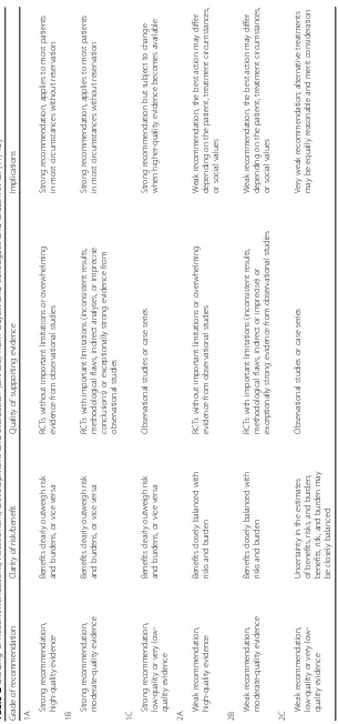
citations were included for the period between January 2000 and December 2016 using the primary search strat-egy: hernia, groin, inguinal, femoral, crural, umbilical, epigastric, spigelian, ventral, incisional, incarcerated, strangulated, acute, emergency, repair, suture, mesh, dir-ect, synthetic, polypropylene, prosthetic, biologic, SSI, wound infection, bowel resection, intestinal resection, complication, morbidity, recurrence, timing, laparoscopy combined with AND/OR. No search restrictions were imposed. The dates were selected to allow comprehen-sive published abstracts of clinical trials, consensus con-ference, comparative studies, congresses, guidelines, government publication, multicenter studies, systematic reviews, meta-analysis, large case series, original articles, and randomized controlled trials. Narrative review arti-cles were also analysed to determine other possible stud-ies. Recommendation guidelines are evaluated according to the Grading of Recommendations, Assessment, De-velopment and Evaluation (GRADE), a hierarchical, evidence-based rubric [11, 12] summarized in Table 2.
The guidelines statements have been issued to each class according to the CDC wound classification (Table 1).
In 2016, the guidelines have been revised and updated by the WSES working group on emergency repair of complicated abdominal wall hernias according to the most recent literature available.
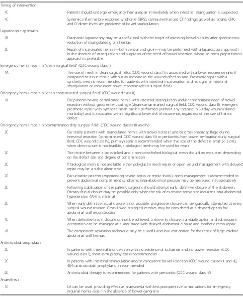
Recommendations Timing of intervention
Patients should undergo emergency hernia repair imme-diately when intestinal strangulation is suspected (grade 1C recommendation).
Systemic inflammatory response syndrome (SIRS), contrast-enhanced CT findings, as well as lactate, serum creatinine phosphokinase (CPK), and D-dimer levels are predictive of bowel strangulation (grade 1C recommendation).
Unfortunately, morbidity and mortality rates remain high for patients who undergo emergency repair of ab-dominal hernias. Early diagnosis of strangulated obstruc-tion may be difficult, and delayed diagnosis can lead to septic complications. However, in the case of suspected bowel strangulation, the benefits outweigh the risks of surgery and patients should undergo immediate surgical intervention.
A recent study performed by Martínez-Serrano et al. prospectively analysed morbidity and mortality rates fol-lowing emergency hernia repair. The study population included 244 patients with complicated abdominal wall hernias requiring surgical repair. In this study, the pa-tients were treated according to standardized protocols with detailed actions taken during the pre-, intra-, and postoperative periods. Clinical outcomes were compared retrospectively to that of 402 patients who had
undergone similar procedures before the development and implementation of the protocols outlined in the study. Results showed higher rates of mortality in pa-tients with acute complication as their first hernia-related symptom and whose treatment was delayed for more than 24 h. Thus, the authors concluded that early detection of complicated abdominal hernias may be the best means of reducing the rate of mortality [13].
Similar results were achieved in the study published in 2014 by Koizumi et al., retrospectively analysing the clinical course and outcomes in 93 patients with strangulated inguinal end femoral hernias. The results demonstrated how the elapsed time from onset to surgery was the most important prognostic factor (P < 0.005) [14].
In 2007, Derici et al. published a retrospective study using univariate and multivariate analyses to investigate factors affecting morbidity and mortality rates in cases of incarcerated abdominal wall hernias [15]. Using the univariate analysis, results showed that symptomatic pe-riods lasting longer than 8 h, the presence of comorbid disease, high American Society of Anesthesiologists (ASA) scores, the use of general anaesthesia, the pres-ence of strangulation, and the prespres-ence of necrosis sig-nificantly affect morbidity rates. In contrast, advanced age, the presence of comorbid diseases, high ASA scores, the presence of strangulation, the presence of necrosis, and hernia repair with graft were found to significantly affect mortality rates by univariate analysis; the presence of necrosis, however, was the only factor that appeared to significantly affect mortality rates based on multivari-ate analysis [16].
A retrospective study evaluated the risk factors associ-ated with bowel resection and treatment outcome in pa-tients with incarcerated groin hernias. The study analysed 182 adult patients with incarcerated groin her-nias who underwent emergency hernia repair in the 10-year period from January 1999 to June 2009. Of these patients, bowel resection was required in 15.4% of cases (28/182). A logistic regression model identified three in-dependent risk factors for bowel resection: lack of health insurance (odds ratio (OR) = 5,P= 0.005), obvious peri-tonitis (OR = 11.52, P = 0.019), and femoral hernia (OR = 8.31,P< 0.001) [17].
pain, fever, tachycardia, and leukocytosis—could not distinguish strangulated from simple obstructions [18]. Furthermore, Shatlla et al. reported a low inci-dence of these classical findings and stated that their presence indicated an advanced stage of strangulation, which would be of limited value for early diagnosis [19]. In 2004, Tsumura et al. published a retrospective study investigating SIRS as a predictor of strangulated small bowel obstruction. Multivariate analysis revealed that the presence of SIRS alongside abdominal muscle guarding was independently predictive of strangulated small bowel obstruction [21].
Among possible diagnostic tests, CPK appears to be a relatively reliable indicator of early intestinal strangulation [22, 23]. Icoz et al. published a prospective study investi-gating the relevance of serum D-dimer measurement as a potential diagnostic indicator of strangulated intestinal hernia. The authors concluded that D-dimer assays should be performed on patients presenting with intestinal emer-gencies to better evaluate and predict ischemic events. Despite having low specificity, elevated D-dimer levels measured upon admission were found to correlate strongly with intestinal ischaemia [24].
In 2012, an interesting retrospective study examin-ing whether various laboratory parameters could pre-dict the viability of strangulation in patients with bowel obstruction was published. Forty patients diag-nosed with bowel strangulation operated within 72 h of the start of symptoms were included in the study. Lactate level was the only laboratory parameter sig-nificantly associated with a lack of viability (P < 0.01, Mann–Whitney U test). Other laboratory data did not show statistically significant associations. The authors concluded that an arterial blood lactate level of 2.0 mmol/L or greater was a useful predictor of non-viable bowel strangulation [25].
Early diagnostic methods to detect bowel strangulation have advanced substantially following the development and refinement of radiological techniques, such as com-puted tomography (CT) scanning [26]. Jancelewicz et al. published a retrospective analysis demonstrating that CT findings of reduced wall enhancement were the most significant independent predictor of bowel strangulation, with 56% sensitivity and 94% specificity. By contrast, ele-vated white blood cell (WBC) count and guarding on physical examination were only moderately predictive. It should be noted, however, that an elevated WBC was the only variable found to be independently predictive of bowel strangulation in patients with small bowel obstruction [27].
In 2014, Kahramanca et al. retrospectively analysed the role of WBC count and fibrinogen as predictive factors of incarcerated abdominal hernia. Comparing 100 pa-tients with incarcerated hernia with 100 papa-tients with
uncomplicated hernia, the results showed that high levels of WBC and fibrinogen were significantly predict-ive of morbidity and cost burden (P< 0.001) [28].
Laparoscopic approach
Diagnostic laparoscopy may be a useful tool with the target of assessing bowel viability after spontaneous reduction of strangulated groin hernias (grade 2B recommendation).
Repair of incarcerated hernias—both ventral and groin—may be performed with a laparoscopic approach in the absence of strangulation and suspicion of the need of bowel resection, where an open pre-peritoneal ap-proach is preferable (grade 2C recommendation).
Few studies have focused on the laparoscopic ap-proach to hernia repair in an emergency setting.
In 2004, Landau and Kyzer published a retrospective study investigating the use of laparoscopy in the repair of incarcerated incisional and ventral hernias. The au-thors argued that laparoscopic repair was feasible and could be safely used to treat patients presenting with in-carcerated incisional and ventral hernias [29].
In 2007, a series of patients with large irreducible groin hernias (omentoceles), treated by laparoscopy without conversions, was published. The authors de-scribed a technique to facilitate complete removal of the hernia contents. A laparoscopic transperitoneal repair for large irreducible scrotal hernias, removing as much omentum as possible, was performed. Then, a small groin incision was made to excise the adherent omen-tum from the distal sac [30].
Another retrospective study published in 2008 investi-gated the role of laparoscopy in the management of in-carcerated (non-reducible) ventral hernias. The authors concluded that laparoscopic repair of ventral abdominal wall hernias could be safely performed with low subse-quent complication rates, even in the event of an
incar-cerated hernia. Careful bowel reduction with
adhesiolysis and mesh repair in an uncontaminated abdomen (without inadvertent enterotomy) using a 5-cm-mesh overlap was an important factor predictive of successful clinical outcome [31].
In 2009, a retrospective study investigating laparo-scopic techniques used to treat incisional hernias in an emergency setting was published. The results of this series also demonstrated the feasibility of laparoscopic surgery to treat incarcerated incisional hernias in an emergency setting [32].
be used to resect bowel, if necessary, or to repair an oc-cult contralateral hernia, present in 11.2–50% of cases. The authors concluded that the laparoscopic repair is a feasible procedure with acceptable results; however, its efficacy needs to be studied further, ideally with larger, multicentre randomized controlled trials [33].
The retrospective 4-year analysis of 188 patients who underwent emergency surgical repair of strangulated groin hernias (57 laparoscopic and 131 open, including one and ten bowel resections, respectively,P= 0.117) re-vealed a significant lower wound infection rate (P < 0.018) in the laparoscopic group, without a higher recurrence rate (P< 0.815) [34].
Hernioscopy is a mixed laparoscopic–open surgical technique for incarcerated inguinal hernias. Specifically, it is effective in evaluating the viability of the herniated loop, thus avoiding unnecessary laparotomy [35].
A prospective randomized study in 2009 aimed to evaluate the impact of hernia sac laparoscopy on the morbidity and mortality of cases with a spontaneous re-duction of the strangulated hernia content before the as-sessment of its viability. Ninety-five patients were randomly assigned to two groups: group A (21 patients managed using hernia sac laparoscopy) and group B (20 patients managed without laparoscopy). The median hospital stay was 28 h for group A and 34 h for group B. Four patients of group B had major complications, whereas there was none observed in group A. Two un-necessary laparotomies and two deaths occurred in group B. The authors concluded that hernia sac laparos-copy seems to be an accurate and safe method of pre-venting unnecessary laparotomy, and in high-risk patients, it contributes to decreased morbidity [36]..
Emergency hernia repair in“clean surgical field”(CDC wound class I)
The use of mesh in clean surgical fields (CDC wound class I) is associated with lower recurrence rate, if compared to tissue repair, without an increase in the wound infection rate. Prosthetic repair with a syn-thetic mesh is recommended for patients with intes-tinal incarceration and no signs of intestinal strangulation or concurrent bowel resection (clean surgical field) (grade 1A recommendation).
Ventral hernias
For patients with intestinal incarceration and no signs of intestinal strangulation or concurrent bowel resection, the surgical field is presumed clean and the infectious risk for synthetic mesh is low. The absence of intestinal wall ischaemia makes patients less prone to bacterial translocation.
Advantages have demonstrated using a mesh for hernia repair in clean fields; such advantages include
low rate of long-term complications and reduction of recurrence [37–42].
A wide variety of small-sized retrospective studies comparing mesh use to suture repair in the treatment of acute irreducible hernias have been published [39, 43, 44]. The prospective randomized trial by Abdel-Baki et al. compared the use of mesh repair (group 1, 21 patients) and tissue repair (group 2, 21 patients) in 42 cases with acute para-umbilical hernia. The wound infection rate be-tween the two groups was not statistically significant. At follow-up (mean 16 ± 5.5 months), there were four recur-rences in group 2 (4/21, 19%) and no recurrecur-rences in group 1 (P< 0.05) [42].
The prospective 6-year study by Abd Ellatif et al. in-cluded 115 patients who underwent acutely incarcerated abdominal wall hernia repair. The results showed low rates of wound infection (4.3%) and recurrence (4.3%), with a mean follow-up of 42 months. The authors there-fore concluded that mesh hernioplasty is crucial to pre-vent recurrence and that it is safe for repairing acutely incarcerated hernias [45].
Groin hernias
The retrospective study by Venara et al. compared the 30-day outcome after acute hernia (inguinal, femoral, and umbilical) repair with or without mesh. The study in-cluded 166 patients, of which 64 were treated with and 102 without mesh repair. Among the 64 patients who underwent mesh repair, four patients had concomitant bowel resection. Among the 102 patients who underwent primary repair, 21 patients had concomitant bowel resec-tion. The mesh repair was neither related to a significant increase of complications (P= 0.89) nor related to surgical site infection (SSI) (P= 0.95), overall morbidity (OR = 1.5, confidence interval (CI) = 95%, P = 0.458), and major complications (OR = 1.2, CI = 95%,P= 0.77) [37].
A recent prospective study included 202 patients with acutely incarcerated groin hernias. The results showed extremely low rates of wound infection, mesh infections, and recurrence. The authors concluded that the use of mesh in incarcerated hernias is safe [46].
Emergency hernia repair in“clean-contaminated surgical field”(CDC wound class II)
The use of prosthetic grafts in clean-contaminated set-tings is seldom described. Most studies on the subject focus on elective repair.
Ventral hernias
In 2000, Mandalà et al. published a series of patients with incisional hernias treated with non-absorbable prostheses and associated visceral surgery. The low inci-dence of suppurative complications, with neither re-moval of the patch nor recurrences in the short term, showed that non-absorbable mesh repair in potentially contaminated fields was safe [47].
Retrospective studies by Vix et al., Birolini et al., and Geisler et al. report wound-related morbidity rates of 10.6, 20, and 7%, respectively, following mesh use in both clean-contaminated and contaminated procedures [48–50].
The retrospective study by Campanelli et al. analysed ten prosthetic hernia repairs in potentially contaminated fields and reported no major or minor complications after a 21-month follow-up period [51].
On the other hand, in 2010, Xourafas et al. retrospect-ively examined the impact of mesh use on ventral hernia repairs with simultaneous bowel resections attributable to either cancer or bowel occlusion. Researchers found a significantly higher incidence of postoperative infection in patients with a prosthetic mesh compared to those without mesh. According to the multivariate regression analysis, prosthetic mesh use was the only significant risk factor, irrespective of other variables such as drain use, defect size, or type of bowel resection [52].
The large-sized US National Surgical Quality Improve-ment Program (NSQIP) study by Choi et al., analysed and compared postoperative outcome following ventral hernia repair, in the 5-year period from 1 January 2005 to 4 April 2010, including 6721 clean-contaminated cases, of which 3879 underwent mesh repair and 2842 underwent non-mesh repair. The results did not show a significant statistical difference in the rate of deep inci-sional SSI and return to OR within 30 days, between the mesh and non-mesh groups [53].
One of the few available studies investigating acute hernia repair is the small-sized retrospective analysis by Nieuwenhuizen et al. including 23 patients who under-went acute hernia repair with intestinal resection, and surprisingly, it revealed a higher incidence of wound in-fection in the primary suture group (5/14, 35%) than in the mesh group (2/9, 22%) [54].
Another retrospective analysis of emergency prosthetic repair of incarcerated incisional hernias with simultan-eous bowel resection in potentially contaminated fields including 60 patients demonstrated that the intestinal resection was associated with high rates of wound infec-tion (38%) [55].
The prospective 6-year study by Abd Ellatif et al. in-cluded 163 patients who underwent acutely incarcerated abdominal wall hernia mesh repair, of which 48 required intestinal resection and anastomosis and 155 did not. No significant difference was found in terms of post-operative morbidities, wound infection, and recur-rence rate between the two groups. The authors therefore concluded that mesh hernia repair is crucial to prevent recurrence and that it is safe for repairing acutely incarcerated hernias, even in case of intestinal resection [45].
In 2013, a prospective study to present a 7-year experi-ence with the use of prosthetic mesh repair in the man-agement of the acutely incarcerated and/or strangulated ventral hernias was published. Resection–anastomosis of non-viable small intestine was performed in 18 patients (23%) and was not regarded as a contraindication for prosthetic repair [43].
Haskins et al. evaluated the outcomes after emer-gency ventral hernia repair in 1357 patients with CDC wound class II from the American College of Surgeons (ACS) NSQIP database and did not find any statistical significance in wound-related or add-itional 30-day patient morbidity or mortality, be-tween mesh and non-mesh emergency ventral hernia repair. The authors concluded that emergency ven-tral hernia repair with a mesh can be safely per-formed without an increase in wound-related or additional early patient morbidity or mortality in CDC wound class II [56].
The randomized trial by Kassem and El-Haddad com-pared the use of onlay polypropylene mesh positioned and supported by omentum and/or peritoneum versus inlay implantation of polypropylene-based composite mesh in 60 patients with complicated wide-defect ven-tral hernias, including 12 bowel resections. Postopera-tively, seven patients developed a wound infection (11.6%) and two patients developed a recurrence (3%), after 3 and 8 months, respectively [57].
Groin hernias
Some studies have asserted that prosthetic repair of abdominal hernias can be safely performed alongside simultaneous colonic operations. Such joint proce-dures, they argue, exhibit acceptable rates of infec-tious complications and recurrence, and consequently, they stated that there is insufficient evidence to advo-cate the avoidance of prosthetic mesh in clean-contaminated fields, assuming that the appropriate technique is used [44, 58].
with mesh and 17 without mesh) did not show any sta-tistically significant differences in terms of morbidity be-tween the two groups and led to the conclusion that strangulated inguinal hernia cannot be considered a contraindication to the mesh repair even in case of in-testinal resection [59].
A recent prospective study by Bessa et al. enrolled 234 patients with acutely or strangulated groin her-nias of which 34 underwent resection and anasto-mosis of non-viable intestine. The results did not show any significant difference (P = 0.7) in the rate of wound or mesh infection between hernias with vi-able versus non-vivi-able contents. The authors con-cluded that the presence of non-viable intestine could not be regarded as a contraindication for prosthetic repair [46].
In the retrospective study by Venara et al. including a subgroup of 25 patients who underwent acute her-nia repair with concomitant bowel resection (four with mesh repair and 21 with primary repair), bowel resection appeared to be a risk factor for overall post-operative complications (P > 0.0001) and major com-plications (P = 0.003), but not for postoperative SSI (P = 0.42). The authors concluded that mesh repair appeared to be safe in the treatment of incarcerated hernia, since after multivariate analysis, mesh place-ment was not a significant predictor of postoperative complication (P = 0.458) [37].
In 2014, a SR and meta-analysis including nine studies has been published, investigating the optimal technique to treat strangulated inguinal hernia (mesh vs non-mesh repair). The wound infection rate has been found to be lower in the mesh group than in the control group (OR = 0.46, CI = 95%, P = 0.07). The recurrence rate was found to be lower in the mesh repair group (OR = 0.2, CI = 95%, P = 0.02). Nonetheless, the authors concluded that the study did not allow to currently recommend the use of mesh in case of bowel resection, despite the finding of similar SSI rates with either mesh repair or non-mesh tech-niques, when comparing bowel resection and no bowel resection (OR = 1.50, P = 0.73) [60].
Emergency hernia repair in“contaminated-dirty surgical field”(CDC wound classes III and IV)
For stable patients with strangulated hernia with bowel necrosis and/or gross enteric spillage during intestinal resection (contaminated, CDC wound class III) or peritonitis from bowel perforation (dirty sur-gical field, CDC wound class IV), primary repair is recommended when the size of the defect is small (< 3 cm); when direct suture is not feasible, a bio-logical mesh may be used for repair (grade 2C recommendation).
The choice between a cross-linked and a non-cross-linked biological mesh should be evaluated depending on the defect size and degree of contamination (grade 2C recommendation).
If a biological mesh is not available, either polyglactin mesh repair or open wound management with delayed
repair may be a viable alternative (grade 2C
recommendation).
In cases of bacterial peritonitis, patients must undergo contaminated surgical intervention, which means that the surgical field is infected and the risk of surgical site infection is very high.
High infection rates are reported after emergency hernia repairs with a polypropylene mesh of CDC wound class III. A retrospective study by Kelly and Behrman reported a 21% infection rate in a series of emergency and elective incisional hernia repairs [61]. Recently, a retrospective study by Carbonell et al. in-vestigated open ventral hernia repairs performed with a polypropylene mesh in the retro-rectus position in clean-contaminated and contaminated fields: the 30-day surgical site infection rate was 7.1 and 19.0%, respectively [62].
Some authors investigated the use of absorbable prosthetic materials [64]. However, the use of ab-sorbable prosthesis exposes the patient to an
inevit-able hernia recurrence. These meshes, once
implanted, induce an inflammatory reaction that, through a hydrolytic reaction, digests and removes and digests the implanted prosthetic material com-pletely. In this case, the high risk of hernia recur-rence is explained by the complete dissolution of the prosthetic support [63].
of either the presence or not of the cross-linking, bio-logical prostheses are divided into two subgroups: the partially remodelling ones (cross-linked) and the com-pletely remodelling ones (not cross-linked). Thanks to the presence of additional links, the partially remodel-ling ones resist better and for a longer period to mechanical stress [64].
Many retrospective studies have explored the prom-ising role of biological mesh in contaminated fields, but most of these investigations did not focus on emergency repair of incarcerated hernias [66–86]. Al-though a biological mesh in these situations is safe, long-term durability has still not been demonstrated [87–89].
A recent multicentre large-sized retrospective study compared suture, synthetic mesh, and biologic matrix in contaminated ventral hernia repair. On multivariate ana-lysis, a biologic matrix was associated with a non-significant reduction in both SSI and recurrences, whereas a synthetic mesh was associated with fewer re-currences compares to suture and non-significant in-crease in SSI [90].
A prospective study by Catena et al. published in 2007 focused on complicated incisional hernia repair using mesh prosthetics made of porcine dermal collagen (PDC). Incisional hernioplasty using PDC grafts was found to be a safe and efficient approach to difficult con-taminated cases [81].
Coccolini et al. published the results of the first 193 patients of the Italian Register of Biological Prosthesis (IRBP) [86]. This prospective multicentre study suggests the usefulness, versatility, and ease of using biological prosthesis in many different situations, including con-taminated surgical fields.
The literature review by Coccolini et al. covered the use of biological meshes for abdominal reconstruction in emergency and elective setting in transplanted patients and reported a complication rate of 9.4% [84].
In 2014, Han et al. published a retrospective study in-cluding 63 patients who underwent emergency surgery for acute incarcerated abdominal wall hernias with hu-man acellular dermal matrix (ADM) repair with a very low rate of infection (1.6%) as well as recurrences (15.9%) in a follow-up of 43 months. Bowel resection, performed in 33 patients, did not significantly affect the bulge and recurrence rate (P = 0.262). Interestingly, multivariate analysis demonstrated three factors to be significantly related to bulge and recurrence: BMI (P= 0.008), defect size (P= 0.016), and numbers of bio-logical meshes used (P= 0.027) [91].
The systematic review by Lee et al. included a total of 32 studies regarding the use of synthetic and biologic materials for abdominal wall reinforcement in contami-nated fields. In contamicontami-nated and/or dirty fields, wound
infection rates were similar, but pooled hernia rates were 27.2% (95% CI = 9.5–44.9) with biological and 3.2% (95% CI = 0.0–11.0) with synthetic non-absorbable meshes. Other outcomes were comparable [92].
The recent multicentre prospective observational study by De Simone et al. included 71 patients who underwent emergency ventral hernia repair with a bio-logical mesh. The surgical field resulted contaminated in 27 patients (38%), potentially contaminated in 19 pa-tients (26.7%), and dirty in 25 papa-tients (35.2%). Early postoperative (3rd–7th postoperative days) wound infec-tion occurred in 21 patients (29.57%). High ASA score (≥3) (OR = 2.82, CI = 1.85–6.43, P = 0.03), smoking (OR = 4.1, CI = 1.73–6.35,P= 0.02), diabetes (OR = 3.23, CI = 1.92–4.38, P = 0.04), chronic immunosuppression (OR = 2.41, CI = 0.33–5.25, P = 0.003), previous hernia repair (OR = 1.99, CI = 1.5–2.9,P= 0.002), dirty surgical field (OR = 1.87, CI = 0.35–4.4, P = 0.04), sublay extra-peritoneal bio-prosthesis placement (OR = 0.45, CI = 0.27–1.13, P = 0.009), and no anterior fascia clos-ure (OR = 0.33, CI = 0.2–2.3, P= 0.04) were associated with wound complications. After a mean follow-up time of 27.2 months, hernia recurrence occurred in 19 pa-tients (26.76%) [93].
Haskins et al. evaluated the outcomes after emergency ventral hernia repair in 1092 patients from the ACS NSQIP database and did not find any statistical signifi-cance in wound-related or additional 30-day patient morbidity or mortality, between mesh and non-mesh emergency ventral hernia repair. The authors concluded that emergency ventral hernia repair with a mesh can be safely performed without an increase in early wound-related or additional 30-day patient morbidity or mortal-ity in CDC wound classes III and IV [56].
The use of biological materials in clinical practice has led to innovative methods of treating abdominal wall de-fects in contaminated surgical fields, although there is still an insufficient level of high-quality evidence on their value, and there is still a very huge price difference be-tween the synthetic and biological meshes [10]. All litera-ture reviews found in the MEDLINE database supported biologic mesh use in the setting of contaminated fields, but the literature included in these reviews consisted of case series and case reports with low levels of evidence [94]. Despite the lack of a cohesive body of evidence, pub-lished studies on biological mesh suggest that cross-linked mesh prosthetics have the lowest failure rate in contami-nated and outright infected fields. To better guide sur-geons, prospective randomized trials should be undertaken to evaluate the short- and long-term out-comes associated with biological meshes [90, 95].
intra-abdominal pressure may be measured intraoperatively (grade 2C recommendation).
A prospective study published by Beltrán et al. ex-amined 81 consecutively unselected patients
present-ing with complicated hernias and intestinal
obstruction. The researchers used intra-abdominal pressure, measured with the intravesicular pressure method, to assess the clinical severity of strangulated hernias and predict intestinal strangulation [96]. Pa-tients with intestinal strangulation and peritonitis are critically ill cases, commonly shocked and at high risk of septic complications; these patients may ex-perience high intraoperative intra-abdominal pres-sure. Such hypertension may be the underlying cause of increased pulmonary pressures, reduced cardiac output, splanchnic hypoperfusion, and oliguria, lead-ing to an abdominal compartment syndrome. In-creased pressure within the constricting abdominal compartment in conjunction with unchanging or more likely disease-induced reduced abdominal com-pliance will also greatly reduce visceral perfusion within the abdominal compartment leading to an acute bowel injury [97–99]. This “acute bowel injury” results in release of pro-inflammatory mediators into the peritoneum and systemic circulation, leading to neutrophil priming, increased intestinal wall permeability, extravasa-tion of fluid into the bowel wall and mesentery, transloca-tion of intestinal bacteria, and absorptransloca-tion of bacterial endotoxin [100–103]. Even relatively mild intra-abdominal hypertension (IAH) (e.g. an IAP of 15 mmHg) has been reported to decrease intestinal microcirculatory blood flow, increase bowel wall permeability, and induce irreversible gut histopathological changes, bacterial trans-location, and multi-organ dysfunction syndrome [103–105]. Prophylactic treatment to avoid abdominal compart-ment syndrome involves refraining from abdominal clos-ure when fascial approximation becomes problematic due to excessive tension (“open abdomen”) [106, 108]. In this setting, negative pressure peritoneal therapy may play a role in mitigating the bio-mediator effects that cause distant organ failure and is an additional potential benefit of an open abdomen.
Even in cases where the abdominal wall can be closed after a laparotomy involving the discovery of diffuse contamination, fulfilling the World Society of Emergency Surgery criteria for severe complicated intra-abdominal sepsis [107, 108], there is controversy as to whether the abdominal wall should be closed or left open. It is financially cheaper and would be pref-erable from a patient’s standpoint to have a single op-eration and to not be submitted to longer critical care unit management if it was possible to primarily close the abdomen [109]. However, there is a now developing biologic rationale with early clinical
evidence that the open abdomen after severe compli-cated intra-abdominal sepsis may be preferable due to its ability to allow negative pressure peritoneal therapy which may modulate the course of systemic inflammation with progressive organ dysfunction [110, 111] and to provide a survival signal that needs to be confirmed in larger studies [112, 113].
Following stabilization of the patient, surgeons should attempt early, definitive closure of the abdomen. Primary fascial closure may be possible only when the risk of ex-cessive tension or recurrent IAH is minimal (grade 2C recommendation).
When early definitive fascial closure is not possible, progressive closure can be gradually attempted at every surgical wound revision. Cross-linked biological meshes may be considered as a delayed option for abdominal wall reconstruction (grade 2C recommendation).
After the patient’s stabilization, the primary object-ive is early and definitobject-ive closure of the abdomen to minimize complications. For many patients, primary fascial closure may be possible within a few days of the first operation. In other patients, early definitive fascial closure may not be possible. In these cases, surgeons must resort to progressive closure, in which the abdomen is incrementally closed each time the patient undergoes a surgical revision. Many methods of fascial closure have been described in the medical literature [94, 114–117].
In 2012, a retrospective analysis evaluating the use of vacuum-assisted closure and mesh-mediated fascial trac-tion (VACM) as temporary abdominal closure was pub-lished. The study compared 50 patients treated with VACM and 54 using non-traction techniques (control group). VACM resulted in a higher fascial closure rate and lower planned hernia rate than methods that did not provide fascial traction [117].
Occasionally, abdominal closure is only partially achieved, resulting in large, debilitating hernias of the abdominal wall that will eventually require complex sur-gical repair. Bridging meshes will often result in bulging or recurrences [118]. The Italian Biological Prosthesis Working Group (IBPWG) proposed a decisional algo-rithm in using biological meshes to restore abdominal wall defects [64].
When definitive fascial closure cannot be achieved, a skin-only closure is a viable option and subsequent even-tration can be managed at a later stage with delayed ab-dominal closure and synthetic mesh repair (grade 1C recommendation).
with enteric spillage due to a complicated abdominal wall hernia. These patients are often considered critic-ally ill due to septic complications. Ordonez et al. de-scribed a series of 217 non-trauma patients with severe peritonitis and who were managed with dam-age control surgery. Definitive fascia closure was achieved in 51% of the patients. Failure of definitive fascia closure occurred in 106 patients; of these, 72 (68%) were managed with skin-only closure. Skin-only closure could be an alternative for patients with fail-ure of definitive fascia closfail-ure, reducing the risk of complications of open abdomen and abdominal com-partmental syndrome. Patients could be deferred for delayed definitive abdominal closure with synthetic mesh repair [119].
The component separation technique may be a use-ful and low-cost option for the repair of large midline abdominal wall hernias (grade 1B recommendation).
The component separation technique (CST) for reconstructing abdominal wall defects without the use of prosthetic material was described in 1990 by Ramirez et al. [120]. The technique is based on en-largement of the abdominal wall surface by transla-tion of the muscular layers without damaging the muscle innervation and blood supply [121]. In most series, several modifications to the original technique have been performed, including the use of prosthetic material [122–125]. In a prospective randomized trial comparing CST with bridging the defect with a pros-thetic material, CST was found to be superior, al-though a similar recurrence rate was found after a 24-month follow-up [126]. However, high recurrence rates (up to 38.7%) after component separation have recently been reported [127].
The microvascular tensor fasciae latae (TFL) flap is a feasible option for reconstruction of exceptionally large abdominal wall defects. This technique can also be combined with other methods of reconstruction. Vascularized flaps provide healthy autologous tissue coverage without implantation of foreign material at the closure site. A close collaboration between plastic
and abdominal surgeons is important for this
reconstruction [128].
Antimicrobial prophylaxis
In patients with intestinal incarceration with no evidence of ischaemia and no bowel resection (CDC wound class I), short-term prophylaxis is recommended (grade 2C recommendation).
In patients with intestinal strangulation and/or con-current bowel resection (CDC wound classes II and III), 48-h antimicrobial prophylaxis is recommended (grade 2C recommendation).
Antimicrobial therapy is recommended for patients with peritonitis (CDC wound class IV, grade 2C recommendation).
In aseptic hernia repair, Staphylococcus aureus from the exogenous environment or the patient’s skin flora is typically the source of infection. In patients with intes-tinal strangulation, the surgical field may be contami-nated by bacterial translocation [8, 9] from intestinal villi of incarcerated ischemic bowel loops as well as by con-comitant bowel resections. In patients with peritonitis, both antimicrobial therapy and surgery are always recommended.
Anaesthesia
Local anaesthesia (LA) can be used, providing effect-ive anaesthesia with less postoperateffect-ive complications for emergency inguinal hernia repair in the absence of bowel gangrene (grade 1C recommendation).
LA is one of the most commonly used anaesthetic methods in inguinal hernia repair [129–131]. However, the role of LA in emergency inguinal hernia repair is still controversial [132–134]. The recent retrospective 5-year experience by Chen et al. reported that LA could provide effective anaesthesia and patient safety in emergency in-guinal hernia repair, with less cardiac complications (P = 0.044) and respiratory complications (P = 0.027), shorter ICU stay (P= 0.035) and hospital stay (P= 0.001), as well as lower cost (P= 0.000) and faster recovery time (P= 0.000) than general anaesthesia [135].
However, general anaesthesia should be preferred in the case of suspected bowel gangrene and need of intestinal resection and always in the case of peritonitis.
Conclusions
Appendix T4
Table 3Resume of recommendation guidelines
GoR Recommendation
Timing of intervention
1C Patients should undergo emergency hernia repair immediately when intestinal strangulation is suspected
1C Systemic inflammatory response syndrome (SIRS), contrast-enhanced CT findings, as well as lactate, CPK, and D-dimer levels are predictive of bowel strangulation
Laparoscopic approach
2B Diagnostic laparoscopy may be a useful tool with the target of assessing bowel viability after spontaneous reduction of strangulated groin hernias
2C Repair of incarcerated hernias—both ventral and groin—may be performed with a laparoscopic approach in the absence of strangulation and suspicion of the need of bowel resection, where an open preperitoneal approach is preferable
Emergency hernia repair in“clean surgical field”(CDC wound class I)
1A The use of mesh in clean surgical fields (CDC wound class I) is associated with a lower recurrence rate, if compared to tissue repair, without an increase in the wound infection rate. Prosthetic repair with a synthetic mesh is recommended for patients with intestinal incarceration and no signs of intestinal strangulation or concurrent bowel resection (clean surgical field)
Emergency hernia repair in“clean-contaminated surgical field”(CDC wound class II)
1A For patients having complicated hernia with intestinal strangulation and/or concomitant need of bowel resection without gross enteric spillage (clean-contaminated surgical field, CDC wound class II), emergent prosthetic repair with synthetic mesh can be performed (without any increase in 30-day wound-related morbidity) and is associated with a significant lower risk of recurrence, regardless of the size of hernia defect
Emergency hernia repair in“contaminated-dirty surgical field”(CDC wound classes III and IV)
2C For stable patients with strangulated hernia with bowel necrosis and/or gross enteric spillage during intestinal resection (contaminated, CDC wound class III) or peritonitis from bowel perforation (dirty surgical field, CDC wound class IV), primary repair is recommended when the size of the defect is small (< 3 cm); when direct suture is not feasible, a biological mesh may be used for repair
2C The choice between a cross-linked and a non-cross-linked biological mesh should be evaluated depending on the defect size and degree of contamination
2C If biological mesh is not available, either polyglactin mesh repair or open wound management with delayed repair may be a viable alternative
2C For unstable patients (experiencing severe sepsis or septic shock), open management is recommended to prevent abdominal compartment syndrome; intra-abdominal pressure may be measured intraoperatively
2C Following stabilization of the patient, surgeons should attempt early, definitive closure of the abdomen. Primary fascial closure may be possible only when the risk of excessive tension or recurrent intra-abdominal hypertension (IAH) is minimal
2C When early definitive fascial closure is not possible, progressive closure can be gradually attempted at every surgical wound revision. Cross-linked biological meshes may be considered as a delayed option for abdominal wall reconstruction
1C When definitive fascial closure cannot be achieved, a skin-only closure is a viable option and subsequent eventration can be managed at a later stage with delayed abdominal closure and synthetic mesh repair
1B The component separation technique may be a useful and low-cost option for the repair of large midline abdominal wall hernias
Antimicrobial prophylaxis
2C In patients with intestinal incarceration with no evidence of ischaemia and no bowel resection (CDC wound class I), short-term prophylaxis is recommended
2C In patients with intestinal strangulation and/or concurrent bowel resection (CDC wound classes II and III), 48-h antimicrobial prophylaxis is recommended
2C Antimicrobial therapy is recommended for patients with peritonitis (CDC wound class IV)
Anaesthesia
Abbreviations
CDC:Centers for Disease Control and Prevention; OR: odds ratio;
RCT: randomized controlled trial; WSES: World Society of Emergency Surgery
Acknowledgements Not applicable.
Availability of data and supporting materials
There are no individual author data that reach the criteria for availability.
Funding
No authors received any funding resource. The paper received a WSES Institutional waiver for this publication.
Authors’contributions
AB, SDS, and MN revised the manuscript. All authors reviewed and approved the final manuscript.
Ethics approval and consent to participate Not applicable.
Consent for publication Not applicable.
Competing interests
The authors declare that they have no competing interests.
Publisher’s Note
Springer Nature remains neutral with regard to jurisdictional claims in published maps and institutional affiliations.
Author details
1Department of Surgery, University of Bologna, Bologna, Italy.2Department of Surgery, Macerata Hospital, Macerata, Italy.3Department of Surgery, Maggiore Hospital, Bologna, Italy.4Department of General Surgery, Papa Giovanni XXIII Hospital, Bergamo, Italy.5Department of Surgery, Red Cross Hospital Beverwijk, Erasmus University Medical Center, Rotterdam,
Netherlands.6Department of Surgical Science, Istituto Clinico Sant’Ambrogio, Milan, Italy.7Department of General Surgery, Mozyr City Hospital, Mazyr, Belarus.8Denver Health, University of Colorado, Denver, CO, USA. 9Department of Surgery, University of Pittsburgh School of Medicine, Pittsburgh, USA.10Division of Trauma, Emergency Surgery and Surgical Critical Care, Massachusetts General Hospital, Harvard Medical School, Boston, MA, USA.11Department of Surgery, University of Florida, Gainesville, FL, USA. 12Department of Abdominal Surgery, University Hospital Meilahti, Helsinki, Finland.13Department of Surgery, University of Hawaii, Honolulu, HI, USA. 14Department of Primary Care and Emergency Medicine, Kyoto University Graduate School of Medicine, Kyoto, Japan.15Division of General Surgery, Rambam Health Care Campus, Haifa, Israel.16Division of Trauma Surgery, Hospital de Clinicas, School of Medical Sciences, University of Campinas, Campinas, Brazil.17Department of Surgery, Universidad del Valle, Fundacion Valle del Lili, Cali, Colombia.18General Surgery Clinic, Azienda ULSS 5 “Polesana”, Adria, RO, Italy.19General Surgery Clinic, University Hospital St. George/Medical University of Plovdiv, Plovdiv, Bulgaria.20Department of Surgery 1, Lviv Regional Hospital, Danylo Halytsky Lviv National Medical University, Lviv, Ukraine.21Department of Surgery, Sheri-Kashmir Institute of Medical Sciences, Srinagar, India.22Griffith Base Hospital, Griffith, NSW, Australia.23Federal University of Juiz de Fora (UFJF), Juiz de Fora, MG, Brazil. 24Faculdade de Ciências Médicas e da Saúde de Juiz de Fora (SUPREMA), Juiz de Fora, MG, Brazil.25Department of General Surgery, Trauma and
Emergency Surgery Division, ABC Medical School, Santo André, SP, Brazil. 26Department of Emergency Surgery, Maggiore Parma Hospital, Parma, Italy. 27Department of Surgery, Tbilisi State Medical University, Tbilisi, Georgia. 28
Department of Surgery, Yonsei University College of Medicine, Seoul, South Korea.29Department of Surgery, Montichiari Hospital, ASST Spedali Civili Brescia, Brescia, Italy.30Department of Surgery, Mazzoni Hospital, Ascoli Piceno, Italy.31Catholic University of the Sacred Heart, Rome, Italy. 32
Department of Surgery, Tianjin Nankai Hospital, Nankai Clinical School of Medicine, Tianjin Medical University, Tianjin, China.33Department of Surgery, RIPAS Hospital, Bandar Seri Begawan, Brunei.34Department of Surgery Mansoura, Faculty of Medicine, Mansoura University, Mansoura, Egypt.
35Department of Surgery, Pt. BDS Post Graduate Institute of Medical Sciences, Rohtak, India.36Department of Laparoscopic and Bariatric Surgery, Health City Cayman Islands, Grand Cayman, Cayman Islands.37Department of Surgery, College of Health Sciences, Obafemi Awolowo University Hospital, Ile-Ife, Nigeria.38II Cátedra de Clínica Quirúrgica, Hospital de Clínicas, Facultad de Ciencias Médicas, Universidad Nacional de Asunción, San Lorenzo, Paraguay.393rd Department of General Surgery, Jagiellonian University Collegium Medium, Krakow, Poland.40Department of Surgery, G. Giglio Hospital Cefalù, Palermo, Italy.41Department of Surgery, Division of Trauma, Surgical Care, Burns and Acute Care Surgery, UC San Diego Medical Center, San Diego, CA, USA.42Academic Department of Surgery, University Hospitals Birmingham NHS Foundation Trust, Edgabaston, Birmingham, UK. 43Department of Colorectal Surgery, New Queen Elizabeth Hospital Birmingham, University Hospitals Birmingham NHS Foundation Trust, Edgbaston, Birmingham, UK.44University of Catania, Catania, Italy. 45
Department of Surgery, Brescia Hospital, Brescia, Italy.46Departments of Critical Care Medicine and Surgery, Foothills Medical Centre, Calgary, AB, Canada.47Letterkenny Hospital, Donegal, Ireland.48Niguarda Hospital, Milan, Italy.
Received: 6 June 2017 Accepted: 31 July 2017
References
1. Helgstrand F, Rosenberg J, Kehlet H, Bisgaard T. Outcomes after emergency versus elective ventral hernia repair: a prospective nationwide study. World J Surg. 2013;37(10):2273–9. doi:10.1007/s00268-013-2123-5.
2. Miserez M, Alexandre JH, Campanelli G, et al. The European hernia society groin hernia classication: simple and easy to remember. Hernia. 2007;11(2): 113–6. doi:10.1007/s10029-007-0198-3.
3. Horan TC, Gaynes RP, Martone WJ, Jarvis WR, Grace ET. CDC definitions of nosocomial surgical site infections, 1992: a modification of CDC definitions of surgical wound infections. AJIC Am J Infect Control. 1992;20(5):271–4. doi: 10.1016/S0196-6553(05)80201-9.
4. Garner J. CDC guideline for prevention of surgical wound infections, 1985. Infect Control. 1986;7(3):193–200.
5. Cruse P, Foord R. The epidemiology of wound infection. A ten-year prospective study of 62,939 wounds. Surg Ckin North Am. 1980;60:27–40. 6. Olson M, O’Connor M, Schwartz M. A 5-year prospective study of 20,193
wounds at the Minneapolis VA Medical Center. Ann Surg. 1984;199:253–9. 7. Breuing K, Butler CE, Ferzoco S, et al. Incisional ventral hernias: review of the
literature and recommendations regarding the grading and technique of repair. Surgery. 2010;148(3):544–58. doi:10.1016/j.surg.2010.01.008. 8. Samel S, Keese M, Kleczka M, et al. Microscopy of bacterial translocation
during small bowel obstruction and ischemia in vivo—a new animal model. BMC Surg. 2002;2:6. http://www.pubmedcentral.nih.gov/articlerender. fcgi?artid=126214&tool=pmcentrez&rendertype=abstract
9. Akcay MN, Capan MY, Gundogdu C, Polat M, Oren D. Bacterial translocation in experimental intestinal obstruction. J Int Med Res. 1996;24(1):17–26. 10. Montgomery A. The battle between biological and synthetic meshes in
ventral hernia repair. Hernia. 2013;17(1):3–11. doi:10.1007/s10029-013-1043-5. 11. Guyatt G, Gutterman D, Baumann MH, et al. Grading strength of
recommendations and quality of evidence in clinical guidelines: report from an American College of Chest Physicians task force. Chest. 2006; 129(1):174–81. doi:10.1378/chest.129.1.174.
12. Brozek JL, Akl EA, Compalati E, et al. Grading quality of evidence and strength of recommendations in clinical practice guidelines. Part 3 of 3. The GRADE approach to developingrecommendations. Allergy. 2011; 64(8):1109–16. doi:10.1111/j.1398-9995.2010.02530.x.
13. Martínez-Serrano MA, Pereira JA, Sancho J, Argudo N, López-Cano M, Grande L. Specific improvement measures to reduce complications and mortality after urgent surgery in complicated abdominal wall hernia. Hernia. 2012;16(2):171–7. doi:10.1007/s10029-011-0875-0.
14. Koizumi M, Sata N, Kaneda Y, et al. Optimal timeline for emergency surgery in patients with strangulated groin hernias. Hernia. 2014;18(6): 845–8. doi:10.1007/s10029-014-1219-7.
15. Derici H, Unalp HR, Bozdag AD, Nazli O, Tansug T, Kamer E. Factors affecting morbidity and mortality in incarcerated abdominal wall hernias. Hernia. 2007;11(4):341–6. doi:10.1007/s10029-007-0226-3.
selection for elective repair. In: Surgery (United States), vol. 160; 2016. p. 1379–91. doi:10.1016/j.surg.2016.06.027.
17. Ge BJ, Huang Q, Liu LM, Bian HP, Fan YZ. Risk factors for bowel resection and outcome in patients with incarcerated groin hernias. Hernia. 2010;14(3): 259–64. doi:10.1007/s10029-009-0602-2.
18. Sarr MG, Bulkley GB, Zuidema GD. Preoperative recognition of intestinal strangulation obstruction. Prospective evaluation of diagnostic capability. Am J Surg. 1983;145(1):176–82. doi:10.1016/0002-9610(83)90186-1. 19. Shatlla A, Chamberlain B, Webb W. Current status of diagnosis and
management of strangulation obstruction of the small bowel. Am J Sur. 1978;132:299–303.
20. Bizer LS, Liebling RW, Delany HM, Gliedman ML. Small bowel obstruction. The role of nonoperative treatment in simple intestinal obstruction and predictive criteria for strangulation obstruction. Surgery. 1981;89(4):407–13. 21. Tsumura H, Ichikawa T, Hiyama E, Murakami Y, Sueda T. Systemic
inflammatory response syndrome (SIRS) as a predictor of strangulated small bowel obstruction. Hepato-Gastroenterology. 2004;51(59):1393–6. 22. Graeber G, O’Neil J, Wolf R, Wukich D, Caffery P, Harman J. Elevated levels of
peritoneal serum creatine phosphokinase with strangulated small bowel obstruction. Arch Surg. 1983;118:837–40.
23. Davies MG, Hagen PO. Systemic inflammatory response syndrome. Br J Surg. 1997;84(7):920–35. doi:10.1080/17415349.2015.1113150. 24. Icoz G, Makay O, Sozbilen M, et al. Is D-dimer a predictor of strangulated intestinal hernia? World J Surg. 2006;30(12):2165–9. doi:10.1007/s00268-006-0138-x.
25. Tanaka K, Hanyu N, Iida T, et al. Lactate levels in the detection of preoperative bowel strangulation. Am Surg. 2012;78(1):86–8.
26. Balthazar EJ. CT of small-bowel obstruction. Am J Roentgenol. 1994;162: 255–61. doi:10.2214/ajr.162.2.8310906.
27. Jancelewicz T, Vu LT, Shawo AE, Yeh B, Gasper WJ, Harris HW. Predicting strangulated small bowel obstruction: an old problem revisited. J Gastrointest Surg. 2009;13(1):93–9. doi:10.1007/s11605-008-0610-z. 28. Kahramanca S, Kaya O, Ozgehan G, et al. Are fibrinogen and complete
blood count parameters predictive in incarcerated abdominal hernia repair? Int Surg. 2014;99(6):723–8. doi:10.9738/INTSURG-D-13-00107.1.
29. Landau O, Kyzer S. Emergent laparoscopic repair of incarcerated incisional and ventral hernia. Surg Endosc Other Interv Tech. 2004;18(9): 1374–6. doi:10.1007/s00464-003-9116-7.
30. Palanivelu C, Rangarajan M, John SJ. Modified technique of laparoscopic intraperitoneal hernioplasty for irreducible scrotal hernias (omentoceles): how to remove the hernial contents. World J Surg. 2007;31(9):1889–91. doi: 10.1007/s00268-007-9157-5.
31. Shah RH, Sharma A, Khullar R, Soni V, Baijal M, Chowbey PK. Laparoscopic repair of incarcerated ventral abdominal wall hernias. Hernia. 2008;12(5): 457–63. doi:10.1007/s10029-008-0374-0.
32. Olmi S, Cesana G, Erba L, Croce E. Emergency laparoscopic treatment of acute incarcerated incisional hernia. Hernia. 2009;13(6):605–8. doi:10.1007/ s10029-009-0525-y.
33. Deeba S, Purkayastha S, Paraskevas P, Athanasiou T, Darzi A, Zacharakis E. Laparoscopic approach to incarcerated and strangulated inguinal hernias. JSLS J Soc Laparoendosc Surg. 2009;13(3):327–31. http://www.ncbi.nlm.nih. gov/pubmed/19793471%5Cn. http://www.pubmedcentral.nih.gov/ articlerender.fcgi?artid=PMC3015964.
34. Yang GPC, Chan CTY, Lai ECH, Chan OCY, Tang CN, Li MKW. Laparoscopic versus open repair for strangulated groin hernias: 188 cases over 4 years. Asian J Endosc Surg. 2012;5(3):131–7. doi:10.1111/j.1758-5910.2012.00138.x. 35. Sajid MS, Ladwa N, Colucci G, Miles WF, Baig MK, Sains P. Diagnostic
laparoscopy through deep inguinal ring: a literature-based review on the forgotten approach to visualize the abdominal cavity during emergency and elective groin hernia repair. Surg Laparosc Endosc Percutan Tech. 2013; 23:251–4. doi:http://dx.doi.org/10.1097/SLE.0b013e31828dacc5
36. Sgourakis G, Radtke A, Sotiropoulos GC, Dedemadi G, Fouzas I, Karaliotas C. Assessment of strangulated content of the spontaneously reduced inguinal hernia via hernia sac laparoscopy: preliminary results of a prospective randomized study. Surg Laparosc Endosc Percutaneous Tech. 2009;19:133–7. doi:http://dx.doi.org/10.1097/SLE.0b013e31819d8b8b
37. Venara A, Hubner M, Le Naoures P, Hamel JF, Hamy A, Demartines N. Surgery for incarcerated hernia: short-term outcome with or without mesh. Langenbeck’s Arch Surg. 2014;399(5):571–7. doi:10.1007/s00423-014-1202-x. 38. Burger JWA, Luijendijk RW, Hop WCJ, Halm JA, Verdaasdonk EGG, Jeekel J.
Long-term follow-up of a randomized controlled trial of suture versus mesh
repair of incisional hernia. Ann Surg. 2004;240(4):575–8. doi:10.1097/01.sla. 0000141193.08524.e7.
39. Luijendijk RW, Hop WCJ, van den Tol MP, et al. A comparison of suture repair with mesh repair for incisional hernia. N Engl J Med. 2000;343(6): 392–8. doi:10.1056/NEJM200008103430603.
40. Korenkov M, Sauerland S, Arndt M, Bograd L, Neugebauer EAM, Troidl H. Randomized clinical trial of suture repair, polypropylene mesh or autodermal hernioplasty for incisional hernia. Br J Surg. 2002;89(1):50–6. doi:10.1046/j.0007-1323.2001.01974.x.
41. Sanjay P, Reid TD, Davies EL, Arumugam PJ, Woodward A. Retrospective comparison of mesh and sutured repair for adult umbilical hernias. Hernia. 2005;9(3):248–51. doi:10.1007/s10029-005-0342-x.
42. Abdel-Baki NA, Bessa SS, Abdel-Razek AH. Comparison of prosthetic mesh repair and tissue repair in the emergency management of incarcerated para-umbilical hernia: a prospective randomized study. Hernia. 2007;11(2): 163–7. doi:10.1007/s10029-007-0189-4.
43. Bessa S, Abdel-Razek A. Results of prosthetic mesh repair in the emergency management of the acutely incarcerated and/or strangulated ventral hernias: a seven years study. Hernia. 2013;17(1):59–65.
44. Lohsiriwat V, Sridermma W, Akaraviputh T, et al. Surgical outcomes of Lichtenstein tension-free hernioplasty for acutely incarcerated inguinal hernia. Surg Today. 2007;37(3):212–4. doi:10.1007/s00595-006-3380-9. 45. Abd Ellatif ME, Negm A, Elmorsy G, Al-Katary M, Yousef AEAM, Ellaithy R.
Feasibility of mesh repair for strangulated abdominal wall hernias. Int J Surg. 2012;10(3):153–6. doi:10.1016/j.ijsu.2012.02.004.
46. Bessa SS, Abdel-fattah MR, Al-Sayes IA, Korayem IT. Results of prosthetic mesh repair in the emergency management of the acutely incarcerated and/or strangulated groin hernias: a 10-year study. Hernia. 2015;19(6): 909–14. doi:10.1007/s10029-015-1360-y.
47. Mandalà V, Bilardo G, Darca F, et al. Some considerations on the use of heterologous prostheses in incisional hernias at risk of infection. Hernia. 2000;4:268–71. 48. Vix J, Meyer C, Rohr S, Bourtoul C. The treatment of incisional and
abdominal hernia with a prosthesis in potentially infected tissues? A series of 47 cases. Hernia. 1997;1(4):157–61. doi:10.1007/BF01234750.
49. Birolini C, Massazo Utiyama E, Junqueira Rodrigues A, Birolini D. Elective colonic operation and prosthetic repair of incisional hernia: does contamination contraindicate abdominal wall prosthesis use? J Am Coll Surg. 2000;191(4):366–72. doi:10.1016/S1072-7515(00)00703-1. 50. Geisler DJ, Reilly JC, Vaughan SG, Glennon EJ, Kondylis PD. Safety and
outcome of use of nonabsorbable mesh for repair of fascial defects in the presence of open bowel. Dis Colon rectum. 2003;46(8):1118–23. doi:10.1007/ s10350-004-7290-x.
51. Campanelli G, Nicolosi F, Pettinari D, Avesani E. Prosthetic repair, intestinal resection, and potentially contaminated areas: safe and feasible? Hernia. 2004;8:190–2. 52. Xourafas D, Lipsitz SR, Negro P, Ashley SW, Tavakkolizadeh A.
Impact of mesh use on morbidity following ventral hernia repair with a simultaneous bowel resection. Arch Surg. 2010;145(8):739–44. doi:10.1001/archsurg.2010.144.
53. Choi JJ, Palaniappa NC, Dallas KB, Rudich TB, Colon MJ, Divino CM. Use of mesh during ventral hernia repair in clean-contaminated and contaminated cases: outcomes of 33,832 cases. Ann Surg. 2012;255(1):176–80. doi:10.1097/ SLA.0b013e31822518e6.
54. Nieuwenhuizen J, Van Ramshorst GH, Ten Brinke JG, et al. The use of mesh in acute hernia: frequency and outcome in 99 cases. Hernia. 2011;15(3):297–300. doi:10.1007/s10029-010-0779-4.
55. Zafar H, Zaidi M, Qadir I, Memon A. Emergency incisional hernia repair: a difficult problem waiting for a solution. Ann Surg Innov Res. 2012;6:1. doi:10.1186/1750-1164-6-1.
56. Haskins IN, Amdur RL, Lin PP, Vaziri K. The use of mesh in emergent ventral hernia repair: effects on early patient morbidity and mortality. J Gastrointest Surg. 2016;20(11):1899–903. doi:10.1007/s11605-016-3207-y.
57. Kassem MI, El-Haddad HM. Polypropylene-based composite mesh versus standard polypropylene mesh in the reconstruction of complicated large abdominal wall hernias: a prospective randomized study. Hernia. 2016;20(5): 691–700. doi:10.1007/s10029-016-1526-2.
58. Wysocki A, Kulawik J, Poźniczek M, Strzałka M. Is the Lichtenstein operation of strangulated groin hernia a safe procedure? World J Surg. 2006;30(11): 2065–70. doi:10.1007/s00268-005-0416-z.
60. Hentati H, Dougaz W, Dziri C. Mesh repair versus non-mesh repair for strangulated inguinal hernia: systematic review with meta-analysis. World J Surg. 2014;
61. Kelly ME, Behrman SW. The safety and efficacy of prosthetic hernia repair in clean-contaminated and contaminated wounds. Am Surg. 2002;68(6):524–8.
62. Carbonell AM, Criss CN, Cobb WS, Novitsky YW, Rosen MJ. Outcomes of synthetic mesh in contaminated ventral hernia repairs. J Am Coll Surg. 2013; 217(6):991–8. doi:10.1016/j.jamcollsurg.2013.07.382.
63. Finan KR, Vick CC, Kiefe CI, Neumayer L, Hawn MT. Predictors of wound infection in ventral hernia repair. Am J Surg. 2005;190(5):676–81. doi:10.1016/j.amjsurg.2005.06.041.
64. Coccolini F, Agresta F, Bassi A, et al. Italian Biological Prosthesis Work-Group (IBPWG): proposal for a decisional model in using biological prosthesis. World J Emerg Surg. 2012;7(1):34. doi:10.1186/1749-7922-7-34.
65. Dunne JR, Malone DL, Tracy JK, Napolitano LM. Abdominal wall hernias: risk factors for infection and resource utilization. J Surg Res. 2003;111(1):78–84. doi:10.1016/S0022.
66. Saettele TM, Bachman SL, Costello CR, et al. Use of porcine dermal collagen as a prosthetic mesh in a contaminated field for ventral hernia repair: a case report. Hernia. 2007;11(3):279–85. doi:10.1007/s10029-006-0186-z.
67. Smart N, Immanuel A, Mercer-Jones M. Laparoscopic repair of a Littre’s hernia with porcine dermal collagen implant (Permacol). Hernia. 2007;11(4): 373–6. doi:10.1007/s10029-007-0197-4.
68. Liyanage SH, Purohit GS, Frye JNR, Giordano P. Anterior abdominal wall reconstruction with a Permacol implant. J Plast Reconstr Aesthet Surg. 2006; 59(5):553–5. http://www.ncbi.nlm.nih.gov/pubmed/16749204
69. Gupta A, Zahriya K, Mullens PL, Salmassi S, Keshishian A. Ventral herniorrhaphy: experience with two different biosynthetic mesh materials. Surgisis and Alloderm Hernia. 2006;10(5):419–25. doi:10.1007/s10029-006-0130-2.
70. Albo D, Awad SS, Berger DH, Bellows CF. Decellularized human cadaveric dermis provides a safe alternative for primary inguinal hernia repair in contaminated surgical fields. Am J Surg. 2006;192(5 SPEC. ISS) doi:10.1016/j. amjsurg.2006.08.029.
71. Schuster R, Singh J, Safadi BY, Wren SM. The use of acellular dermal matrix for contaminated abdominal wall defects: wound status predicts success. Am J Surg. 2006;192(5 SPEC. ISS):594–7. doi:10.1016/j.amjsurg.2006.08.017. 72. Alaedeen DI, Lipman J, Medalie D, Rosen MJ. The single-staged approach to
the surgical management of abdominal wall hernias in contaminated fields. Hernia. 2007;11(1):41–5. doi:10.1007/s10029-006-0164-5.
73. Kim H, Bruen K, Vargo D. Acellular dermal matrix in the management of high-risk abdominal wall defects. Am J Surg. 2006;192(6):705–9. doi:10.1016/ j.amjsurg.2006.09.003.
74. Diaz JJ, Guy J, Berkes MB, Guillamondegui O, Miller RS. Acellular dermal allograft for ventral hernia repair in the compromised surgical field. Am Surg. 2006;72(12):1181–7. doi:10.1680/udap.2010.163.
75. Patton JH, Berry S, Kralovich KA. Use of human acellular dermal matrix in complex and contaminated abdominal wall reconstructions. Am J Surg. 2007;193(3):360–3. doi:10.1016/j.amjsurg.2006.09.021.
76. Bellows CF, Albo D, Berger DH, Awad SS. Abdominal wall repair using human acellular dermis. Am J Surg. 2007;194(2):192–8. doi:10.1016/j.amjsurg. 2006.11.012.
77. Jin J, Rosen MJ, Blatnik J, et al. Use of acellular dermal matrix for complicated ventral hernia repair: does technique affect outcomes? J Am Coll Surg. 2007;205(5):654–60. doi:10.1016/j.jamcollsurg.2007.06.012. 78. Franklin ME, Gonzalez JJ, Michaelson RP, Glass JL, Chock DA. Preliminary
experience with new bioactive prosthetic material for repair of hernias in infected fields. Hernia. 2002;6(4):171–4. doi:10.1007/s10029-002-0078-9. 79. Jr MEF, Jr ÆJJG, Franklin ME, Gonzalez JJ, Glass JL. Use of porcine small
intestinal submucosa as a prosthetic device for laparoscopic repair of hernias in contaminated fields: 2-year follow-up. Hernia. 2004;8(3):186–9. doi:10.1007/s10029-004-0208-7.
80. Helton WS, Fisichella PM, Berger R, Horgan S, Espat NJ, Abcarian H. Short-term outcomes with small intestinal submucosa for ventral abdominal hernia. Arch Surg. 2005;140(6):549-560-562. doi:10.1001/archsurg.140.6.549. 81. Catena F, Ansaloni L, Gazzotti F, et al. Use of porcine dermal collagen graft
(Permacol) for hernia repair in contaminated fields. Hernia. 2007;11(1):57–60. doi:10.1007/s10029-006-0171-6.
82. Treviño JM, Franklin ME, Berghoff KR, Glass JL, Jaramillo EJ. Preliminary results of a two-layered prosthetic repair for recurrent inguinal and ventral
hernias combining open and laparoscopic techniques. Hernia. 2006;10(3): 253–7. doi:10.1007/s10029-006-0085-3.
83. Shaikh FM, Giri SK, Durrani S, Waldron D, Grace PA. Experience with porcine acellular dermal collagen implant in one-stage tension-free reconstruction of acute and chronic abdominal wall defects. World J Surg. 2007;31(10): 1966–72. doi:10.1007/s00268-007-9174-4.
84. Coccolini F, Catena F, Bertuzzo VR, Ercolani G, Pinna A, Ansaloni L. Abdominal wall defect repair with biological prosthesis in transplanted patients: single center retrospective analysis and review of the literature. Updat Surg. 2013;65(3):191–6. doi:10.1007/s13304-013-0212-5. 85. Cavallaro A, Menzo E Lo, di Vita M, et al. Use of biological meshes for
abdominal wall reconstruction in highly contaminated fields. World J Gastroenterol 2010;16(15):1928-1933. doi:10.3748/wjg.v16.i15.1928. 86. Coccolini F, Poiasina E, Bertoli P, et al. The Italian Register of Biological
Prostheses. Eur Surg Res. 2013;50(3–4)
![Table 1 Surgical wound classification [4]](https://thumb-us.123doks.com/thumbv2/123dok_us/807711.1578060/2.595.61.541.573.732/table-surgical-wound-classification.webp)