INTRODUCTION
Gender differences, represented in large measure by the differential effects of sex-specific hormones, exist in a variety of cardiovascular,(1–4) cardiopulmonary, (5,6) neurodegenerative, (7–9) endocrine (10) and metabolic bone diseases such as osteoporosis (11–13). Indeed, the role of sex steroids in cardiovascular disease has been studied extensively (14–27). Clini-cally, female patients show relative car-diac protection from acute infarctions and better outcome following myocardial in-farction compared with males (21). Such gender dimorphisms may be due to the beneficial effects of estrogens or to the absence of the deleterious effects of an-drogens (28,29). Differences in estrogen receptor (ER) signaling also may play a significant role in outcome following
cardiovascular diseases (21,30–32). In ad-dition, gender differences in proinflam-matory signaling and immune responses have been described (33,34). Elegant work by Chaudry (4) and other investiga-tors demonstrate that alteration of im-mune function by sex steroids can lead to therapeutic interventions and im-proved outcomes. A better understanding of sex hormone regulation from a cell bi-ology perspective will be critical therefore in improving patient outcomes.
Stem cell transplantation has revolu-tionized the treatment of hematological disorders such as myelodysplastic syn-drome and acute myeloid leukemia (35,36). In recent years, stem cell therapy has been used to improve postmyocardial infarction, ventricular repair, and remod-eling mechanisms (37). In this context,
stem cell therapy may be associated with better functional recovery of the infarcted ventricles in treated patients (38–40).
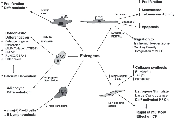
Recent studies reported the presence of estrogen and testosterone receptors on stem cells (41–43), suggesting that estro-gen and testosterone may modify the function of those cells (18,19,44,45) (Figures 1 and 2). 17β-estradiol en-hances the proliferation and migration of endothelial progenitor cells (EPCs) to the injured vessels, or ischemic myocar-dial tissues, which, through the process of homing, help in repair and regeneration to compensate for the lost tissue. It also has been postulated that vascular en-dothelial growth factor (VEGF) might be responsible for EPC migration in re-sponse to 17β-estradiol (44). On the con-trary, the role of androgens on stem cells is controversial, but most studies sug-gest that androgens have inhibitory ef-fects on stem cell functions, and anti-androgens could negate these effects (41). Thus, modification of the function of stem cells through estrogenic or anti-androgenic stimulus may help formulate “super stem
Rinki Ray,
1Nathan M Novotny,
1Paul R Crisostomo,
1Tim Lahm,
2Aaron Abarbanell,
1and Daniel R Meldrum
1,3,4Address correspondence and reprint requests toDaniel R Meldrum, 2017 Van Nuys Med-ical Science Building, 635 Barnhill Drive, Indianapolis, IN 46202. Phone: 317-313-5217; Fax: 317-274-2940; E-mail: [email protected].
Submitted January 12, 2008; Accepted for publication April 30, 2008; Epub (www.molmed. org) ahead of print May 5, 2008.
1Departments of Surgery, 2Pulmonary and Critical Care Medicine, 3Cellular and Integrative Physiology, and the 4Center for
Immunobiology, Indiana University School of Medicine, Indianapolis, Indiana, United States of America
Gender dimorphisms exist in the pathogenesis of a variety of cardiovascular, cardiopulmonary, neurodegenerative, and en-docrine disorders. Estrogens exert immense influence on myocardial remodeling following ischemic insult, partially through paracrine growth hormone production by bone marrow mesenchymal stem cells (MSCs) and endothelial progenitor cells. Estro-gens also facilitate the mobilization of endothelial progenitor cells to the ischemic myocardium and enhance neovascularization at the ischemic border zone. Moreover, estrogens limit pathological myocardial remodeling through the inhibitory effects on the proliferation of the cardiac fibroblasts. Androgens also may stimulate endothelial progenitor cell migration from the bone marrow, yet the larger role of androgens in disease pathogenesis is not well characterized. The beneficial effects of sex steroids include alteration of lipid metabolism in preadipocytes, modulation of bone metabolism and skeletal maturation, and prevention of os-teoporosis through their effects on osteogenic precursors. In an example of sex steroid-specific effects, neural stem cells exhibit enhanced proliferation in response to estrogens, whereas androgens mediate inhibitory effects on their proliferation. Although stem cells can offer significant therapeutic benefits in various cardiovascular, neurodegenerative, endocrine disorders, and dis-orders of bone metabolism, a greater understanding of sex hormones on diverse stem cell populations is required to improve their ultimate clinical efficacy. In this review, we focus on the effects of estrogen and testosterone on various stem and progenitor cell types, and their relevant intracellular mechanisms.
cells” with better therapeutic efficacy. To achieve this goal, the role of sex hormones on stem cell function must be elucidated. It is the purpose of this review to summa-rize the current knowledge of the effects of estrogens and androgens on various stem cell populations.
Embryonic Stem Cells
The expression of estrogen receptors ER-αand ER-βin embryoid bodies takes place as early as d 2, suggesting the in-fluence of estrogen on the differentiation and proliferation of human embryonic stem cells (ESCs) and embryoid bodies (43). The effects of estrogens on ESC proliferation have been demonstrated by Han et al. 17β-estradiol induces an in-crease of ([3] H) thymidine incorporation by murine ESCs and enhances DNA syn-thesis, which is in turn inhibited by anti-estrogen tamoxifen. Estrogens upregulate the expression of ERαand ERβprotein levels and increase mRNA expression of the proto-oncogenes c-fos, c-jun, and c-myc. In addition, 17β-estradiol acti-vates the MAPK cascade as well as cyclin-dependent kinases, with associ-ated increases in cyclins D1 and E, which are important intermediaries in cell cycle progression (42).
Although it is known that estrogens en-hance the proliferation of embryonic stem cells, the role of androgens is not well un-derstood. The presence of androgen re-ceptors (AR) in ESCs has been detected as early as 4.5 d in mice and 5 d in humans, and it also was observed that the concen-tration of AR increases during differentia-tion of ESCs in a stage-dependent manner (46). Testosterone or dihydrotestosterone treatment is not associated with any sig-nificant change in androgen receptor mRNA expression level. But, in a contrast that suggests a testosterone depletion ef-fect, nilutamide, a nonsteroidal antiandro-gen, causes proliferation of ESCs through an increase of Akt protein expression and decreased cell-cycle inhibitor p27 (Kip1) expression (41). These findings suggest a definite role of both sex steroids in the differentiation and proliferation of embry-onic stem cells.
Mesenchymal Stem Cells
MSCs are novel therapeutic agents for organ protection, and estrogens may en-hance the protective function of MSCs by increasing or decreasing cytokine and growth factor production in these cells (47). Our previous study proved that gender differences exist in activated MSC function. In particular, lipopolysaccha-ride- (LPS) and hypoxia-induced VEGF production was significantly greater in female MSCs compared with male MSCs. Female MSCs express significantly less pro-inflammatory cytokines, TNF-αand IL-6, compared with male MSCs in re-sponse to acute LPS and hypoxia, sug-gesting their ability to limit inflamma-tory reactions (18). In males, TNFR1 regulates VEGF, TNF, and IL-6 produc-tion, whereas TNFR expression status does not affect cytokine and growth hormone production in females (19). We also observed gender differences in stem cell-mediated protection in a Langen-dorff preparation. Rat hearts were sub-jected to 25 min of warm global ischemia followed by 40 min of reperfusion and
were assigned randomly to one of three groups: (a) vehicle treated; (b) male MSC treated; and (c) female MSC treated. Fe-male MSC-treated hearts exhibited sig-nificantly improved contractility and compliance as compared with hearts treated with male MSC or vehicle (48).
Regarding the role of estrogens in osteogenic differentiation of MSCs, there is evidence that 17β-estradiol sup-ports growth and differentiation mostly through the ERαreceptor (49). This re-ceptor bias may be attributed to in-terindividual variability and gender differences of osteoblast responses of MSCs to estrogen manifested by ERα polymorphism. In terms of male sex hormone effects, testosterone decreases the specific alkaline phosphatase activ-ity in male MSCs but does not affect calcium deposition in either sex (50). Bone marrow MSCs, when exposed to osteogenic differentiation medium sup-plemented with 17β-estradiol, increase the expression of bone morphogenetic protein (BMP) and osteocalcin, and significantly increase the deposition of
calcium (51,52). 17β-estradiol also stim-ulates the expression of osteogenic genes for ALP, collagen I, and TGF-β1 by MSCs (12). These observations sug-gest the bulk of the heavy lifting in bone metabolism/physiology is han-dled by the female sex steroids.
Clues to the molecular mechanisms underlying the role of sex hormones in MSC differentiation may be found in the ERK pathway. Resveratrol, a phy-toestrogen found in red wine, stimu-lates the expression of osteoblastic markers such as RUNX2/CBFA1, os-terix, and osteocalcin in human bone marrow mesenchymal stem cell cul-tures. This effect is associated with a rapid activation of ERK1 and ERK2, and also can be inhibited by the ERK inhibitor PD98059. Resveratrol en-hances osteoblastic maturation and cal-cium deposition into the extracellular matrix (53) and its effects on osteoblas-tic differentiation are mediated through NO/cGMP pathway (54). Sex hormone involvement in MSC function also in-cludes leptin- and vitamin D-linked increases in aromatase activity during osteogenesis, leading to osteogenic
differentiation of MSCs (55) and signifi-cant E2 linked increases in the lipid stores of differentiated adipocytes (52).
Hematopoietic Stem Cells
There are gender differences in hematopoietic progenitor cell concentra-tions in cord blood samples collected from infants. Specifically, male infants had significantly higher median CD34+ cell concentrations than female infants (31.8/μL compared with 30.2/μL, respec-tively; P= 0.03). This relative increase is reflected in adult males, who, when com-pared with age-matched females, have an increased number of colony-forming cells, erythroblastic colonies, and granulocyte-macrophage colonies in their peripheral blood (56). This signifies that gender may affect the hematopoietic potential of cord blood transplants (57).
The effect of sex steroids on B lym-phopoiesis is the subject of extensive, continuing study. Sex steroids suppress B lymphocyte production in murine bone marrow. Pre B-lymphocytes produce the heavy chain of IgM (μchain) (58), under the influence of the IgM constant region gene (cμ) (59), which can act as a marker
of pre-B cells. V(D)J recombination con-fers the ability of the immune system to respond to a vast number of foreign anti-gens, which occurs particularly in imma-ture lymphocytes and is mediated by the recombination activating gene prod-ucts Rag1 and Rag2 (60,61). 17β-estradiol treatment reduces cμ+ pre-B cells, as-sociated with a decrease in Ig gene re-arrangements and rag1 transcripts (62). It also has been demonstrated that ERα is predominantly responsible for mediat-ing 17β-estradiol induced changes in B-cell precursors (63,64). These findings suggest that 17β-estradiol exerts negative influence on the production of B-lineage cells by modifying the differentiation, proliferation, and survival of early B-cell precursors.
A novel system for expansion of hematopoietic stem cells utilizes “selec-tive amplifier genes” that encode fusion proteins (granulocyte colony-stimulating factor [G-CSF] mutant receptor and delta G-CSF mutant receptor) between the granulocyte colony-stimulating factor receptor (G-CSF-R) and the hormone-binding domain of estrogen receptor (ER). ERs were replaced with a mutant recep-tor (TmR), which specifically binds to 4-hydroxytamoxifen (Tm). Interleukin-3 (IL-3)-dependent Ba/F3 (mouse periph-eral blood pro-B) cells and hematopoietic progenitor cells transduced with the fu-sion proteins showed IL-3-independent growth in response to Tm, whereas, the cells were insensitive to estrogen at con-centrations up to10(–7) M to 10(–6)M. Murine bone marrow cells transduced with G-CSF-TmR and delta G-CSF-TmR formed colonies in methyl-cellulose medium in response to Tm, but no colonies appeared with 10(–7)M estrogen or without cytokines. These results sug-gest that the influences of endogenous estrogen can be ablated by using the G-CSF-TmR/Tm or delta G-CSF-TmR/ Tm system to expand hematopoietic stem cells with potential therapeutic ap-plication (65).
Androgens exert an inhibitory effect on B lymphopoiesis (66,67), but enhance erythropoietic differentiation (68) and
thrombocytopoiesis (69). Cultures of human erythropoietic precursor cells col-lected from children’s normal marrow in the presence of erythropoietin demon-strated a significant increase in the num-ber of colony-forming units (CFU-E) and burst-forming units (BFU-E) of derived colonies in the presence of androgens (10(–8)M or 10(–7)M). These colonies also showed increased uroporphyrinogen I synthase activity, indicating increased heme synthesis (68). Androgens also can rescue mature erythroid colony-forming cells from apoptosis induced by serum and growth factor deprivation (70), thus increasing erythrocyte population. Inter-estingly, castration of normal male mice leads to splenic enlargement and expan-sion of the B cell population, which is mediated via androgen receptors present in both immature B cells and marrow stromal cells. These effects can be re-versed with androgen replacement (66), further elucidating the role of the male hormone in lymphopoiesis.
Cardiac Fibroblasts
Gender may affect the healing of ische-mic myocardium through changes in the function of cardiac fibroblasts (CFs). These progenitor cells play a significant role in the remodeling of ischemic my-ocardium, and the signal transduction pathways controlling the proliferation of CFs under hypoxia-induced stress reveal significant gender differences. These studies found females to be resistant to hypoxia-induced inhibition of DNA syn-thesis associated with decreased expres-sion of NFκB and increased expres-sions of p53 and bcl-2 in comparison to males (71,72). Estrogens exert modula-tory effects on cardiac fibroblast func-tion. 17β-estradiol inhibits prolifera-tion and collagen synthesis (3H-proline incorporation) in male and female CFs in a similar way, and facilitates benefi-cial cardiac remodeling following is-chemia. Consequently, hormone re-placement therapy using 17β-estradiol may exert protective effects on post menopausal women against cardiovas-cular events (73).
Cardiac fibroblasts express estrogen receptor protein, and stimulation of CFs with 17β-estradiol causes nuclear translo-cation of these proteins, indicating one effect of estrogen on gene regulation (74). In cardiac fibroblasts, 17β-estradiol plays an inhibitory role on renin-angiotensin system-induced gene expression, signal transduction, and ECM remodeling. An-giotensin II increases fibroblast prolifera-tion and synthesis of collagen types I and III through the upregulation of ex-pression of the angiotensin AT(1) recep-tor gene and β1 integrins. 17β-estradiol can prevent these increases in prolifera-tion and AT(1) receptor mRNA levels and can attenuate the collagen synthesis in response to angiotensin II. 17β -estra-diol inhibits AngII-stimulated expression of β1 integrins significantly and attenu-ates collagen gel contraction (75).
Estrogens can improve cardiac fibroblast-mediated remodeling of ischemic myo-cardium through both genomic and nongenomic mechanisms. 17β-estradiol exerts an inhibitory effect on the growth of cardiac fibroblasts through both ERα and ERβ(76). Underlying molecular mechanisms include increased mitogen-activated protein (MAP) kinase p42/44 activation and decreased p38 activation (77). In addition, 17β-estradiol increases the steady-state mRNA level of trans-forming growth factor-β3and fibronectin
in these cells (78). A recent study has demonstrated that the selective estrogen receptor agonists PPT (4,4’,4’’-[4-propyl-([1]H)-pyrazole-1,3,5-triyl] tris-phenol) for ER-αand DPN (2,3-bis[4-hydroxyphenyl]-propionitrile) for ER-β, stimulate the large-conductance Ca++-activated K+ (BK[Ca]) channels in cultured human cardiac fibroblasts (HCFs). In whole-cell configuration, depolarizing pulses evoked large outward currents (Ik)with
an outward rectification, the amplitude of which was increased in the presence of DPN or PPT. Paxilline, a selective blocker of BK(Ca) channels, could re-verse the DPN- or PPT-induced ampli-tude of Ik. However, no change in the
tran-scriptional level of the BK(Ca)-channel α-subunit was observed by RT-PCR
analysis in chronic treatment with these two compounds. These findings suggest that estrogen induces a rapid stimulatory effect on human cardiac fibroblasts via a nongenomic mechanism through the ac-tivation of BK(Ca)-channels (79). Build-ing on this understandBuild-ing of the molecu-lar mechanisms in cardiac fibroblasts may enable us to modify the functions of these cells.
Endothelial Progenitor Cells
Blood contains endothelial progenitor cells (EPC), which can differentiate into endothelial cells and modulate healing of injured vessels. In one study on a healthy middle-aged population without known cardiovascular risk factors, it has been demonstrated that women exhib-ited a distinctly higher EPC colony-forming capacity (approximately 150%) and greater migratory activity (40%) compared with men (80). Moreover, human women with a higher plasma estrogen concentration showed a signifi-cantly higher level of circulating EPCs. Increase in the number of EPCs by 17β -estradiol is mediated by decreased rate of apoptosis through a caspase-8-dependent pathway (81). The effects of estrogen on EPCs are mediated via ERαreceptor (82). However, another similar study failed to demonstrate significant gender-specific differences in the frequency of colony formation (83).
mobilizes EPCs via endothelial nitric oxide synthase–mediated activation of matrix metalloproteinase-9 (84). Upregu-lation of MMP-9 results in the release of soluble Kit-ligand (sKitL), which facili-tates the transfer of endothelial cells from the quiescent to proliferative pool (85).
Disease models in animals also have been employed to examine the effects of sex hormones on progenitor cell func-tionality. In spontaneously hypertensive rats, the number of differentiated and adherent EPCs derived from bone mar-row was lower compared with age-matched normotensive rats. Treatment with 17β-estradiol significantly increases the number of EPCs. EPCs derived from hypertensive rats show low telomerase activity and early senescence. Estrogen treatment delays senescence and aug-ments telomerase activity through PI3-K/ Akt pathway (86,87).
Regarding male sex hormone effects, recent clinical studies suggest that an-drogens increase the number of circulat-ing EPCs through a possible effect on bone marrow. It has been revealed that hypogonadotrophic hypogonadal men have low circulating EPCs that increase significantly after androgen treatment (88). A direct effect of testosterone also was suggested by expression of andro-gen receptor (AR) mRNA and protein in human EPCs. Synthetic androgen, methyltrienolone (R1881), causes AR translocation in the nucleus, suggesting its activation increases proliferation, migration, and colony formation activ-ity of these cells. Proliferation, migra-tion, and colony formation activities of the EPCs could be abolished by pre-treatment with flutamide (89). A greater understanding of these molecular mech-anisms will yield insights into how gen-der differences affect the healing process in patients.
Preadipocytes
Regulation of lipid metabolism plays a key role in atherosclerosis and ische-mic cardiac diseases. Estrogens con-trol the expression of lipogenic genes such as leptin, perilipin, peroxisome
proliferation activator receptor-delta, and lipoprotein lipase in adipocytes and estrogen supplementation helps to reduce adipose mass and adipocyte size and prevents development of obe-sity in postmenopausal state (90–92) by both genomic and nongenomic mecha-nisms (93). Genomic effects of estrogen are limited to the regulation of leptin and lipoprotein lipase expression whereas nongenomic effects are medi-ated via the s messenger systems, namely cAMP cascade and the phos-phoinositide cascade. Activation of the cAMP cascade by estrogen is followed by activation of hormone-sensitive li-pase leading to lipolysis in adipose tissues. Activation of protein kinase C through the PI3K cascade, controls the proliferation and differentiation of preadipocytes (93). Human pre-adipocytes (PAs) possess ERαprotein and express ERαgene, but do not ex-press ERβreceptors, indicating that effect of estrogen on adipogenesis is mediated through the ERαreceptor (52,94,95). The effects of estrogens on ERαis site specific (95).
In humans, development of abdomi-nal fat deposition is inversely propor-tionate to blood testosterone levels (96). Androgen receptors are found on preadipocytes (97,98) and the effects of testosterone on these cells also are site-specific. Castration is associated with increased proliferation and differen-tiation of epididymal and perirenal preadipocytes in male rats; whereas, peripubertal testosterone supplementa-tion reduces inguinal and retroperi-toneal fat depots of ovariectomized (OVX) rats. Testosterone decreases adipocyte proliferation without affect-ing adipocyte mean cell size or the size distribution profiles (99). Androgens act directly on fat cells by upregulating α2-AR expression (100). Androgens also exert their modulatory effects on the transcription factor C/EBPα, which is a key regulator of the expression of adipogenic genes (101), providing mo-lecular context for gender-based effects on adipocyte physiology.
Osteogenic Progenitors
Estrogen and testosterone play crucial roles in bone metabolism. Even in males, estrogen is critical for the pubertal growth spurt characterized by skeletal maturation, accrual of peak bone mass, and the maintenance of bone mass in the adult through its effects on remodeling and bone turnover (102).
In OVX rats, estrogen deficiency causes osteopenia and induces bone turnover. Endosteal bone formation in OVX rats is associated with an increased proliferation of both osteoblast precursor cells present in the marrow stroma and along the endosteal bone surface. The os-teoclast surface (percent of the bone tra-becular surface covered with osteoclasts) also increases in OVX rats following ovariectomy, suggesting that bone for-mation increases in correlation with bone resorption. Estradiol supplementa-tion reverses both the increase in resorp-tion and formaresorp-tion indices (103).
with increased VEGF production and in-creased osteoclastic activity (113).
Testosterone also has beneficial effects on bone metabolism in adult males me-diated by androgen receptors. Different genomic and nongenomic pathways are believed to be involved in mediating the effects of testosterone on bone metabo-lism. The nongenomic effects are medi-ated via Akt activation (114) through stimulation of src kinase (115,116). MAP kinase signaling cascade also is activated with testosterone treatment resulting in increased expression of Raf-1 and ERK-2 (115). Insulin like growth factor-I (IGF-I), and insulin like growth factor binding proteins (IGFBP) also play a significant part. Androgen decreases insulin like growth factor binding protein IGFBP-4 which is inhibitory for osteoblasts, and increases IGFBP-2 and IGFBP-3 mRNA and protein levels, which have stimula-tory effects on osteoblasts (117).
The genomic effect of testosterone is mediated by the increased osteoprote-gerin (OPG) expression. Osteoproteosteoprote-gerin is a receptor activator of NF-κB ligand, which inhibits the differentiation of the osteoclast precursor into a mature osteo-clast (118). However, the effect of testos-terone on osteoprotegerin expression is controversial. Some authors have demon-strated that 5α-dihydrotestosterone (DHT) reduces OPG in a dose-dependent manner (119). In total, these observations underscore the importance of understand-ing the differential effects of sex hormones on bone metabolism and physiology.
Neural Stem Cells
Estrogens modulate neurogenesis dur-ing embryonic development. Estrogens induce the neuronal phenotype in embry-onic stem cell culture and enhance prolif-eration of embryonic neural stem cells, increasing the ratio of neurons to glial cells (120,121). In combination with poly-L-ornithine/fibronectin, estrogens also have been shown to accelerate differenti-ation and maturdifferenti-ation of neurons (121). Furthermore, estrogens also enhance dif-ferentiation and survival of dopaminergic neurons harvested from human neural
stem cells, suggesting a possible role of estrogens in the transplantation of neural stem cells as a therapeutic approach for Parkinson’s disease (122). Like the previ-ously discussed cell types, beneficial ef-fects of estrogens on neurons are medi-ated through both genomic and nongenomic pathways (123,124).
Testosterone has a negative influence on neural stem cell proliferation. Nan-drolone (19-Nortestosterone) reduces cell proliferation in neural stem cells stimu-lated with epidermal growth factor, which can be reversed by flutamide, a receptor antagonist. Nandrolone also de-creases the BrdU labeling of neural stem cells in the dentate gyrus, indicating re-duced cell proliferation in vivo(125). These observations serve to emphasize the differential role of gender specific hormones on neural cell ontogeny.
CONCLUSION
Sexual dimorphism clearly influences the function of various stem cell types through-out the body. A better understanding of the effects of estrogen and testosterone on these cells will allow investigators and clinicians to modulate the functions of these cells di-rectly, with the ultimate goal of generating more potent stem cell applications for the treatment of human disease.
ACKNOWLEDGMENTS
This work was supported in part by NIH R01GM070628, NIH R01HL085595, NIH K99/R00 HL0876077, NIH
F32HL085982, AHA Grant-in-aid, and AHA Post-doctoral Fellowship 0725663Z.
REFERENCES
1. Kher Aet al. (2005) Sex differences in the myocar-dial inflammatory response to acute injury. Shock. 23:1-10.
2. Deitch EAet al. (2007) Hormonally active women tolerate shock-trauma better than do men: a pro-spective study of over 4000 trauma patients. Ann. Surg. 246:447-53; discussion 453–5.
3. Choudhry MAet al. (2005) Gender differences in acute response to trauma-hemorrhage. Shock. 24 Suppl 1:101–6.
4. Choudhry MA, Bland KI, Chaudry IH. (2006) Gender and susceptibility to sepsis following trauma. Endocr. Metab. Immune Disord. Drug Targets. 6:127–35.
5. Lahm T et al. (2007) Endogenous estrogen attenu-ates pulmonary artery vasoreactivity and acute hypoxic pulmonary vasoconstriction: the effects of sex and menstrual cycle. Am. J. Physiol. En-docrinol. Metab. 293:E865–71.
6. Deitch EAet al. (2008) Resistance of the female, as opposed to the male, intestine to I/R-medi-ated injury is associI/R-medi-ated with increased resist-ance to gut-induced distant organ injury. Shock. 29:78–83.
7. Schwendimann RN, Alekseeva N. (2007) Gender issues in multiple sclerosis. Int. Rev. Neurobiol. 79:377–92.
8. Shulman LM. (2007) Gender differences in Parkinson’s disease. Gend. Med. 4:8–18. 9. Lobo RA. (2007) Menopause and stroke and the
effects of hormonal therapy. Climacteric. 10 Suppl 2:27–31.
10. Eugene D, Djemli A, Van Vliet G. (2005) Sexual dimorphism of thyroid function in newborns with congenital hypothyroidism. J. Clin. En-docrinol. Metab. 90:2696–700.
11. Feng W et al. (2007) Prevention of osteoporosis and hypogonadism by allogeneic ovarian trans-plantation in conjunction with intra-bone mar-row-bone marrow transplantation. Transplanta-tion. 84:1459–66.
12. Zhou S et al. (2001) Estrogen modulates estrogen receptor alpha and beta expression, osteogenic activity, and apoptosis in mesenchymal stem cells (MSCs) of osteoporotic mice. J. Cell. Biochem. Suppl. Suppl 36:144–55.
13. DiSilvio L, Jameson J, Gamie Z, Giannoudis PV, Tsiridis E. (2006) In vitroevaluation of the direct effect of estradiol on human osteoblasts (HOB) and human mesenchymal stem cells (h-MSCs). Injury. 37 Suppl 3:S33–42.
14. Vaccarino V, Krumholz HM, Berkman LF, Horwitz RI. (1995) Sex differences in mortality after myocardial infarction. Is there evidence for an increased risk for women? Circulation. 91: 1861–71.
15. Hodis HN, Mack WJ. (2002) Atherosclerosis im-aging methods: assessing cardiovascular disease and evaluating the role of estrogen in the preven-tion of atherosclerosis. Am. J. Cardiol. 89:19E–27E; discussion 27E.
16. Paroo Z, Haist JV, Karmazyn M, Noble EG. (2002) Exercise improves postischemic cardiac function in males but not females: consequences of a novel sex-specific heat shock protein 70 re-sponse. Circ. Res. 90:911–7.
17. Herrington DM et al. (2000) Effects of estrogen replacement on the progression of coronary-artery atherosclerosis. N. Engl. J. Med. 343: 522-529.
18. Crisostomo PR et al. (2006) Sex dimorphisms in activated mesenchymal stem cell function. Shock. 26:571–4.
20. Pitcher JM et al. (2006) Endogenous estrogen me-diates a higher threshold for endotoxin-induced myocardial protection in females. Am. J. Physiol. Regul. Integr. Comp. Physiol. 290:R27–33. 21. Wang M, Crisostomo P, Wairiuko GM, Meldrum
DR. (2006) Estrogen receptor-alpha mediates acute myocardial protection in females. Am. J. Physiol. Heart Circ. Physiol. 290:H2204–9. 22. Baker Let al. (2003) The role of estrogen in
car-diovascular disease. J. Surg. Res. 115:325–44. 23. Wang M et al. (2005) Role of endogenous
testos-terone in myocardial proinflammatory and proapoptotic signaling after acute ischemia-reperfusion. Am. J. Physiol. Heart Circ. Physiol. 288:H221–6.
24. Nelson NT et al. (2006) Does endogenous testos-terone mediate the lower preconditioning thresh-old in males? J. Surg. Res. 131:86–90.
25. Crisostomo PR, Wang M, Wairiuko GM, Morrell ED, Meldrum DR. (2006) Brief exposure to ex-ogenous testosterone increases death signaling and adversely affects myocardial function after ischemia. Am. J. Physiol. Regul. Integr. Comp. Physiol. 290:R1168–74.
26. Nam UH et al. (2007) The effect of chronic exoge-nous androgen on myocardial function following acute ischemia-reperfusion in hosts with differ-ent baseline levels of sex steroids. J. Surg. Res. 142:113–8.
27. Meldrum DR. (2006) Estrogen increases protective proteins following trauma and hemorrhage. Am. J. Physiol. Regul. Integr. Comp. Physiol. 290:R809–11. 28. Cavasin MA, Tao Z, Menon S, Yang XP. (2004)
Gender differences in cardiac function during early remodeling after acute myocardial infarc-tion in mice. Life Sci. 75:2181–92.
29. Cavasin MA, Tao ZY, Yu AL, Yang XP. (2006) Testosterone enhances early cardiac remodeling after myocardial infarction, causing rupture and degrading cardiac function. Am. J. Physiol. Heart Circ. Physiol. 290:H2043–50.
30. Pelzer T et al. (2005) The estrogen receptor-alpha agonist 16alpha-LE2 inhibits cardiac hypertrophy and improves hemodynamic function in estrogen-deficient spontaneously hypertensive rats. Car-diovasc. Res. 67:604–12.
31. Booth EA, Obeid NR, Lucchesi BR. (2005) Activa-tion of estrogen receptor-alpha protects the in vivorabbit heart from ischemia-reperfusion in-jury. Am. J. Physiol. Heart Circ. Physiol. 289: H2039–47.
32. Shearman AM et al. (2003) Association between estrogen receptor alpha gene variation and car-diovascular disease. JAMA. 290:2263–70. 33. McMurray RW, Ndebele K, Hardy KJ, Jenkins JK.
(2001) 17-beta-estradiol suppresses IL-2 and IL-2 receptor. Cytokine. 14:324–33.
34. Crane-Godreau MA, Wira CR. (2005) Effects of estradiol on lipopolysaccharide and Pam3Cys stimulation of CCL20/macrophage inflammatory protein 3 alpha and tumor necrosis factor alpha production by uterine epithelial cells in culture. Infect. Immun. 73:4231–7.
35. de Witte T, Suciu S, Brand R, Muus P, Kroger N.
(2007) Autologous stem cell transplantation in myelodysplastic syndromes. Semin. Hematol. 44:274-277.
36. Breems DA, Lowenberg B. (2007) Acute myeloid leukemia and the position of autologous stem cell transplantation. Semin. Hematol. 44:259–66. 37. Crisostomo PR, Meldrum DR. (2007) Stem cell
delivery to the heart: clarifying methodology and mechanism. Crit. Care Med. 35:2654–6.
38. Mangi AAet al. (2003) Mesenchymal stem cells modified with Akt prevent remodeling and re-store performance of infarcted hearts. Nat. Med. 9:1195–201.
39. Assmus B et al. (2002) Transplantation of progen-itor cells and regeneration enhancement in acute myocardial infarction (TOPCARE-AMI). Circula-tion. 106:3009–17.
40. Erbs S et al. (2007) Restoration of microvascular function in the infarct-related artery by intracoro-nary transplantation of bone marrow progenitor cells in patients with acute myocardial infarction: the Doppler Substudy of the Reinfusion of En-riched Progenitor Cells and Infarct Remodeling in Acute Myocardial Infarction (REPAIR-AMI) trial. Circulation. 116:366–74.
41. Chang CYet al. (2006) Androgenic and antian-drogenic effects and expression of androgen re-ceptor in mouse embryonic stem cells. Fertil. Steril. 85 Suppl 1:1195–203.
42. Han HJ, Heo JS, Lee YJ. (2006) Estradiol-17beta stimulates proliferation of mouse embryonic stem cells: involvement of MAPKs and CDKs as well as protooncogenes. Am. J. Physiol. Cell Phys-iol. 290:C1067–75.
43. Hong SH et al. (2004) Expression of estrogen re-ceptor-alpha and -beta, glucocorticoid receptor, and progesterone receptor genes in human em-bryonic stem cells and embryoid bodies. Mol. Cells. 18:320–5.
44. Hamada H et al. (2006) Estrogen receptors alpha and beta mediate contribution of bone marrow-derived endothelial progenitor cells to functional recovery after myocardial infarction. Circulation. 114:2261–70.
45. Marin-Husstege M, Muggironi M, Raban D, Skoff RP, Casaccia-Bonnefil P. (2004) Oligoden-drocyte progenitor proliferation and maturation is differentially regulated by male and female sex steroid hormones. Dev. Neurosci. 26:245–54. 46. Bremner WJ, Millar MR, Sharpe RM, Saunders
PT. (1994) Immunohistochemical localization of androgen receptors in the rat testis: evidence for stage-dependent expression and regulation by androgens. Endocrinology. 135:1227–34. 47. Crisostomo PR et al. (2008) Human mesenchymal
stem cells stimulated by TNF-alpha, LPS, or hy-poxia produce growth factors by an NF kappa B-but not JNK-dependent mechanism. Am. J. Phys-iol. Cell. Physiol. 294:C675–82.
48. Crisostomo PR et al. (2007) In the adult mes-enchymal stem cell population, source gender is a biologically relevant aspect of protective power. Surgery. 142:215–21.
49. Wang Q et al. (2006) Temporal expression of es-trogen receptor alpha in rat bone marrow mes-enchymal stem cells. Biochem. Biophys. Res. Com-mun. 347:117–23.
50. Leskela HV et al. (2006) Estrogen receptor alpha genotype confers interindividual variability of response to estrogen and testosterone in mes-enchymal-stem-cell-derived osteoblasts. Bone. 39:1026–34.
51. Fawell SE et al. (1990) Inhibition of estrogen receptor-DNA binding by the “pure” antiestro-gen ICI 164,384 appears to be mediated by im-paired receptor dimerization. Proc. Natl. Acad. Sci. U. S. A. 87:6883–7.
52. Hong L, Colpan A, Peptan IA. (2006) Modula-tions of 17-beta estradiol on osteogenic and adi-pogenic differentiations of human mesenchymal stem cells. Tissue Eng. 12:2747–53.
53. Dai Z et al. (2007) Resveratrol enhances prolifera-tion and osteoblastic differentiaprolifera-tion in human mesenchymal stem cells via ER-dependent ERK1/2 activation. Phytomedicine. 14:806–14. 54. Song LH et al. (2006) Resveratrol prevents CsA
inhibition of proliferation and osteoblastic differ-entiation of mouse bone marrow-derived mes-enchymal stem cells through an ER/NO/cGMP pathway. Toxicol. In Vitro. 20:915–22.
55. Pino AM et al. (2006) Aromatase activity of human mesenchymal stem cells is stimulated by early differentiation, vitamin D and leptin. J. En-docrinol. 191:715–25.
56. Horner S, Pasternak G, Hehlmann R. (1997) A statistically significant sex difference in the num-ber of colony-forming cells from human periph-eral blood. Ann. Hematol. 74:259–63.
57. Aroviita P, Teramo K, Hiilesmaa V, Kekomaki R. (2005) Cord blood hematopoietic progenitor cell concentration and infant sex. Transfusion. 45: 613–21.
58. Kitamura D, Roes J, Kuhn R, Rajewsky K. (1991) A B cell-deficient mouse by targeted disruption of the membrane exon of the immunoglobulin mu chain gene. Nature. 350:423–6.
59. Schrader CE, Linehan EK, Mochegova SN, Woodland RT, Stavnezer J. (2005) Inducible DNA breaks in Ig S regions are dependent on AID and UNG. J. Exp. Med. 202:561–8.
60. Oettinger MA, Schatz DG, Gorka C, Baltimore D. (1990) RAG-1 and RAG-2, adjacent genes that synergistically activate V(D)J recombination. Science. 248:1517–23.
61. Mombaerts Pet al. (1992) RAG-1-deficient mice have no mature B and T lymphocytes. Cell. 68: 869–77.
62. Medina KL, Strasser A, Kincade PW. (2000) Es-trogen influences the differentiation, prolifera-tion, and survival of early B-lineage precursors. Blood. 95:2059–67.
63. Thurmond TS et al. (2000) Role of estrogen recep-tor alpha in hematopoietic stem cell develop-ment and B lymphocyte maturation in the male mouse. Endocrinology. 141:2309–18.
Kincade PW. (1998) The role of estrogen recep-tors and androgen receprecep-tors in sex steroid regu-lation of B lymphopoiesis. J. Immunol. 161:27–34. 65. Xu R et al. (1999) A selective amplifier gene for
tamoxifen-inducible expansion of hematopoietic cells. J. Gene Med. 1:236–44.
66. Viselli SM, Reese KR, Fan J, Kovacs WJ, Olsen NJ. (1997) Androgens alter B cell development in normal male mice. Cell. Immunol. 182:99–104. 67. Erben RG, Eberle J, Stangassinger M. (2001) B
lymphopoiesis is upregulated after orchiectomy and is correlated with estradiol but not testos-terone serum levels in aged male rats. Horm. Metab. Res. 33:491–8.
68. Claustres M, Sultan C. (1986) Stimulatory effects of androgens on normal children’s bone marrow in culture: effects on BFU-E, CFU-E, and uropor-phyrinogen I synthase activity. Horm. Res. 23: 91–8.
69. Sullivan PS, Jackson CW, McDonald TP. (1995) Castration decreases thrombocytopoiesis and testosterone restores platelet production in cas-trated BALB/c mice: evidence that testosterone acts on a bipotential hematopoietic precursor cell. J. Lab. Clin. Med. 125:326–33.
70. Kim SW et al. (2005) Direct and indirect effects of androgens on survival of hematopoietic progeni-tor cells in vitro. J. Korean Med. Sci. 20:409–16. 71. Griffin M, Lee HW, Zhao L, Eghbali-Webb M.
(2000) Gender-related differences in proliferative response of cardiac fibroblasts to hypoxia: effects of estrogen. Mol. Cell. Biochem. 215:21–30. 72. Zhao X, Eghbali-Webb M. (2002) Gender-related
differences in basal and hypoxia-induced activa-tion of signal transducactiva-tion pathways controlling cell cycle progression and apoptosis, in cardiac fibroblasts. Endocrine. 18:137–45.
73. Dubey RK, Gillespie DG, Jackson EK, Keller PJ. (1998) 17Beta-estradiol, its metabolites, and prog-esterone inhibit cardiac fibroblast growth. Hyper-tension. 31:522–8.
74. Grohe C et al. (1997) Cardiac myocytes and fi-broblasts contain functional estrogen receptors. FEBS Lett. 416:107–12.
75. Zhou L, Shao Y, Huang Y, Yao T, Lu LM. (2007) 17beta-estradiol inhibits angiotensin II-induced collagen synthesis of cultured rat cardiac fibrob-lasts via modulating angiotensin II receptors. Eur. J. Pharmacol. 567:186–92.
76. Watanabe T et al. (2003) 17 beta-estradiol inhibits cardiac fibroblast growth through both subtypes of estrogen receptor. Biochem. Biophys. Res. Com-mun. 311:454–9.
77. Stewart JA Jr, Cashatt DO, Borck AC, Brown JE, Carver WE. (2006) 17beta-estradiol modulation of angiotensin II-stimulated response in cardiac fibroblasts. J. Mol. Cell. Cardiol. 41:97–107. 78. Mercier I, Colombo F, Mader S, Calderone A.
(2002) Ovarian hormones induce TGF-beta(3) and fibronectin mRNAs but exhibit a disparate action on cardiac fibroblast proliferation. Cardio-vasc. Res. 53:728–39.
79. Wang YJ, Lin MW, Wu SN, Sung RJ. (2007) The activation by estrogen receptor agonists of the
BK(Ca)-channel in human cardiac fibroblasts. Biochem. Pharmacol. 73:1347–57.
80. Hoetzer GLet al. (2007) Gender differences in circulating endothelial progenitor cell colony-forming capacity and migratory activity in middle-aged adults. Am. J. Cardiol. 99:46–8.
81. Strehlow K et al. (2003) Estrogen increases bone marrow-derived endothelial progenitor cell pro-duction and diminishes neointima formation. Circulation. 107:3059–65.
82. Foresta C et al. (2007) Oestrogen stimulates en-dothelial progenitor cells via oestrogen receptor-alpha. Clin. Endocrinol. (Oxf). 67:520–5. 83. Ciulla MM et al. (2006) Endothelial colony
form-ing capacity is related to C-reactive protein levels in healthy subjects. Curr. Neurovasc. Res. 3:99–106. 84. Iwakura Aet al. (2006) Estradiol enhances recov-ery after myocardial infarction by augmenting incorporation of bone marrow-derived endothe-lial progenitor cells into sites of ischemia-induced neovascularization via endothelial nitric oxide synthase-mediated activation of matrix metalloproteinase-9. Circulation. 113:1605–14. 85. Heissig B et al. (2002) Recruitment of stem and
progenitor cells from the bone marrow niche re-quires MMP-9 mediated release of kit-ligand. Cell. 109:625–37.
86. Imanishi T, Kobayashi K, Hano T, Nishio I. (2005) Effect of estrogen on differentiation and senes-cence in endothelial progenitor cells derived from bone marrow in spontaneously hyperten-sive rats. Hypertens. Res. 28:763–72.
87. Imanishi T, Hano T, Nishio I. (2005) Estrogen reduces endothelial progenitor cell senescence through augmentation of telomerase activity. J.Hypertens. 23:1699–706.
88. Foresta C et al. (2006) Reduced number of circu-lating endothelial progenitor cells in hypogo-nadal men. J. Clin. Endocrinol. Metab. 91:4599–602. 89. Foresta C et al. (2007) Androgens stimulate
en-dothelial progenitor cells through an androgen receptor-mediated pathway. Clin. Endocrinol. (Oxf). 68:284–9.
90. D’Eon TM et al. (2005) Estrogen regulation of adi-posity and fuel partitioning. Evidence of genomic and non-genomic regulation of lipogenic and ox-idative pathways. J. Biol. Chem. 280:35983–91. 91. Jaubert AM et al. (2007) Nongenomic estrogen
effects on nitric oxide synthase activity in rat adipocytes. Endocrinology. 148:2444–52. 92. Enerback S, Gimble JM. (1993) Lipoprotein lipase
gene expression: physiological regulators at the transcriptional and post-transcriptional level. Biochim. Biophys. Acta. 1169:107–25.
93. Mayes JS, Watson GH. (2004) Direct effects of sex steroid hormones on adipose tissues and obesity. Obes. Rev. 5:197–216.
94. Joyner JM, Hutley LJ, Cameron DP. (2001) Estro-gen receptors in human preadipocytes. Endocrine. 15:225–30.
95. Shinozaki S et al. (2007) Site-specific effect of estradiol on gene expression in the adipose tissue of ob/ob mice. Horm. Metab. Res. 39:192–6.
96. Bjorntorp P. (1991) Metabolic implications of body fat distribution. Diabetes Care. 14:1132–43. 97. Xu X, De Pergola G, Bjorntorp P. (1990) The ef-fects of androgens on the regulation of lipolysis in adipose precursor cells. Endocrinology. 126: 1229–34.
98. Dieudonne MN, Pecquery R, Boumediene A, Leneveu MC, Giudicelli Y. (1998) Androgen re-ceptors in human preadipocytes and
adipocytes: regional specificities and regulation by sex steroids. Am. J. Physiol. 274:C1645–52. 99. James RG, Krakower GR, Kissebah AH. (1996)
Influence of androgenicity on adipocytes and precursor cells in female rats. Obes. Res. 4:463–70.
100. Bouloumie A, Valet P, Dauzats M, Lafontan M, Saulnier-Blache JS. (1994) In vivoupregulation of adipocyte alpha 2-adrenoceptors by andro-gens is consequence of direct action on fat cells. Am. J. Physiol. 267:C926–31.
101. Garcia E, Lacasa M, Agli B, Giudicelli Y, Lacasa D. (1999) Modulation of rat preadipocyte adi-pose conversion by androgenic status: involve-ment of C/EBPs transcription factors. J. En-docrinol. 161:89–97.
102. Grumbach MM. (2000) Estrogen, bone, growth and sex: a sea change in conventional wisdom. J. Pediatr. Endocrinol. Metab. 13 Suppl 6:1439–55.
103. Modrowski D, Miravet L, Feuga M, Marie PJ. (1993) Increased proliferation of osteoblast pre-cursor cells in estrogen-deficient rats. Am. J. Physiol. 264:E190–6.
104. Di Gregorio GB et al. (2001) Attenuation of the self-renewal of transit-amplifying osteoblast progenitors in the murine bone marrow by 17 beta-estradiol. J. Clin. Invest. 107:803–12. 105. Oreffo RO, Kusec V, Romberg S, Triffitt JT.
(1999) Human bone marrow osteoprogenitors express estrogen receptor-alpha and bone mor-phogenetic proteins 2 and 4 mRNA during os-teoblastic differentiation. J. Cell. Biochem. 75:382–92.
106. Gao Yet al. (2004) Estrogen prevents bone loss through transforming growth factor beta signal-ing in T cells. Proc. Natl. Acad. Sci. U. S. A. 101: 16618–23.
107. Cenci S et al. (2003) Estrogen deficiency induces bone loss by increasing T cell proliferation and lifespan through IFN-gamma-induced class II transactivator. Proc. Natl. Acad. Sci. U. S. A. 100: 10405–10.
108. Ohmori S, Kanda K, Kawano S, Kambe F, Seo H. (2001) Effects of estrogen on tail suspen-sion-induced disuse atrophy in ovariectomized rats: evaluation of the expression of interleukin-6 mRNA in the femur. Environ. Med. 45:12–4. 109. Masiukiewicz US, Mitnick M, Gulanski BI,
110. Jilka RLet al. (1992) Increased osteoclast devel-opment after estrogen loss: mediation by inter-leukin-6. Science. 257:88–91.
111. Ryan MR et al. (2005) An IL-7-dependent re-bound in thymic T cell output contributes to the bone loss induced by estrogen deficiency. Proc. Natl. Acad. Sci. U. S. A. 102:16735–40. 112. Weitzmann MN, Roggia C, Toraldo G,
Weitzmann L, Pacifici R. (2002) Increased pro-duction of IL-7 uncouples bone formation from bone resorption during estrogen defi-ciency. J. Clin. Invest. 110:1643–50. 113. Kodama I et al. (2004) Estrogen regulates the
production of VEGF for osteoclast formation and activity in op/op mice. J. Bone Miner. Res. 19:200–6.
114. Kang HYet al. (2004) Nongenomic androgen ac-tivation of phosphatidylinositol 3-kinase/Akt signaling pathway in MC3T3-E1 osteoblasts. J. Bone Miner. Res. 19:1181–90.
115. Kousteni S et al. (2001) Nongenotropic, sex-nonspecific signaling through the estrogen or androgen receptors: dissociation from transcrip-tional activity. Cell. 104:719–30.
116. Migliaccio Aet al. (2000) Steroid-induced andro-gen receptor-oestradiol receptor beta-Src com-plex triggers prostate cancer cell proliferation. Embo. J. 19:5406–17.
117. Gori F, Hofbauer LC, Conover CA, Khosla S. (1999): Effects of androgens on the insulin-like growth factor system in an androgen-respon-sive human osteoblastic cell line. Endocrinology. 140:5579–86.
118. Chen Q, Kaji H, Kanatani M, Sugimoto T, Chihara K. (2004) Testosterone increases osteo-protegerin mRNA expression in mouse os-teoblast cells. Horm. Metab. Res. 36:674–8. 119. Hofbauer LC, Hicok KC, Chen D, Khosla S.
(2002) Regulation of osteoprotegerin production by androgens and anti-androgens in human osteoblastic lineage cells. Eur. J. Endocrinol. 147:269–73.
120. Brannvall K, Korhonen L, Lindholm D. (2002) Estrogen-receptor-dependent regulation of neu-ral stem cell proliferation and differentiation. Mol. Cell. Neurosci. 21:512–20.
121. Murashov AK, Pak ES, Hendricks WA, Tatko LM. (2004) 17beta-Estradiol enhances neuronal differentiation of mouse embryonic stem cells. FEBS Lett. 569:165–8.
122. Kishi Yet al. (2005) Estrogen promotes differen-tiation and survival of dopaminergic neurons derived from human neural stem cells. J. Neu-rosci. Res. 79:279–86.
123. Liao SL, Chen WY, Chen CJ. (2002) Estrogen at-tenuates tumor necrosis factor-alpha expression to provide ischemic neuroprotection in female rats. Neurosci. Lett. 330:159–62.
124. Segars JH, Driggers PH. (2002) Estrogen action and cytoplasmic signaling cascades. Part I: membrane-associated signaling complexes. Trends Endocrinol. Metab. 13:349–54. 125. Brannvall K, Bogdanovic N, Korhonen L,