M E T H O D O L O G Y A R T I C L E
Open Access
Optimized production of transgenic buffalo
embryos and offspring by cytoplasmic
zygote injection
Fanli Meng
1,2, Hui Li
1, Xiaoli Wang
1, Guangsheng Qin
1, Björn Oback
2*and Deshun Shi
1*Abstract
Background:Cytoplasmic injection of exogenous DNA into zygotes is a promising technique to generate transgenic livestock. However, it is still relatively inefficient and has not yet been demonstrated to work in buffalo. We sought to improve two key technical parameters of the procedure, namely i) how much linear DNA to inject and ii) when to inject it. For this, we introduced a constitutively expressed enhanced green fluorescent protein (EGFP) plasmid into buffalo zygotes.
Results:First, we found that the proportion of EGFP-expressing blastocysts derived from zygotes injected with 20 or 50 ng/μL DNA was significantly higher than from those injected with 5μg/mL. However, 50 ng/μL exogenous DNA compromised blastocyst development compared to non-injected IVF controls. Therefore the highest net yield of EGFP-positive blastocysts was achieved at 20 ng/μL DNA. Second, zygotes injected early (7–8 h post-insemination [hpi]) developed better than those injected at mid (12–13 hpi) or late (18–19 hpi) time points. Blastocysts derived from early injections were also more frequently EGFP-positive. As a consequence, the net yield of EGFP-expressing
blastocysts was more than doubled using early vs late injections (16.4 % vs 7.7 %). With respect to blastocyst quality, we found no significant difference in cell numbers of EGFP-positive blastocysts vs non-injected blastocysts. Following embryo transfer of six EGFP-positive blastocysts into four recipient animals, two viable buffalo calves were born. Biopsied ear tissues from both buffalo calves were analyzed for transgene presence and expression by Southern blot, PCR and confocal laser scanning microscopy, respectively. This confirmed that both calves were transgenic.
Conclusions:Our cytoplasmic injection protocol improved generation of transgenic embryos and resulted in the first transgenic buffalo calves produced by this method.
Keywords:Buffalo, Cytoplasmic injection, Plasmid, Transgenic, Zygote
Background
Livestock transgenesis is a tool for elucidating gene function and can also play a key role in many biotechno-logical applications, such as establishing genetic disease models and producing new animal products [1]. In the past, transgenic farm animals have been generated by pronuclear microinjection (reviewed in [2]), somatic nu-clear transfer cloning (SCNT) and lentiviral infection (reviewed in [3]). Gene transfer by pronuclear
microinjection has a low success rate for generating transgenic livestock with 100 % germline transmission [4, 5]. It has been reported that transgene expression in bovine and porcine embryos is only approximately 3 % and 20 %, respectively, with the majority of embryos be-ing mosaics [6, 7]. Overall, only about 1 % of livestock embryos from pronuclear injection develop into trans-genic founders, posing a major obstacle in transtrans-genic animal production. Likewise, SCNT only results in about 1–5 % of transgenic embryos developing into fertile live offspring, mainly owing to genetic and epigenetic abnor-malities associated with the cloning procedure [8]. Lenti-viral infection of early embryos has increased the efficiency of transgenesis to 10–30 % [9]. However, these vectors have other drawbacks, including i) restricted
* Correspondence:[email protected];[email protected] 2
Present address: AgResearch Ltd., Ruakura Research Centre, Reproductive Technologies, 10 Bisley Road, Private Bag 3123, Hamilton, New Zealand
1
Animal Reproduction Institute, State Key Laboratory for Conservation and Utilization of Subtropical Agro-bioresources, Guangxi University, 75 Xiuling Road, Nanning 530005, P.R China
transgene size, ii) silencing of viral DNA, and iii) cre-ation of mosaic animals, in which only some cells carry the transgene [10].
Cytoplasmic DNA injection has emerged as an alterna-tive method to introduce foreign genes into zygotes. Com-pared to traditional pronuclear injection, this technique does not require visualization of the male and female pro-nuclei. This is an advantage for zygotes whose high lipid contents disguise the pronuclei, such as in cattle, sheep and pig [11]. In these species, zygotes are centrifuged to visualize the pronuclei, an approach which may compromise their developmental capacity [5]. Using cytoplasmic injection, transgenic mice [12–15], rats [16], cynomolgus monkey [17], cattle [18] and pigs [9–21] have been produced. Using condensed DNA in combination with electroporation, 2.4 % of injected mouse zygotes devel-oped into transgene-expressing blastocysts [13]. In order to further increase efficiency, high concentrations (625 ng/μL) of divalent cation-complexed DNA was injected, resulting in a net yield of up to 7.4 % of EGFP-positive murine morulae/blastocysts [13]. In cattle, injection of naked DNA into IVF zygotes did first not yield EGFP-positive blastocysts, however, by injecting DNA– lipo-some complexes the rate increased to 12 % [22].
In the present study, we introduced linear DNA into buffalo zygotes by cytoplasmic injection. By varying the amount of exogenous DNA and time point of injection, we achieved a doubling in the net production of transgene-expressing blastocysts. Following blastocyst transfer into surrogate recipients, we obtained viable transgenic buffalo calves. Taken together, we established a robust technique that resulted in the first transgenic buffalo using an opti-mized cytoplasmic injection protocol.
Methods
Reagents and media
Chemicals were purchased from Sigma-Aldrich (St. Louis, MO, USA) and all embryo manipulations were carried out on a warm stage (38.5 °C), unless indicated otherwise.
DNA preparation
The transgenic cassette, a 4.7 kb pEGFP-N1 plasmid encoding the EGFP gene driven by the CMV promoter (GenBank Accession #U55762, Clontech, USA), was purified by using an endotoxin-free kit (QIAGEN, USA), according to the manufacturer’s instructions. The puri-fied plasmid was digested by ApaL I at position 4361, resulting in linear molecules with similar staggered (“sticky”) ends, and the gel extract purified by using the QIAEX II Extraction Kit. DNA concentration was deter-mined by using a NanoDrop™ 1000 spectrophotometer (Thermo Fisher Scientific Inc., Waltham, MA, USA). Linearized DNA was diluted to 100 ng/μL in MilliQ® water, aliquoted and stored at−20 °C until use.
In vitro maturation (IVM) and fertilization (IVF) of embryos
Water buffalo ovaries (Bubalus bubalis) were collected from a local abattoir within 20–30 min after slaughter and transported to the laboratory in a thermos con-taining phosphate-buffered saline (PBS) at 30–35 °C within 4–6 h. Buffalo cumulus-oocyte complexes (COCs) were recovered by aspiration of buffalo folli-cles (diameter 2–6 mm) using a 10 mL disposable syringe with 18-gauge needle. Only oocytes with com-pact, non-atretic cumulus oophorus-corona radiata, and a homogenous ooplasm were selected for IVM. The IVM medium comprised TCM-199, supplemented with 26.2 mmol/L NaHCO3, 5 mmol/L HEPES, 5 %
es-trous cow serum (OCS, self-preparation), 2 % bovine fol-licular fluid (BFF) and 0.1 ng/μL FSH). COCs were transferred to a 35 mm glass dish containing 1.5 mL IVM medium and cultured for 20–22 h under a hu-midified atmosphere of 5 % CO2 in air at 38.5 °C.
After IVM, buffalo COCs were fertilized with proven water buffalo sperm using our standard IVF proced-ure [23].
Cytoplasmic injection
Presumptive buffalo zygotes which had extruded the sec-ond polar body were selected and transferred to a 50μL drop of culture medium in a 60 mm dish overlaid by mineral oil. pEGFP-N1 plasmid was loaded into a micro-injection needle (inner tip diameter 4–5 μm). Using manual micromanipulators (NT88-V3, Narishige, Japan) and micro-injectors (CellTram® Oil, Eppendorf, Germany) mounted to an inverted microscope (Nikon T300, Japan), approximately 12 pl plasmid was injected into the zygote cytoplasm. The estimated injected volume (V) was calculated from the average inner radius of the injection needle (r= 4.5 μm) and length of the injected liquid column (h = 200 μm) by using the formula V =πr2 * h. The basic micromanipulation medium was TCM-199, supplemented with 5 mmol/L NaHCO3, 5 mmol/L
HEPES and 5 % OCS. After injection, the zygotes were washed twice in TCM-199, supplemented with 3 % OCS culture medium and transferred into culture drops.
In vitro culture (IVC)
Following cytoplasmic injection, 10–15 zygotes were transferred to a 30 μL drop of culture medium and co-cultured with primary cumulus cells. All cultures were overlaid with mineral oil and done in an incubator with 5 % CO2 in humidified air at 38.5 °C. The culture
EGFP expression in pre-implantation embryos
EGFP expression was observed under an epifluorescence inverted microscope (NikonT300, Japan) on D7. Briefly, different stages of implantation embryos were exposed to blue light (excitation wavelength 488 nm, emission wavelength 530 nm), the EGFP expression signal was observed and fluorescent photos acquired with a CCD camera (DS-5Mc, Nikon, Japan).
Determination of embryo cell numbers
Embryos were stained with 1 mg/mL Hoechst 33342 (B2261) for 10 min, washed twice in PBS and single blastocysts mounted into a drop of acid solution (50 mL MilliQ® H2O + 100 μL 5 N HCl + 50 μL Tween-20) on
glass slides. Images were acquired as described above and total nuclei numbers were manually counted.
Embryo transfer and pregnancy monitoring
Embryo development into blastocysts was assessed seven days after insemination (D7). EGFP-positive blastocysts were identified using an inverted fluorescence micro-scope as described above. EGFP-positive blastocysts were scored, and morphological grade 1 and 2 blasto-cysts (i.e. with a symmetrical and spherical ICM of uniform size, color and density) were selected for embryo transfer. Recipient hybrid buffalo, derived from crossing local swamp buffalo breeds with Murrah river buffalos and purchased from local farmers, were synchronized as described [24]. On D7 following estrus (estrus = D0 = day of IVF), EGFP-positive blastocysts were transferred non-surgically into the uterine lumen ipsilateral to the corpus luteum. The pregnancy status of recipient cows was deter-mined on D40 of gestation by using ultrasonography (Aloka SSD-500 scanner with a 5 MHz linear rectal probe, Aloka Co Ltd, Tokyo, Japan).
Transgene detection
Buffalo calves were delivered after gestation. To detect the integration of exogenous genes in these transgenic buffalo calves, genomic DNA from ear tissue was extracted and used as template to set up polymerase chain reaction (PCR). EGFP- specific amplification was performed by using the following primer pair: Forward: 5'- CTGGTC GAGCTGGACGGCGAC (724–744 in pEGFP-N1: within EGFP coding sequence) -3'; Reverse: 5'-CTACAAAT GTGGTATGGCTGA- (1443–1423 in pEGFP-N1: be-tween EGFP coding sequence and SV40 poly A) 3′; PCR conditions were: pre-denature 95 °C for 5 min, and then 95 °C for 45 s, 60 °C for 45 s, 72 °C for 45 s for 35 cycles, and a last extension at 72 °C for 7 min; the PCR product size was 720 bp.
Integration of the EGFP gene was also determined by Southern blot. Briefly, 20 μg of genomic DNA from ear tissue was digested with BamH I, separated on a 1 %
agarose gel and blotted onto a nylon membrane. An EGFP probe fragment was amplified by using the same primers as for PCR. Genomic DNA extracted from non-transgenic buffalo ear tissue was used as a negative con-trol. Random-primed DIG-11-dUTP labeling of the DNA probe was carried out using the‘High Prime DNA Labeling and Detection Starter Kit II’ (Roche, USA), according to the manufacturer’s instructions.
Detection of EGFP expression
Buffalo ear tissue was harvested by removing the hairs with a scalpel blade, rinsing thoroughly in cold PBS, and fixing the cells in 4 % fresh paraformaldehyde at 4 °C overnight. The tissues were washed and perfused in gradient concentration of sucrose solution (5 %, 10 %, 15 %, 30 %) at 4 °C, before embedding in CRYO-OCT Tissue-Tek™ (Fisher Scientific, USA). Cryo-sections were cut at 15 μm thickness and observed under a confocal laser scanning microscope (Zeiss LSM 510META, Germany) to identify EGFP expression. Images were acquired with an AxioCam (Zeiss), keeping all microscope and laser settings kept constant between different groups and replicates. Brightfield and fluorescent images were digitally enhanced for brightness and contrast in Corel Paint Shop Pro XI (‘Histogram adjustment’). The same settings were used for images of all three groups.
Statistical analysis
Statistical significance was accepted at P< 0.05 and de-termined using the two-tailed Fisher exact test for inde-pendence in 2 x 2 tables for developmental data and transgene expression (Tables 1, 2) or the paired two-tailed Student t-test for cell counts. All values are pre-sented as mean ± SD, unless indicated otherwise.
Results and discussion
We focused on two parameters for optimization, namely i) DNA concentration and ii) injection time point.
Optimizing amount of injected DNA
compared to 5 ng/μL (68.2 ± 5.8 % or 80.0 ± 18.0 % vs. 28.6 ± 5.6 %, P< 0.05). These findings are comparable to the 44–71 % fluorescent blastocysts previously achieved by injecting 10 ng/μL plasmid into bovine zygotes [25]. However, embryo development was compromised at 50 ng/μL compared to non-injected IVF controls (14.2 ± 3.8 % vs. 31.6 ± 4.1 %, P< 0.05). A similar inverse rela-tionship between efficiency of transgenesis and embryo development was previously observed in mouse pro-nuclear [26] and cytoplasmic injection [12]. We there-fore used 20 ng/μL DNA for optimizing the injection time point in the next set of experiments.
Optimizing time point of injection
For efficient transgene integration, the embryonic gen-ome has to be accessible to the exogenous DNA. There-fore, the timing of DNA injection with respect to the period of pronuclear formation and chromatin remodel-ing is critical. We next compared different time points of injection with respect to embryo development and frequency of transgene expression (Table 2). Buffalo zygotes were cytoplasmically injected at 7–8 hpi, 12–13 hpi and 18–19 hpi. In analogy to bovine, these time points should roughly correspond to early (pronuclei stages PN1-3), medium (PN3-6) and late (PN5-6) pro-nuclei formation, respectively [27]. Non-injected IVF zygotes served as control. There were no significant differences in cleavage rates between these time points. Blastocyst development tended to be better at earlier time points but these differences were not significant. Accord-ingly, the proportion of EGFP-positive blastocysts was sig-nificantly higher at 7–8 hpi vs 18–19 hpi (63.3 ± 5.9 % vs
37.5 ± 2.9 %,P< 0.05) and their net yield was more than doubled between these two time points (16.4 ± 5.9 % vs 7.7 ± 2.9 %,P= 0.12). A similar increase in EGFP-positive blastocysts was previously observed when DNA-liposome complexes were injected into bovine oocytes vs zygotes at 16 hpi, resulting in net efficiencies of 0 % vs 12 %, respect-ively [22]. Our finding is consistent with the chronology of early subcellular events following fertilization. At the earli-est injection time point, both maternal and paternal genomes are not yet fully enclosed by their respective pro-nuclear membranes and therefore accessible. The male genome is also undergoing chromatin de-condensation, protamine removal and histone exchange [27–29]. Col-lectively, these events should facilitate transgene insertion. At the latest time point, when pronuclear membranes have fully formed and the pronuclei have reached their maximal size, access to the genome will be more restricted and the chances of integration reduced. Post-replicative transgene insertion into S-phase chromatin at this stage will result in mosaicism if only one of the two daughter cells inherits the transgenic chromosomes, whereas trans-gene insertion prior to DNA replication will decrease the likelihood of mosaicism. Taken together, we settled on injecting 20 ng/μL DNA at 7–8 hpi as optimized condi-tions for subsequent experiments.
Cell counts of transgene-expressing blastocysts
We next characterized the morphological quality of blastocysts derived from our optimized injection con-ditions. There was no significant difference between randomly selected EGFP-positive and non-injected IVF blastocysts with respect to total cell counts (70 ±
Table 2Effect of injection timing on embryo development and transgene expression
Time point of injection
n N Cleavage (% ± SD)
Blastocyst development EGFP+Blastocysts Net yield EGFP+ (% ± SD)a (% ± SD)b Blastocysts (% ± SD)c No injection 3 71 59 (83.1 ± 2.6) 25 (35.2 ± 5.5) 0 0
7–8 hpi 3 116 100 (86.2 ± 3.6) 30 (25.9 ± 3.9) 19 (63.3 ± 5.9)d 16.4 ± 5.9
12–13 hpi 3 102 83 (81.4 ± 3.8) 23 (22.5 ± 1.4) 12 (52.2 ± 2.9)d, e 11.8 ± 2.9
18–19 hpi 3 78 62 (79.5 ± 5.5) 16 (20.5 ± 9.1) 6 (37.5 ± 2.9)e 7.7 ± 2.9 nnumber of independent experiments;a
proportion of embryos placed into IVC (N) that developed into D7 blastocysts (B) grade 1–3;b
proportion of EGFP+ blastocysts out of total D7 blastocysts;c
proportion of EGFP+
blastocysts out of N;d, e
rows with different superscripts within a column differP< 0.05 from non-injected control
Table 1Effect of injected DNA concentration on embryo development and transgene expression
DNA Concentration
n N Cleavage (% ± SD)
Blastocyst development EGFP+Blastocysts Net yield EGFP+ (% ± SD)a (% ± SD)b Blastocysts (% ± SD)c 0 ng/μL 3 57 49 (86.0 ± 4.9) 18 (31.6 ± 4.1)d 0 0
5 ng/μL 3 110 83 (75.5 ± 2.1) 28 (25.5 ± 1.7)de 8 (28.6 ± 5.6)d 7.3 ± 1.6
20 ng/μL 3 98 72 (73.5 ± 1.8) 22 (22.4 ± 6.8)de 15 (68.2 ± 5.8)e 15.3 ± 3.7
50 ng/μL 3 106 76 (71.7 ± 5.6) 15 (14.2 ± 3.8)e 12 (80.0 ± 18.0)e 11.3 ± 3.8 nnumber of independent experiments;a
proportion of embryos placed into IVC (N) that developed into D7 blastocysts (B) grade 1–3;b
proportion of EGFP+
blastocysts out of total D7 blastocysts;c
proportion of EGFP+
blastocysts out of N;d, e
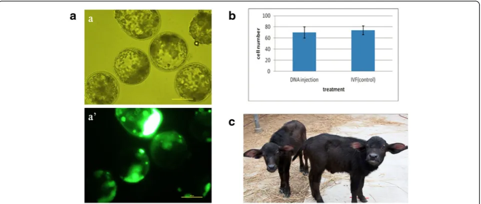
10 vs. 74 ± 8, P> 0.05, Fig. 1b). This indicated that transgene-expressing blastocysts were not compro-mised in terms of overall morphological quality.
Embryo transfer and detection of transgenic buffalo calves
To determine their in vivo viability and capacity for full term development, six EGFP-positive blastocysts were transferred into the uterine horns of four cross-bred buf-falo recipients (Table 3). One recipient was pregnant at D40 and two buffalo calves, one male and one female, were born on 2 December 2010 (Table 3, Fig. 1c). Since the GFP expression in embryos could reflect transient transcription of non-integrated DNA, both animals were analyzed for EGFP gene integration (Fig. 2a). Genomic DNA, extracted from ear biopsies of the two calves and a wild-type control calf, was used as a template. Both PCR and Southern blot specifically detected presence of the EGFP transgene in the two buffalo calves that devel-oped from EGFP-positive blastocysts (Fig. 2b, c). For single-copy integration, the minimal size of the resulting BamH I fragment recognized by the Southern probe
would be 3.7 kb. For multi-copy integrations (head-to-head, head-to-tail or mixed), the next bigger fragment size would be 3.7 + 1.0 = 4.7 kb. Multi-copy insertion would also result in increased signal intensity. The ob-served fragment sizes and signal intensities are consistent with head-to-head multi-copy and single-copy integration in the male and female calf, respectively (Fig. 2c).
We further used confocal laser scanning microscopy to directly observe EGFP expression in transgenic buf-falo ear tissue cultures. EGFP signal was specifically ob-served in all cells from presumptive transgenic primary tissue cultures, indicating that the randomly integrated transgene was expressed and functional (Fig. 2d).
The male and female transgenic calves were born after twin embryo transfer. In cattle, the majority of females from mixed-sex dizygotic twin pregnancies are freemar-tins [30]. This condition is due to exchange of cellular material and hormones between the vasculary connected twin placentas. As a result, over 90 % of female twins have abnormally masculinized reproductive organs and are infertile. This phenomenon also occurs in buffalo [31]. In our case, the female transgenic calf was diag-nosed as freemartin by ultrasound analysis, precluding it from subsequent mating. The male calf has so far not been mated and analyzed for transgene segregation in the offspring.
Conclusions
We demonstrate that injecting 20 ng/μL exogenous DNA into buffalo zygotes at 7–8 hpi reproducibly results in >15 % transgene-expressing blastocysts. This net effi-ciency compares favorably to studies in mice and other Fig. 1Characterization of EGFP-positive buffalo blastocysts.a. Microscopic evaluation by brightfield (a) and wide-field epifluorescence (a’); scale
bar =100μm.b. Nuclei numbers in EGFP-positive and non-injected IVF blastocysts.c. Mixed-sex twin buffalo calves born in December 2010
Table 3Embryo transfer summary
Recipient nET Pregnancies at D40 Calves born
1 1 0 0
2 2 2 2
3 1 1 0
4 2 0 0
livestock species, perhaps in part due to species-specific differences that affect stability and processing of exogen-ous DNA [32]. Using these optimized parameters, we produced the first transgenic buffalos from cytoplasmic injection. This technique would also be applicable to non-reporter transgenes, which could be detected on biopsies prior to embryo transfer [33, 34].
Abbreviations
IVM:In vitro maturation; IVF: In vitro fertilization; hpi: Hours post-insemination.
Competing interests
All authors declare that they have no conflict of interest that could inappropriately influence, or be perceived to influence, the submitted work.
Authors’contributions
FM executed the microinjection experiments, analyzed the data and drafted the manuscript. HL performed the PCR and Southern analyses, XW took confocal images, GQ performed the embryo transfers, BO analyzed the data, performed the statistical analysis and revised the manuscript. DS conceived the study and participated in its design. All authors read and approved the final manuscript.
Acknowledgements
We thank Dr Jingwei Wei for help with drafting the manuscript, Dr Stefan Wagner for help with Southern blot analysis and Drs Fenghua Lu and Qingyou Li for preparing the media and EGFP plasmids, respectively. This work was supported by the National Transgenic Project (2009ZX08007-009B, 2011ZX08007-003), Guangxi natural science funding (2012GXNSFCB053002) and funding of State Key Laboratory for Conservation and Utilization of Subtropical Agro-bioresources (KSL-CUSAb-2012-02). BO was supported by AgResearch core funding.
Received: 8 June 2015 Accepted: 29 September 2015
References
1. Laible G, Wei J, Wagner S. Improving livestock for agriculture–technological
progress from random transgenesis to precision genome editing heralds a
new era. Biotechnol J. 2015;10(1):109–20.
2. Wall RJ. Pronuclear microinjection. Cloning Stem Cells. 2001;3(4):209–20.
3. Clark J, Whitelaw B. A future for transgenic livestock. Nat Rev Genet.
2003;4(10):825–33.
4. Eyestone W. Production and breeding of transgenic cattle using in vitro
embryo production technology. Theriogenology. 1999;51(2):509–17.
5. Hammer RE, Pursel VG, Rexroad Jr CE, Wall RJ, Bolt DJ, Ebert KM, et al.
Production of transgenic rabbits, sheep and pigs by microinjection. Nature.
1985;315(6021):680–3.
6. Kubisch H, Hernandez-Ledezma J, Larson M, Sikes J, Roberts R. Expression of
two transgenes in in vitro matured and fertilized bovine zygotes after DNA
microinjection. J Reprod Fertil. 1995;104(1):133–9.
7. Kubisch H-M, Larson M, Funahashi H, Day B, Roberts R. Pronuclear visibility,
development and transgene expression in IVM/IVF-derived porcine
embryos. Theriogenology. 1995;44(3):391–401.
8. Oback B. Climbing mount efficiency–small steps, not giant leaps towards higher
cloning success in farm animals. Reprod Domest Anim. 2008;43 Suppl 2:407–16.
9. Hofmann A, Kessler B, Ewerling S, Weppert M, Vogg B, Ludwig H, et al. Efficient
transgenesis in farm animals by lentiviral vectors. EMBO Rep. 2003;4(11):1054–60.
10. Ritchie WA, King T, Neil C, Carlisle AJ, Lillico S, McLachlan G, et al. Transgenic
sheep designed for transplantation studies. Mol Reprod Dev. 2009;76(1):61–4.
11. McEvoy T, Coull G, Broadbent P, Hutchinson J, Speake B. Fatty acid
composition of lipids in immature cattle, pig and sheep oocytes with intact
zona pellucida. J Reprod Fertil. 2000;118(1):163–70.
12. Page RL, Butler SP, Subramanian A, Gwazdauskas FC, Johnson JL, Velander
WH. Transgenesis in mice by cytoplasmic injection of polylysine/DNA
mixtures. Transgenic Res. 1995;4(6):353–60.
13. Schmotzer CA, Butler SP, Pearson RE, Velander WH, Gwazdauskas FC. Assessment
of DNA Expression Following Cytoplasmic Microinjection of Condensed DNA
into Murine Embryos Using Electropulsation. Transgenics. 2003;4(1):55–63.
14. Brinster RL, Chen HY, Trumbauer ME, Yagle MK, Palmiter RD. Factors
affecting the efficiency of introducing foreign DNA into mice by
microinjecting eggs. Proc Natl Acad Sci U S A. 1985;82(13):4438–42.
15. Dunlap-Brown M, Butler SP, Velander WH, Gwazdauskas FC. Murine embryo
development following cytoplasmic injection of linear and condensed DNA.
Open J Anim Sci. 2012;2(4):244–52.
Fig. 2Characterization of transgenic buffalo calves derived from cytoplasmic zygote injection.a. Main features of injected pEGFP-N1 plasmid, showing restriction positions (in brackets), PCR primer binding sites (single arrows), Southern probe location (double block arrow) and expected
amplicon sizes. p = promoter, pA = polyA site, R = resistance;b,c. Detection of EGFP by PCR (b) and Southern blot (c) in genomic DNA extracted
from biopsied ear tissues of transgenic buffalo calves (male, female) and wild-type buffalo ear tissue (WT). Water provided a no template control.
d. Ear tissue sections from transgenic calves (male, female) and wild-type control calf (WT) observed by confocal laser scanning microscopy
16. Ma Y, Zhang X, Shen B, Lu Y, Chen W, Ma J, et al. Generating rats with
conditional alleles using CRISPR/Cas9. Cell Res. 2014;24(1):122–5.
17. Niu Y, Shen B, Cui Y, Chen Y, Wang J, Wang L, et al. Generation of
gene-modified cynomolgus monkey via Cas9/RNA-mediated gene
targeting in one-cell embryos. Cell. 2014;156(4):836–43.
18. Bevacqua R, Canel N, Hiriart M, Sipowicz P, Rozenblum G, Vitullo A, et al. Simple
gene transfer technique based on I-SceI meganuclease and cytoplasmic
injection in IVF bovine embryos. Theriogenology. 2013;80(2):104–13. e29.
19. Garrels W, Mátés L, Holler S, Dalda A, Taylor U, Petersen B, et al. Germline
transgenic pigs by Sleeping Beauty transposition in porcine zygotes and targeted integration in the pig genome. PLoS One. 2011;6(8), e23573.
20. Ivics Z, Garrels W, Mátés L, Yau TY, Bashir S, Zidek V, et al. Germline
transgenesis in pigs by cytoplasmic microinjection of Sleeping Beauty
transposons. Nature Protoc. 2014;9(4):810–27.
21. Li Z, Zeng F, Meng F, Xu Z, Zhang X, Huang X, et al. Generation of
Transgenic Pigs by Cytoplasmic Injection of piggyBac Transposase Based pmGENIE-3 Plasmids. Biol Reprod. 2014;90(5):93.
22. Vichera G, Moro L, Salamone D. Efficient transgene expression in IVF
and parthenogenetic bovine embryos by intracytoplasmic injection of
DNA–liposome complexes. Reprod Domest Anim. 2011;46(2):214–20.
23. Shi D, Avery B, Greve T. Effects of temperature gradients on in vitro
maturation of bovine oocytes. Theriogenology. 1998;50(4):667–74.
24. Yang CY, Pang CY, Yang BZ, Li RC, Lu YQ, Liang XW. Optimization of
cryopreservation of buffalo (Bubalus bubalis) blastocysts produced by in vitro fertilization and somatic cell nuclear transfer. Theriogenology.
2012;78(7):1437–45.
25. Iqbal K, Barg-Kues B, Broll S, Bode J, Niemann H, Kues W. Cytoplasmic
injection of circular plasmids allows targeted expression in mammalian
embryos. Biotechniques. 2009;47(5):959–68.
26. Wu GY, Wu CH. Receptor-mediated in vitro gene transformation by a
soluble DNA carrier system. J Biol Chem. 1987;262(10):4429–32.
27. Xu K, Greve T. A detailed analysis of early events during in-vitro fertilization
of bovine follicular oocytes. J Reprod Fertil. 1988;82(1):127–34.
28. McLay DW, Clarke HJ. Remodelling the paternal chromatin at fertilization in
mammals. Reproduction. 2003;125(5):625–33.
29. Liu L, Yang X. Interplay of maturation-promoting factor and mitogen-activated
protein kinase inactivation during metaphase-to-interphase transition of
activated bovine oocytes. Biol Reprod. 1999;61(1):1–7.
30. Padula AM. The freemartin syndrome: an update. Anim Reprod Sci.
2005;87(1–2):93–109.
31. Iannuzzi L, Di Meo GP, Perucatti A, Ciotola F, Incarnato D, Di Palo R, et al.
Freemartinism in river buffalo: clinical and cytogenetic observations.
Cytogenet Genome Res. 2005;108(4):355–8.
32. Powell DJ, Galli C, Moor RM. The fate of DNA injected into mammalian
oocytes and zygotes at different stages of the cell cycle. J Reprod Fertil.
1992;95(1):211–20.
33. Hyttinen JM, Peura T, Tolvanen M, Aalto J, Alhonen L, Sinervirta R, et al.
Generation of transgenic dairy cattle from transgene-analyzed and sexed
embryos produced in vitro. Biotechnology (N Y). 1994;12(6):606–8.
34. Chen SH, Vaught TD, Monahan JA, Boone J, Emslie E, Jobst PM, et al.
Efficient production of transgenic cloned calves using preimplantation
screening. Biol Reprod. 2002;67(5):1488–92.
Submit your next manuscript to BioMed Central and take full advantage of:
• Convenient online submission
• Thorough peer review
• No space constraints or color figure charges
• Immediate publication on acceptance
• Inclusion in PubMed, CAS, Scopus and Google Scholar
• Research which is freely available for redistribution