DOI 10.1007/s10152-002-0104-4
Abstract The photophores of Meganyctiphanes were in-vestigated with regard to the control of light production and with respect to their role in a hitherto unknown com-munication system using light flashes which became evi-dent from observation of specialised signalling behav-iour. To that purpose the light production was recorded during presentation of a range of stimuli delivered to the intact, tethered shrimp. Stimuli used were changes in ambient light, water turbulence, simulated predator ap-proach and light flashes, as well as electric shocks and serotonin injections. Strong negative light gradients, ex-aggerating the natural sunset signal, reliably elicited light production, the peak of which lasted on average 2 min. In the late phase of this light production, low fre-quency water oscillations and turbulent flow (assumed intraspecific communication signals at close range) elic-ited transient increases in light production. Artificial light flashes presented to a group of shrimp evoked a signalling behaviour in which the animal points the light of its photophore beamers (positioned at the ventral side and normally directed downwards) for a fraction of a second at observers within the same depth level. The re-sponses produced by the signalling behaviour indicate a fixed delay with respect to the triggering flash. Electric stimulation of the ventral nerve cord via implanted elec-trodes resulted in a strong light production with a latency of 160 ms. Injection of serotonin, resulting in haemo-lymph concentrations of 10–5 M and higher, initiated in-creasingly strong and inin-creasingly long-lasting continu-ous light production. Implications for the control of the photophores are discussed.
Keywords Euphausiacea · Meganyctiphanes · Bioluminescence · Serotonin · Control of photophores
Introduction
The present study investigates the control of the photo-phores of the euphausiid Meganyctiphanes norvegica (North Atlantic krill; Fig. 1). Our present knowledge of euphausiid photophores (Peterson 1968; for a review, see Herring and Locket 1978) is predominantly based on their morphology, while no information exists on their assumed role in intraspecific communication. In contrast, the lantern organs of luminescent insects stimulated re-search on structure, control (for a review, see Case and Strauss 1978), and on their role in the corresponding light flash communication system (for a review, see Lloyd 1971). A fairly comprehensive treatise on all as-pects of bioluminescence was published by Hastings and Morin (1991).
The lantern organs of the insects Photuris versicolor and Photinus greenii are innervated by segmental octo-paminergic nerve cells (Christensen and Carlson 1982), called DUM (dorsal unpaired medial) neurons. The re-ported strong effect of serotonin on light production in the photophores of Meganyctiphanes (Doyle and Kay 1967) suggests a corresponding aminergic innervation, since serotonin in the crustacean nervous system is com-monly assumed to represent the functional analogue of octopamine in the insect nervous system.
Potential candidates for the innervation of the photo-phores are serotonergic cells such as those identified by serotonin-like immunoreactivity (Beltz and Kravitz 1983; for a review, see Beltz 1999) in the ganglia of lobsters, in the crayfish ventral nerve cord (Real and Czternasty 1990) and in the crayfish brain (Sandeman et al. 1988, 1995), proved that such cells also exist in eu-phausiids. Serotonin may act as a regular, ionotrope transmitter like glutamate at the neuromuscular synapse of arthropods or it may affect the target cells via a sec-ond messenger system (metabotropic) – usually with longer latencies and longer lasting effects (Bermudez et al. 1992; Roeder 1994, 1999). Finally, serotonin was also found to act as a hormone, because it accelerates the heartbeat frequency in Astacus at bath-applied doses Communicated by F. Buchholz
T. Fregin · K. Wiese (
✉
)Zoologisches Institut und Zoologisches Museum der Universität, Martin-Luther-King-Platz 3, 20146 Hamburg, Germany
e-mail: kwiese@zoologie.uni-hamburg.de Fax: +49-40-428383937
O R I G I N A L A R T I C L E
Torsten Fregin · Konrad Wiese
The photophores of Meganyctiphanes norvegica (M. Sars)
(Euphausiacea): mode of operation
characteristic of hormones – as low as 10–8 M (Florey and Rathmayer 1978). Light and TEM microscopy re-vealed that the photophores of Meganyctiphanes are in-nervated (Peterson 1968; Harvey 1977; Herring and Locket 1978); however, the innervation is inconspicuous and it is not known whether the terminals contain seroto-nin.
In conclusion, three considerations motivated the present study of the photophores in Meganyctiphanes: firstly, the influence of serotonin on the process of light production possibly provides information on the func-tional role of the amine in the special context; secondly the photophores are part of an as yet uninvestigated system of intraspecific communication using light sig-nals; thirdly, the possibility of measuring light produc-tion by sensitive photomultiplier tubes in an intact shrimp may elucidate the origin of commands control-ling light production.
Materials and methods
Freshly caught shrimp M. norvegica (krill) were provided by the crew of the ships of Kristinebergs Marina Forskningsstation, Fiskebäckskil, Gulmarfjord, Sweden, where the experiments were performed. The shrimp, caught during daytime, arrived on shore in a 50-l thermos container at 0–5°C and, to provide optimal con-ditions, were directly transferred to a 1-m3tank (fibreglass,
paint-ed in light grey, non-reflecting, supplipaint-ed with flow-through seawa-ter at 8°C) in a constant temperature dark room. If the shrimp were not fed, after about 3 days the photophores could no longer be ac-tivated by normally effective triggering signals (see below). As a consequence, homogenised Mytilus were suspended in the tank water, which preserved the readiness of the shrimp to light the photophores for another few days. The shrimp had to be handled with extreme care in order to prevent the delicate flagella of the antennules from breaking off, which leaves the shrimp without sensitivity to flow events.
Triggering light production
Depending on the duration of the light period which the shrimp have experienced previously, a negative light gradient (dimming or switching off room lights) resulted in an activation of the pho-tophores with a latency of about 20 s. For measurements individu-al shrimp were preselected based on their readiness to activate the photophores. The IC-sensor TSL 235 (Texas Instruments) was used to measure irradiance. It has a sensitivity of 0.2 nW/cm2
and provides a square-wave frequency at the output, which is proportional to irradiance (1 kHz at the output corresponds to 1 mW/cm2). When room lights were on an irradiance of
60 µW/cm2was measured, in the condition with room lights off it
was 0.2 nW/cm2, the situation in which most photophore light
measurements were made. The spectral sensitivity of the eyes of
M. norvegica is centred around a maximum at 480 nm (Kampa
1955; Frank and Case 1988; for fireflies see Cronin et al. 2000).
Tethering the shrimp for measurement of light production
A cubelet (4×4×4 mm) cut from perspex and fitted with a 1.5 mm hole was glued (cyanoacrylate: Pattex-Sekundenkleber) to the ani-mal’s carapace, the hole in parallel with the long axis of the body. During the 30 s required for the glue to harden, the animal had to lie on its side in a Petri dish with only the ventral side and
swim-Fig. 1 A Meganyctiphanes norvegica Sars (Arctic krill), body
length about 40 mm. The skeleton is transparent and allows in-spection of most internal structures from the outside. The ante-nnular flagella in this specimen are damaged, showing about half the intact length. Note the large eyes, the folded filter basket formed from bristle-lined, double branched pereopods for sifting plankton, and the set of five, continuously and metachronically beating pairs of swimmerets. (Photograph by Torsten Fregin.)
B Meganyctiphanes, in tethered condition, as viewed ventrally
merets partly immersed in a film of water. The shrimp, immersed in seawater, was then attached to the tip of a toothpick fitted to a wooden stick, the other end of which was clamped above the cu-vette. By turning the cubelet around the toothpick, the ventral side of the shrimp with all ten photophores could be turned sideways to face the lens and photomultiplier tube (PMT). The temperature of the water in the experimental dish was kept identical to that in the storage tank to avoid stress to the “stenothermal” organism.
Injections of serotonin
Once the perspex cubicle had been glued to the carapace, freshly prepared solutions, not more than 20 µl mostly 5 µl in volume, of serotonin in distilled water were injected by Hamilton syringe into the pleon in a ventrocaudal direction. The volume of haemolymph within the shrimp was determined to be one-third of the water dis-placement (total body volume) of the shrimp (H. Dircksen, per-sonal communication).
Measurement of light from the photophores
A suitable photomultiplier tube type 1P28 [manufacturer, RCA; distributor, BURLE, Milwaukee, Wis., USA; maximum sensitivity at 350 nm, 50% of maximum sensitivity at 480 nm; the wave-length of 480 nm corresponds to the centre of the spectrum of light emitted by Meganyctiphanes: Widder et al. (1983)] was fitted into a light-proof housing, which contained a wheel of neutral density filters and a mechanical shutter. The voltage generated across the ends of the resistor (1–100 kohms was used), which connected the anode to ground was amplified by an inverting op-erational amplifier with variable gain (typically 30×). To reduce the high frequency noise of the anode current of the PMT and that of the signal amplifier, a low-pass filter was built by connecting the output of the signal amplifier (Ri = 80 ohms) to ground by a capacity of 1 microfarad (µF). The upper frequency limit under that condition was 2,000 Hz, calculated according to Flimit=1/2π RC.
Three arrangements were used to measure light production by the photophores:
1. The PMT fitted with a collimator lens was directed from the side onto a glass aquarium (1–2 m distance) holding a popula-tion of swimming shrimp, or from above (1 m distance) onto the shrimp swimming in the 1-m3tank.
2. A tethered swimming shrimp was rolled sideways so that the set of ten photophores faced the lens and PMT, positioned about 30 cm from the cuvette containing the shrimp.
3. A tethered swimming shrimp inside the experimental cuvette was positioned above the objective (10 mm working distance) of an inverted microscope. The posterior thoracal photophores or the most anterior photophore of the abdominal segments were closest to the attachment of the shrimp and hence moved very little in the visual field of the microscope. An optical beam-splitter allowed us to direct the light from the photo-phore onto the PMT attached to the microscope.
Water displacements
The shrimp were stimulated with water oscillations at frequencies of 3–150 Hz produced by a dipole source (sphere of 12 mm diam-eter) oscillating along an axis oriented frontodorsally and orthogo-nally onto the ventrally directed antennular flagellum. The sphere was pointed at the midpoint of the length of the flagellum. If 12 mm away from the centre of the sphere, this point on the fla-gellum receives water displacements attenuated by a factor 0.125 of the peak-to-peak amplitude of the sphere (van Bergeijk 1967). The oscillations, set to a uniform level of velocity at 1 mm/s at the flagellum, had controlled rising flanks (200 ms duration), formed by a simple device built from a flashlight bulb and a photoresistor (Wiese et al. 1980). Nearly laminar, as well as characteristically
turbulent flow signals were applied from a jet nozzle 5 cm away from the shrimp, directed frontally onto its head. Turbulence was generated by a piston moving up and down in a side branch of the tube close to the jet nozzle.
Visual stimuli
A light-emitting diode (LED) (470 nm) mounted at the end of a perspex tube of 6 mm diameter was positioned in the centre of the large tank (1 m3water volume) and driven by square-wave pulses
(5 V) to emit light flashes from inside the water body and hence directly from within the population of shrimp. Flash duration was varied between 1 and 10 ms.
Stimulation by electric shock
Electrodes were positioned to the right and left of the shrimp’s body in the water of the cuvette and were connected to a 4.5 V battery (zinc–carbon, a fraction of the voltage was used for a pro-tocol of the stimulus). The stimuli were controlled manually by push-button, leading to current flows of approximately 1 s dura-tion. Alternatively, a pair of lacquer-insulated stainless steel wires, the cut ends serving as electrodes, were implanted under micro-scopic control into a tethered shrimp (M. norvegica is almost com-pletely transparent) and positioned near the dorsal surface of the ventral cord within the first two abdominal segments of the shrimp. The stimulus voltage had to be very high (about 50 V) because of the very small electrode surfaces (80 µm diameter) and thin steel wires, which constitute in-series resistors (25 ohms/10 cm) and cause a voltage drop. Effective stimuli were trains (200 Hz repetition) of monopolar rectangular pulses, 0.5–1.0 ms in duration. Trains were produced by the gating mode of the stimulator.
Recording of data
Data were recorded using a notebook computer (Highscreen LeBook) fitted with A/D converter card and in connection with the program LabView (National Instruments, Houston, Tex., USA). Data were collected at a rate of 250 samples per second; only in experiments requiring very long periods of recording was the rate of sampling reduced to 80 samples per second. The dis-plays on the screen of the computer were edited based on the re-quirements of the particular measurements and were then printed.
Results
down-welling light by the posture control system (Land 1980) which detects a maximum of irradiation in the water to establish top and bottom (Lythgoe 1988). To summarise, the light of photophores is not visible unless the shrimp presents its ventral side to the observer.
In all our experiments the photophores were activated by strong, negative light gradients as observed by Mauchline (1960). The earliest light emissions in the 40-l aquarium became visible about 20 s after the onset of darkness, and lasted for 50–120 s. Essentially, a se-quence of light emissions (Fig. 2), each of about 1 s du-ration, were produced by some individuals of the popula-tion, which swim conspicuously in a fast and slightly curved trajectory. Others rested at the bottom of the aquarium and occasionally presented the light of their photophores for 10 or more seconds continuously. This behaviour raised the level of recorded light production significantly (see Fig. 2 with two phases of increased light level).
A large tank of 1×1 m floor size (used in the record-ings shown in the Figures) obviously provided the neces-sary space for a normal signalling behaviour of the shrimp. Either spontaneously or in response to a light signal received, a first shrimp started to swim forward in a curved trajectory of about 1 m length, at maximum speed, while simultaneously rolling around its
longitudi-nal body axis, thereby briefly presenting the ventral side and the light of the photophores to the observer. At the end of the trajectory, the shrimp engaged in a dodging sideways movement and then stopped. Only during pre-sentation, that is for less than a second, did the observer experience the impression of a flash; before and after this the light of the photophores was concealed. Since every signalling triggers a response from more than one conspecific, a sequence of “flashing” occurs which final-ly subsides after about 2 min if no other signals are pro-vided.
The flash seen by the observer was the result of a fast sweep of focused and continuously active light beams through the respective direction. Continuous light pro-duction during the triggered period of activity is shown in Fig. 3.
The shrimp was tethered in a cuvette, with the ventral side and photophores turned towards the PMT, which was about 30 cm away. During the measurement, the shrimp periodically flexed its abdomen somewhat. The movement shifted the optical axis of the photophores away from the PMT simultaneously, which resulted in strong amplitude modulation of the light recorded. Both shifts in pointing of photophores by the posture control system and by movements of the body need to be sub-tracted before changes in amplitude of light recorded can be attributed to the process of light production.
Single light flashes not produced by the rolling be-haviour, but instead probably following neural command only, is assumed to occur, because the recording of flash-ing activity in the big tank showed more flashes than
Fig. 2 Protocol of light flashing emerging from about 100
Meg-anyctiphanes inside an aquarium 35×35×60 cm in size. The photo-multiplier tube (PMT) fitted with a collimator lens records light events within the aquarium from 2 m distance. After the triggering event for light production in the photophores (switching off room lights), the recording is started (0 s), then the PMT power supply and signal amplifier are switched on and lastly a mechanical shut-ter is opened. The recording trace jumps up and shows noise. Flashes represented by small needle type peaks in the recording are generated by the signalling behaviour of the shrimp (see text) distorted here by the small size of the aquarium. The numerous shrimp with activated photophores generate a continuous glow in the aquarium, which elevates the DC-level of the recording. The three sharp peaks between 105 and 170 s of the recording are light signals of a light-emitting diode (LED) used in an attempt to cali-brate the light recorded
Fig. 3 Continuous light production in all ten photophores of a
could be attributed to individuals which were seen dis-playing the signalling behaviour. An appropriately de-signed experimental arrangement is required in order to prove this hypothesis.
The number of shrimps producing flash events was small compared with the total number of shrimps in the tank. However, a longer lights-on period before the trig-gering lights-off signal tended to increase the number of animals activating their photophores.
The light produced by the photophores during the normal, continuous mode of operation became invisible to human eyes if a background light stronger than 10–2µW/cm2was maintained.
Experimental analysis of photophore control
Signals of water displacement stimulate light production
Meganyctiphanes continuously uses the swimmerets at an average 6 Hz beat frequency to generate lift and pro-pulsion. In this functional context it generates a strong and characteristically modulated propulsion jet (Wiese and Ebina 1995; Markl 1983). Meganyctiphanes, adapt-ed to a life in continuous darkness, presumably re-cognises the characteristic turbulent flow produced by conspecifics. These considerations led us to test whether quantified 6 Hz water vibrations, applied to the antenn-ules, would have an effect on the light production of the photophores.
A 6 Hz water vibration of calculated 27 µm at the an-tennular flagellum of M. norvegica evoked increases in light production correlated with stimulus presentation with a latency of 0.5–1.0 s (Fig. 4). The figure also shows responses to two more water displacement and flow signals, which also led to activation of the photo-phores: water flow from a jet nozzle (modulated at 6 Hz and directed head-on to the antennules of a tethered shrimp) and a strong propeller (5 cm diameter) generated turbulence. Each of the tests was made following a tran-sition from light to darkness – towards the end of the triggered photophore activity. The consistently observed increases in light production in response to low frequen-cy water vibration depended on intact antennular flagel-la.
Visual stimuli increase light production
The specific pattern of ten photophores. The peculiar and characteristic pattern of the ten photophores on the ventral body surface of Meganyctiphanes (Fig. 1B, C) is resolved by arthropod eyes only from a few centimetres distance (see also Discussion).
Response to a model of an approaching predator. An in-crease in the size of a black square in the visual field of the tethered shrimp, mimicking the approach of a preda-tor, stopped the spontaneous pointing movements of the
photophores, which were resumed once the size of the black square was decreased again (Fig. 5). This reaction is tentatively interpreted as an attempt by the shrimp to hide the light of its photophores from the approaching enemy. Response of the photophores to natural and artificial light flashes. The PMT fitted with a collimator lens was
Fig. 4 Light production in the photophores in response to signals
of low frequency water turbulence of 6 Hz and of a calculated 27 µm (peak to peak displacement at a point at half the length of the antennular flagellum). The stimulus record (lower trace) uses the electric signal driving the dipole source (see Methods). Light production increases transiently (upper trace) whenever the 6 Hz signal is presented
Fig. 5 A model of an approaching predator (see Methods) inhibits
fluctuations in pointing of the photophores. The black cardboard square (see small inset at top left) was gradually exposed (the
low-er trace, which writes the stimulus, moves upward; noise is due to
pointed at an angle of 45° downwards into the centre of the 1-m3 tank with non-reflecting walls. About 70 shrimp inside the tank were adapted to light for 30 min before the room was darkened completely (see Meth-ods).
Figure 6A shows a PMT recording of light events in the tank at high temporal resolution and a sufficient sig-nal-to-noise ratio. A shrimp resting on the bottom of the tank with all photophores activated and pointing toward the PMT was probably responsible for the baseline shift in the light recording (Fig. 6A, right side). The recording showed a large number of peaks in the light protocol. Al-though the observation of the shrimps in the tank sug-gests that each flash stimulates flashes from other shrimps, call and response are impossible to discriminate in this constellation of recording. Differences in the am-plitude of the flashes recorded resulted from changes in the actual alignment of the photophore optics with the orientation of the PMT, as the shrimps presented their photophores at different locations in the tank.
In several cases, individual flash events seemed to be followed by flashes which often showed delays of be-tween 0.5 and 2.5 s. Attempts to correlate peaks in the re-cording with observed events in the tank tentatively led to the hypothesis that it is flashes produced by the signalling behaviour in response to flashes observed previously which generates the trailing peaks in the recording.
In Fig. 6B another scene of flashing is shown at low gain of the light recording. Nevertheless, the record shows at least four peaks (flashes, arrows) which are trailed by another (asterisks) at a delay of 1.5–2 s. A large number of shrimps with photophores alight were in the tank during this recording, raising the general light level considerably.
Figure 7A, B shows sample records of artificial and natural dialogue situations from the 1 m3 tank holding about 70 shrimps. Monopolar square pulses (5 V/1–10 ms) were used to drive an LED (470 nm) which was posi-tioned in midwater. Single and repeated artificial flashes were produced, identifiable in the recording by their sharply rising flank (indicated by arrows) in contrast to the response flashes produced by the shrimps, which have distinctly slower rise times. Again, in several cases the artificial flashes as well as the natural flashes are fol-lowed by small or large flash events produced by some of the shrimps in the tank. This was most obvious if the LED flashes were given only every 6 s and in the late phase of the 2-min photophore activity period (Fig. 7B). Under the condition of reduced dialogue activity, the re-ply of one or two shrimps directly in response to LED flashes was elicited. Again, the timing of the flash re-sponses showed a delay of 0.5–2.5 s after the leading flash.
Response of photophores to electric stimulation
Stimulation of the entire animal. Wire electrodes were positioned in the water to the right and left of the thorax
of the shrimp. Contact to a 4.5 V battery was made by a hand-operated pushbutton.
The recording shown in Fig. 8A indicates that the first shock produced a baseline shift in the light recording (Fig. 8A), interpreted as a shift in the photophore posi-tion relative to the optics of microscope and PMT. The shift was probably produced by a tail flip. After a latency
Fig. 6A, B Recording of light flash activity within a population of
Meganyctiphanes started by the triggering event. About 70 Meg-anyctiphanes populate a large 1-m3 rectangular tank with
of 1 s, a light production with slowly rising flank is ob-served. The irregular intervals and durations of the stim-ulus pulses reveal that the addressed “slow” light re-sponses are triggered by the onset and not by the end of the current pulse. The latency of the slow response changes from about 1 s for the first response to approxi-mately 0.5 s for the fifth response, indicating response sensitisation.
It is remarkable that for the second through to fifth stimulation a fast response – right at the onset of the cur-rent pulses – is observed in addition to the slow re-sponse. The latency of these fast responses amounts to about 20 ms. However, close examination has identified
this fast response as due to a jerk in the muscular system positioning the photophore.
Selective stimulation of the ventral nerve cord. A pair of insulated wire electrodes (steel wire, 80 µm diameter)
Fig. 7A, B Light-flash responses of Meganyctiphanes following
ar-tificial flashes produced by a blue LED (see Methods). The arar-tificial flashes (arrows) differ from the biogenic flashes by their shorter rise time. A An episode from an early phase of trigger-induced signal-ling, with three artificial flashes (thin arrows) interspersed. Note es-pecially the two shrimp-made intense flashes at 38–40 s after start of the recording. It is likely that the second constitutes the response to the first flash. The larger of the flashes produced by the shrimps are marked by triangles. B An episode from a late phase of trigger-induced signalling, showing that three artificial flashes (thin arrows, spaced 6 s apart) each evoke a flash response (marked by triangles) from one or two shrimp at 1–2 s of delay
Fig. 8 Fast and slow response in an individual photophore to electric
were implanted into the tethered shrimp and due to the transparency of M. norvegica were easily positioned next to the dorsal surface of the ventral nerve cord. A train of monopolar electric pulses of 1 ms duration, repeated at 200 Hz (designed to produce a strong stimulus) and of estimated 6 V net amplitude at the nerve (see Methods) regularly evoked flash events following the onset of the stimulus with a latency of 160–200 ms (Fig. 8B). The re-sponse to the second stimulus (applied only 6 s after the first) is strongly attenuated, whereas the third stimulus (again only 6 s after the second) nevertheless elicited a strong flash of 0.7 s duration. Please note that the third
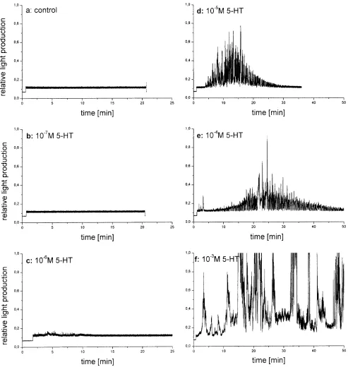
Fig. 9 A Photophore light production following injection of
sero-tonin into the haemolymph of the shrimp. Control (a): injection of 5 µl seawater; recordings b through f: injection of µl of serotonin dissolved in seawater calculated to produce the indicated concen-trations of serotonin (5-HT) in the haemolymph. Depending on the applied dosage, the duration (2 min to several hours) as well as the intensity (relative units: 1–20) of light production increase. Modu-lations of recorded light are due to changes in the alignment of photophores and PMT optics, caused by movements of the shrimp and the photophores. B Statistics of several trials: increasing con-centrations of 5 HT in the haemolymph of Meganyctiphanes in-crease the duration of continuous activity of the photophore. Note that 5-HT doses lower than 10–6M are not effective and that
Meganyctiphanes and to explore the recently observed elaborate signalling behaviour shown by Meganyctip-hanes when stimulated by a preceding light flash. Al-though we are only at the beginning of understanding this communication, the flashing of Meganyctiphanes is not a stress signal or produced to frighten predators, as seen in some copepods when they are caught in a net.
It was found that strong negative gradients of illumi-nation start a phase of light production which lasts on average for 100 s. During this time, light signals pro-duced by conspecifics as well as artificial light flashes evoke a distinct signalling behaviour during which a light-flash response is produced. With respect to the trig-gering flash, this light response is delayed by 1–2 s. The signalling includes protective measures to evade signal-intercepting predators. Ongoing submaximal light pro-duction was found to increase whenever stimuli similar to presumed conspecific communication signals (e.g. 6 Hz modulated turbulent water flow and light signals) were applied. Light flashes can be evoked by neural command, experimentally either by repetitive electric stimulation of the entire animal or by direct stimulation of the giant fibres and the ventral nerve cord. The short-est latencies of flashes generated were at 160 ms. Seroto-nin, highly effective in starting and maintaining light production, is only effective at haemolymph concentra-tions 10–5 M or more, indicating focused release at the site of action. The results of the study have encouraged us to investigate the function of serotonin in the control of light production in more detail.
On the biology of Meganyctiphanes
M. norvegica undergoes extended diel vertical migra-tions (Firth of Clyde: Mauchline 1960; Mauchline and Fisher 1969; Kattegat: Boysen and Buchholz 1984; Gulf of Maine: Frank and Widder 1997; Oslo Fjord: Onsrud and Kaartvedt 1998; Gulmarsfjord: Bergström and Strömberg 1997; Mediterranean Sea: Tarling et al. 1998), most commonly interpreted as attempts to evade visually hunting predators (Poulsen 1926). Most obvious during bright days, the shrimp assemble in the darkness of such deep waters (>100 m below the surface) that visually hunting fish presumably stay above the shrimp in order to remain oriented. At night, the shrimp prefer to re-as-cend to graze on plankton in the most productive water layer (about 20 m below the surface), which is character-ised by a strong temperature gradient (Bergström and Strömberg 1997). The response of Meganyctiphanes to low oxygen, temperatures higher than 5°C, often chang-es in salinity, and changchang-es in light conditions have been described (Tarling et al. 1999; van den Thillart et al. 1999; Spicer et al. 1999).
The facts that the individuals of Meganyctiphanes have to be assumed to often swim dispersedly, spend their life in continued darkness, and the considerable dis-tances covered during diel migration, require a system of intraspecific communication to make themselves noticed stimulus is very short, the elicited flash, however, has a
distinctly longer duration (compare Figs. 8B and 7A).
Effect of injected serotonin on light production by the photophores
Figure 9A, B shows the effects of different amounts of serotonin injected into the haemolymph of Meganyctip-hanes, which in this experiment were in normal posture and pointed the light of all photophores at the PMT posi-tioned below the cuvette from a distance of just 5 cm. Strong effects of the injected serotonin on the duration of light production as well as on the intensity of the light produced were observed if the concentration of the agent in the haemolymph was 10–5M or more. No interpreta-tion is as yet available of the rise and decay funcinterpreta-tions of light production during one experiment (Fig. 9Ad,e,f). The strong modulations of the light measured result from body movements, slight and total flexion of the abdomen and corresponding shifts of the optical axis of the photo-phores with respect to the PMT. The body movements become more and more erratic as the concentration of in-jected serotonin increases.
Following an injection – as a rule – all ten photopho-res of the shrimp started to produce light more or less si-multaneously. In only two cases, where the injection nee-dle was driven postrad into the abdominal musculature, the photophores of the eyestalks were activated first, fol-lowed by a slow progression of activation from the fron-tal photophores to the last ones in the pleon, this process taking an estimated 2 min to completion.
Discussion
The experiments reported here were aimed at investigat-ing the control of light production in the photophores of
and at least occasionally to detect the position of conspe-cifics in the surrounding water. The characteristically turbulent propulsion jet, about 10 cm/s at the start, produced by the shrimp for locomotion (Wiese and Marschall 1990; Wiese and Ebina 1995; Ebina and Miki 1995/1996) is a suitable and specific signal, however, it attenuates realistically to the sensory threshold (range 5 – 40 Hz: 100 µm/s; M.P. Partria, unpublished) within a range of 1 m (Schlichting 1982). Light flashes, on the other hand, are assumed to carry through 10 or more metres of clear water.
Signalling by light in a dark environment invariably attracts predators, i.e. fish, which need some light to see their prey. The shrimp, on the other hand, are likely to hide below the fish, because they are oriented to dark-ness for better protection. Due to continuous life in the dark they are likely to be better adapted to low light lev-els. The fact that the photophores are pointed downwards (Land 1980), if no signalling is intended, indicates that the light of photophores is hidden from predators lurking above the shrimp.
The behaviour of Euphausia superba, the Antarctic krill, which feed even during the day in the thermocline layer (Everson and Bone 1986a, b), is exceptional be-cause it is the result of an advanced social behaviour, swimming in formation, in which the individual has lost the normal measures to protect itself. This exceptional behaviour has led to the hypothesis that photophores are arranged ventrally and point away from the light to re-duce the shadow (by supplying light of the required quality), which the animal’s body produces against the lighter water surface if a predator looks at it from below (“countershading”, Clarke et al 1962; Warner et al 1979; Grinell et al 1988).
Specific signals of water movement increase light production in the photophores
Numerous interneurons of the ventral nerve cord of a crustacean are activated by water movement (Wiersma 1958). In addition to being directionally selective (head-ward/tailward: Wilkens and Larimer 1972; Wiese et al. 1976), the respective interneurons are tuned to respond to two frequency bands of water vibration (low pass 1–40 Hz and high pass 40–400 Hz), complemented by some interneurons activated by signals of both frequency ranges (Plummer et al. 1986). Low pass and high pass interneurons were found by these authors to be distin-guished by inhibitory contrast enhancement. Such inhibi-tory circuitry indicates that significant discriminations are made for the respective signal parameter (Wiese 1988). Bleckmann et al. (1991) and Bleckmann (1994) have pointed out that most spectra of biogenic water dis-placements do not contain frequencies higher than 40 Hz. In Meganyctiphanes, water vibrations and turbu-lent flow of a frequency content of 5–40 Hz, not only 6 Hz as shown in Fig. 4, were observed to increase the light production, whereas vibration above 40 Hz tended
to result in decreases. Detailed measurements are needed to prove and to characterise a corresponding selectivity of response.
The medial (MG) and lateral giant (LG) neurons of the crustacean ventral nerve cord are well known to be collectors of mechanosensory input to the nervous system. Action potentials only, not graded potentials, of the giant fibres equivalent to commands for escape be-haviour, are able to activate the serotonergic nerve cells of the ventral nerve cord of lobsters (Hörner et al. 1997). As the photophores are activated by mechanosensory in-put not only in escape situations, the stores of serotonin in charge of controlling the photophores are likely to have connection to mechanosensory interneurons inde-pendent from giant fibres. Water turbulence was similar-ly found to elicit bioluminescence in a copepod (Hartline et al. 1999).
The optics of the eyes of M. norvegica (Land et al. 1979) resolve the species pattern of photophores (Fig. 1B, C) which is identical in both sexes (Rickert, unpublished results) from not more than 12 cm distance (Kirschfeld, personal communication). Presentation of patterns made of blue LEDs indicate an effect on the si-multaneously recorded light production of the photopho-res. Experiments are required in order to test responses to changed patterns shown at close range.
Black objects in the visual field induced Euphausia superba to keep a large distance away from these (Strand and Hamner 1990). Presentation of a model of an ap-proaching predator was used in this study (Fig. 5). In re-sponse to a black square, which was made to increase in size, the tethered shrimp instantaneously arrested the on-going pointing movement of photophores, a corollary of the animal’s attempts to correct position in space, which is in disarray due to the tethering. The “freeze” reaction of the photophore movement is interpreted as an attempt of the shrimp not to betray its position to the approach-ing predator by the movement of, and perhaps resultapproach-ing traces of light from, the photophores.
The behaviour of the shrimp in response to natural and artificial light flashes leaves no doubt that the appar-ent flash is produced by a sweeping movemappar-ent of the photophores through the direction towards the observer.
A direct neural command for flashing needs to be in-vestigated in more detail. Perhaps such neural command is exclusively established in the photophores of the eye stalk, which differ from the other photophores by their construction, by their colour shade of the screening pig-ment and by their larger diameter.
A direct neural command for flashing is definitively established in the flash beetle (Case and Strauss 1978). The published work on the physiology of visual inter-neurons in the crayfish (Glantz 1977; Kondrashev 1993) does not include measurements of their response to light flashes and of the projections of these interneurons into the abdominal ventral nerve cord.
flashes (short delay of response is characteristic for males, longer delay of the response is characteristic for females) is one known method by which the flasher identifies him- or herself.
On the motor side, a delay of 1–2 s in the responses with respect to the triggering flash requires that Meg-anyctiphanes is capable of an instantaneous fast start and high speed of swimming and rolling at about 40 cm/s, as has been reported in Euphausia superba (Kils 1982).
The control of photophores
Strong negative light gradients are the most reliable trig-gers of light production in the photophores of Meganyc-tiphanes. However, imitations of the naturally occurring slower light gradients at dusk (Gulf of Maine, around sunset: 0.1–0.3% /s reduction of irradiation, Frank and Widder 1997) did not work as triggers. Tentatively, we suggest that 12 h of continuous daylight (and corre-sponding hours of darkness) may be required to render slow light gradients sufficiently strong to activate the photophores or – in another context – to generate Zeitgeber trigger signals in specialised parts of the visual pathway so as to set the internal clocks of the organism (Fleissner and Fleissner 1993).
Hormones (for hormonal control systems in crustaceans see Cooke and Sullivan 1983; Keller 1996), for example those released from the X-organ/sinus-gland complex (Mangerich et al. 1988; Dircksen 1992) in the eyestalk of the shrimp, were at first assumed to control photophore ac-tivity. Red pigment concentrating hormone (RPCH), pig-ment dispersing hormone (PDH) and crustacean cardioac-tive peptide (CCAP) were tentacardioac-tively injected; however, they had no consistent effect on the photophores.
Light production in M. norvegica usually started 20 s after the light gradient and lasted for about 2 min. It is striking that transmembrane currents, elicited by micro-jet-applied serotonin in isolated motoneuron somata of the locust have a similar temporal contour (Bermudez et al. 1992).
Injections of serotonin into Meganyctiphanes had at threshold to produce haemolymph concentrations 10–5to 10–3M to start and maintain light production in the pho-tophores. This fact indicates at a focused release from nerve endings close to the photocytes or similarly allo-cated cellular stores of serotonin in the control of photo-phores. Concentrations typical for hormonal control (e.g. of the salivary gland of Calliphora: 3–30 nM serotonin; Zimmermann and Walz 1999) are not effective.
Upon electric stimulation of the entire shrimp, a strongly sensitising, distinctly delayed response of slow-rising light production is observed (Fig. 8A). Stimulation of the giant fibres of the ventral nerve cord produced a light flash of about 1 s duration at a latency of 160 ms. The electric stimulations used thus far were not specific enough and need to be replaced in future experiments by more selective and lower amplitude stimuli. In particular, direct application of an electric stimulus to the
photo-phore cells or, better, the photocytes would help to com-pare effects with the situation described in the lantern or-gans of the insects Photuris und Photinus by Buck and Case (1961). If there is an analogy between both systems of luminescence, then we are led to hypothesise that neu-ral commands of unidentified interneurons in the ventneu-ral nerve cord stimulate already active light production to higher levels in a fashion similar to the tetanising effect of repeated electric stimuli on the contraction of skeletal muscles (lantern control in fireflies: Buck et al. 1963).
Acknowledgements This work was made possible by the
profes-sional support received from the staff of Kristinebergs Marina Forskningsstation, Fiskebäckskil, Gulmar Fjord, Sweden. The TMR program of the European commission contributed financial help to cover the cost of travel and accommodation. K.W. further acknowledges the hospitality received from Prof. Buchholz (aboard RV Heinke, Helgoland) and from Prof. Flügel (aboard RV Alkor, Kiel) who some years ago introduced him to North Atlantic krill. Dr. B. Schmitz, Zoology, TU Munich, has corrected the English.
References
Beltz BS (1999) Distribution and functional anatomy of amine-containing neurons in decapod crustaceans. Microsc Res Tech 44:105–120
Beltz BS, Kravitz EA (1983) Mapping of serotonin-like immuno-reactivity in the lobster nervous system. J Neurosci 3:365–402 Bergeijk W van (1967) Introductory comments on lateral line
function. In: Cahn P (ed) Lateral line detectors. Indiana Uni-versity Press, Bloomington, pp 73–81
Bergström B, Strömberg JO (1997) Behavioural differences in relation to pycnoclines during vertical migration of the euphausiids Meganyctiphanes norvegica (M. Sars) and
Thysanoessa raschii (M. Sars). J Plankton Res 19:255–261
Bermudez I, Beadle DJ, Benson J (1992) Multiple serotonin-acti-vated currents in isolated neuronal somata from locust thoracic ganglia. J Exp Biol 165:43–60
Bleckmann H (1994) Reception of hydrodynamic stimuli in aquat-ic and semiaquataquat-ic animals. Prog Zool 41:115–210
Bleckmann H, Breithaupt T, Blickhan R, Tautz J (1991) The time course and frequency content of hydrodynamic events caused by moving fish, frogs and crustaceans. J Comp Physiol A 168:749–757
Boysen E, Buchholz F (1984) Meganyctiphanes norvegica in the Kattegat. Studies on the development of a pelagic population. Mar Biol 79:195–207
Buck JB, Case JF (1961) Control of flashing in fireflies. I. The lantern as a neuroeffector organ. Biol Bull 121:234–256 Buck JB, Case JF, Hanson FE (1963) Control of flashing in
fire-flies. III. Peripheral excitation. Biol Bull 125:251–269 Case JF, Strauss LG (1978) Neurally controlled luminescent
sys-tems. In: Herring PJ (ed) Bioluminescence in action. Academ-ic Press, London, pp 331–365
Christensen TA, Carlson AD (1982) The neurophysiology of lar-val firefly luminescence: direct activation through four bifur-cating (DUM) neurons in Photuris versicolor. J Comp Physiol A 148:503–514
Clarke GL, Conover RJ, David CN, Nicol JAC (1962) Compara-tive studies of luminescence in copepods and other pelagic marine animals. J Mar Biol Assoc UK 42:541–564
Cooke IM, Sullivan RE (1983) Hormones and neurosecretion. In: Bliss DE (ed) The biology of crustacea, vol 3. Academic Press, London, pp 205–290
Dircksen H (1992) Fine structure of the neurohemal sinus gland of the shore crab Carcinus maenas and immuno-electron-micro-scopic identification of neurosecretory endings according to their neuropeptide contents. Cell Tissue Res 269:249–262 Doyle JD, Kay RH (1967) Some studies on the bioluminescence
of the euphausiids Meganyctiphanes norvegica and
Tysanoessa raschii. J Mar Biol Assoc UK 47:555–563
Ebina Y, Miki T (1995/1996) Range and biological significance of characteristic water currents produced by the shrimp
Euphausia superba and Metapenaeus intermedius. Zoology
99:163–174
Everson I, Bone DG (1986a) Detection of krill (Euphausia
sup-erba) near the sea surface: preliminary results using a towed
upward-looking echo-sounder. Br Antarct Surv Bull 72:61–70 Everson I, Bone DG (1986b) Effectiveness of the RMT 8 system
for sampling krill (Euphausia superba) swarms. Polar Biol 6:83–90
Fleissner G, Fleissner G (1993) Seeing time. In: Wiese K, et al. (eds) Sensory systems of arthropods. Birkhäuser, Basel, pp 288–306
Florey E, Rathmayer M (1978) The effects of octopamine and oth-er amines on the heart and on neuromuscular transmission in decapod crustaceans: further evidence for a role as neurohor-mone. Comp Biochem Physiol 61C:229–237
Frank TM, Case JF (1988) Visual spectral sensitivities of biolumi-nescent deep-sea crustaceans. Biol Bull 175:261–273
Frank TM, Widder EA (1997) The correlation of downwelling ir-radiance and staggered vertical migration patterns of zoo-plankton in Wilkinson Basin, Gulf of Maine. J Plankton Res 19:1975–1991
Glantz RM (1977) Visual input and motor output of command in-terneurons of the defense reflex pathway in the crayfish. In: Hoyle G (ed) Identified neurons and behavior in arthropods. Plenum Press, New York, pp 259–274
Grinell AD, Narins PM, Awbrey FT, Hamner WM, Hamner PP (1988) Eye/photophore coordination and light following in krill Euphausia superba. J Exp Biol 134:61–77
Hartline DK, Buskey EJ, Lenz PH (1999) Rapid jumps and biolu-minescence elicited by controlled hydrodynamic stimuli in a mesopelagic copepod, Pleuromamma xiphias. Biol Bull 197: 132–143
Harvey BJ (1977) Circulation and dioptric apparatus in the photo-phores of Euphausia pacifica. Can J Zool 55: 884–889 Hastings JW, Morin JG (1991) Bioluminescence. In: Prosser CL
(ed) Neural and integrative animal physiology: comparative physiology, 4th edn. Wiley, New York, pp 131–170
Herring PJ, Locket NA (1978) The luminescence and photophores of euphausiid crustaceans. J Zool 186:431–462
Hörner M, Weiger W, Edwards D, Kravitz EA (1997) Excitation of identified serotonergic neurons by escape command neu-rons in lobsters. J Exp Biol 200:2017–2033
Kampa EM (1955) Euphausiopsin, a new photosensitive pigment from the eyes of euphausiid crustaceans. Nature 175:996–998 Keller R (1996) Neurohormonale Systeme bei Invertebraten. In:
Dudel J, et al (eds) Neurowissenschaft. Springer, Berlin Hei-delberg New York, pp 243–260
Kils U (1982) The swimming behaviour, swimming performance and energy balance of Antarctic krill, Euphausia superba. Bio-mass, Scientific Research Series 3:1–121
Kondrashev SL (1993) Properties of visual neurons from crab op-tic ganglia. In: Wiese K, FG Gribakin FG, Popov AV, Rennin-ger G (eds) Sensory systems of arthropods. Birkhäuser, Basel, pp 145–158
Land MF (1980) Eye movements and the mechanisms of vertical steering in euphausiid crustacea. J Comp Physiol A 137:255– 265
Land MF, Burton FA, Meyer-Rochow VB (1979) The optical ge-ometry of euphausiid eyes. J Comp Physiol A 130:49–62 Landa SB, Drobchenko EA, Boshakov VYU (1993) Mechanism
of flash recognition in light communication of fireflies Luciola
mingrelica Coleoptera, Lampyridae. In: Wiese K, et al (eds)
Sensory systems of arthropods. Birkhäuser, Basel, pp 252–265
Lloyd JE (1971) Bioluminescent communication in insects. Annu Rev Entomol 16:97–122
Lythgoe JN (1988) Light and vision in the aquatic environment. In: Atema J, et al (eds) Springer, Berlin Heidelberg New York, pp 57–82
Mangerich S, Keller R, Dircksen H (1986) Immunocytochemical identification of structures containing putative red pigment concentrating hormone in two species of decapod crustaceans. Cell Tissue Res 245:377–386
Markl H (1983) Vibrational communication. In: Huber F, Markl H (eds) Neuroethology and behavioural physiology. Springer, Berlin Heidelberg New York, pp 332–353
Mauchline J (1960) The biology of the euphausiid
Meganyctip-hanes norvegica (M. Sars). Proc R Soc Edinburgh 67B:141–
179
Mauchline J, Fisher LR (1969) The biology of Euphausiids. Adv Mar Biol 7:1–421
Onsrud MSR, Kaartvedt S (1998) Diel vertical migration of krill
Meganyctiphanes norvegica in relation to physical
environ-ment, food and predators. Mar Ecol Prog Ser 171:209–219 Peterson G (1968) Studies on photophores in the Euphausiacea.
Sarsia 36:1–39
Plummer MR, Tautz J, Wine JJ (1986) Frequency coding of water borne vibrations by abdominal mechanosensory in interneu-rons in the crayfish Procambarus clarkii. J Comp Physiol A 158:751–764
Poulsen EM (1926) Om den store lyskrebs betydning som fiske-foed i Skagerak. Dan Fisk Tidsskr 24:286–289
Real D, Czternasty G (1990) Mapping of serotonin-like immuno-reactivity in the ventral nerve cord of crayfish. Brain Res 521:203–212
Roeder T (1994) Biogenic amines and their receptors in insects. Comp Biochem Physiol 107C:1–12
Roeder T (1999) Octopamine in invertebrates. Prog Neurobiol 59:353–361
Sandeman DC, Sandeman RE, Aitken AR (1988) Atlas of seroto-nin contaiseroto-ning neurons in the optic lobes and brain of the cray-fish Cherax destructor. J Comp Neurol 269:465–478
Sandeman RE, Watson AHD, Sandeman DC (1995) Ultrastructure of the synaptic terminals of the dorsal giant serotonin immu-noreactive neuron and deutocerebral commissure interneurons in the accessory and olfactory lobes of crayfish. J Comp Neu-rol 361:617–632
Schlichting H (1982) Grenzschicht-Theorie. Braun Verlag, Karls-ruhe, Germany, pp 754
Spicer JI, Thomasson MA, Strömberg JO (1999) Possessing a poor anaerobic capacity does not prevent the diel vertical mi-gration of Nordic krill Meganyctiphanes norvegica into hyp-oxic waters. Mar Ecol Prog Ser 185:181–187
Strand SW, Hamner WM (1990) Schooling behavior of Antarctic krill (Euphausia superba) in laboratory aquaria: reactions to chemical and visual stimuli. Mar Biol 106:355–359
Tarling GA, Matthews JBL, Saborowski R, Buchholz F (1998) Vertical migratory behaviour of the euphausiid
Meganyctip-hanes norvegica and its dispersion in the Kattegat Channel.
Hydrobiologia 375/376:331–141
Tarling GA, Matthews JBL, Buchholz F (1999) The effect of a lu-nar eclipse on the vertical migration behaviour of
Meganyctip-hanes norvegica (Crustacea: Euphausiacea) in the Ligurian
Sea. J Plankton Res 21:1475–1488
Thillart G van den, George RY, Strömberg J-O (1999) Hypoxia sensitivity and respiration of the euphausiid crustacean
Meg-anyctiphanes norvegica from Gulmar Fjord, Sweden. Sarsia
84:105–109
Warner JA, Latz MI, Case JF (1979) Cryptic bioluminescence in a midwater shrimp. Science 203:1109–1110
Widder EA, Latz MT, Case JF (1983) Marine bioluminescence spectra measured with an optical multichannel detector system. Biol Bull 165:791–810
Wiersma CAG (1958) On the functional connections of single units in the central nervous system of the crayfish
Wiese K, Wollnik F, Jebram D (1980) The protective reflex of
Bowerbankia (Bryozoa): calibration and use to indicate
move-ment below a capillary surface wave. J Comp Physiol A 137: 297–303
Wilkens LA, Larimer JL (1972) The CNS photoreceptor of cray-fish: morphology and synaptic activity J Comp Physiol 80: 389–407
Zimmermann B, Walz B (1999) The mechanism mediating regen-erative intercellular Ca2+waves in the blowfly salivary gland.
EMBO J 18:3222–3231 Wiese K (1988) The representation of hydrodynamic parameters
in the CNS of the crayfish Procambarus. In: Atema J, Fay RR, Popper AN, Tavolga WN (eds) Sensory biology of aquatic ani-mals. Springer, Berlin Heidelberg New York, pp 665–683 Wiese K, Ebina Y (1995) The propulsion jet of Euphausia
sup-erba (Antarctic krill) as potential communication signal
among conspecifics. J Mar Biol Assoc UK 75:43–54
Wiese K, Marschall HP (1990) Sensitivity to vibration and turbu-lence of water in context with schooling in Antarctic krill (Euphausia superba) In: Wiese K, et al (eds) Frontiers in crus-tacean neurobiology. Birkhäuser, Basel, pp 121–130